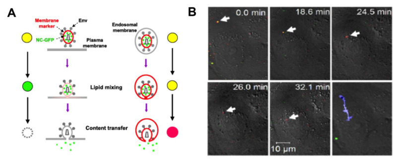
Figure 2.
Fluorescence microscopy imaging of viral fusion: (A) Schematic of doubly labeled HIV-1 viral particles entering into the cell by fusion with the plasma membrane (left) or with the endosomal membrane (right). (B) A complete fusion of HIV-1 (JRFL strain) with endosomal membrane is evidenced by the loss of the green fluorescence signal due to the content release, and the incorporation of the red membrane stain into the endosomal membrane. The blue trace on the last image represents the intracellular trajectory of a viral particle. (Reproduced with permission from [45], copyright 2009, Elsevier).

