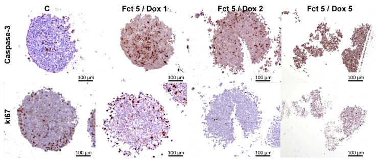
Figure 13.
Representative histological images of MCAs immunostained against caspase-3 and ki67 after exposure to fucosterol (Fct) at 5 µM in combination with doxorubicin (Dox) at 1, 2 and 5 µM. Cells treated with 0.1% DMSO correspond to the control (C). Brown color-diaminobenzidine (DAB) indicates positive staining.

