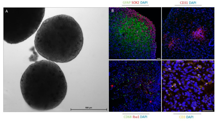
Figure 2.
Glioblastoma organoids after 4 weeks in culture preserve specific elements of the tumor microenvironment. (A) Phase-contrast image of glioblastoma organoids in culture. Scale bar: 500 µm. (B) Immunofluorescence staining of paraffin-embedded glioblastoma organoids for glioblastoma stem cell marker (SOX2), differentiated glioblastoma cell and astrocyte marker (GFAP), endothelial cell marker (CD31), macrophage marker (CD68), microglia marker (Iba1), and T cell marker (CD3). Cell nuclei were stained with DAPl (blue). Scale bars: 50 µm.

