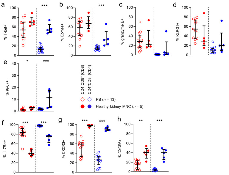
Figure 2.
CD8 and CD4 T cells in kidney tissue are nearly all CXCR3-positive.(a–h) Comparison of expression of T-bet (a), Eomes (b), granzyme B (c) KLRG1 (d), ki-67 (e), IL-7Rα (f), CXCR3 (g) and CXCR6 (h) in CD8 (red) and CD4 (blue) T cells derived from peripheral blood (PB) and healthy kidney. (median with IQR in black). Mann-Whitney U-test was used for statistical comparison. Only significant p-values are displayed: * p < 0.05, ** p ≤ 0.01, *** p ≤ 0.001.

