Abstract
TiO2 of controlled morphologies have been successfully prepared hydrothermally using TiO2 aggregates of different sizes. Different techniques were used to characterize the prepared TiO2 powder such as XRD, XPS, FEGSEM, EDS, and HRTEM. It was illustrated that the prepared TiO2 powders are of high crystallinity with different morphologies such as nanobelt, nanourchin, and nanotube depending on the synthesis conditions of temperature, time, and additives. The mechanism behind the formation of prepared morphologies is proposed involving nanosheet intermediate formation. Furthermore, it was found that the nanoparticle properties were governed by those of TiO2 nanoparticles aggregate used as a precursor. For example, the size of prepared nanobelts was proven to be influenced by the aggregates size used as a precursor for the synthesis.
Keywords: TiO2 nanoparticles, aggregates, morphologies
1. Introduction
Recently, tremendous efforts have been devoted to developing innovative strategies to synthesize nanomaterials with the desired morphologies and properties. Particularly the one-dimensional (1D) structure of TiO2 nanomaterials exhibits interesting properties compared to other TiO2 nanoparticles: it has lower carrier recombination rate and higher charge carrier mobility, thanks to the grain boundaries and junctions absence. In fact, the electron diffusion takes place through the junctions between nanoparticles, inducing slower charge transfer by several orders of magnitude [1]. In addition, it favors light scattering in the photoanode, which increases the light harvesting [2]. Among the studied morphologies and materials, semi conducting nanostructured materials such as nanowires, nanobelts, and nanotube have received particular attention, due to their use as photoanaode for potential applications in different areas such as photovoltaic [2], photo catalysis [3], gas sensing [4], and water photo-splitting [5].
Tuning the size and the morphology of materials is becoming a challenging goal in materials science. Over the past few years, various synthesis methods and protocols have been developed to control the semi-conducting nanomaterials morphology, including vapor–liquid–solid (VLS) [6], solution–liquid–solid (SLS) [7], template-based synthetic approaches [8,9], arc discharge [10], laser ablation [11], chemical vapor deposition [12], microwave [13,14], and sol–gel [15]. Among these synthesis methods, which mostly brought contamination to the synthesis products, the hydrothermal technique has been proven to be a simple and straightforward method using noncomplex apparatus, scalable for large production, with high chemical purity, allowing a large rang of nanomaterial sizes and morphologies [16,17]. Furthermore, the morphology of prepared TiO2 nanomaterials by using hydrothermal method was demonstrated to depend on the concentration of alkaline solution, the synthesis temperature and time, the material precursor used [17,18], additives, Pressure, pH, and the reaction medium [19,20,21,22,23,24,25].
Additionally, the hydrothermal method allows the control of the nanoparticles aggregation [26]. The most reported strategy to control the morphology of oxide nanomaterials is based on using organic surfactant, which adsorbs on a selected crystallographic plan of growing nucleus, leading to a change of its orientation and growth rate. This results in controlling the morphology of the obtained nanomaterial at the final growth stage [27,28]. Additionally, strategies based on aggregation/coalescence of nanomaterials were reported and demonstrated to be efficient in controlling the morphology of the final synthesized powder [29]. The exfoliation step was also reported to be a crucial step in the formation mechanism of prepared morphologies [29]. Most of the studies are based on nanomaterials aggregation/coalescence processes, and to the best of our knowledge, very few are based on exfoliation/aggregation/coalescence processes to explain synthesized morphologies. In the case of TiO2 nanomaterials, there is still a misunderstanding of the mechanism behind the formation of reported morphologies and particularly nanotube, nanobelt, and nanourchin. Some authors claimed that the Na2Ti3O7 nanosheets exfoliation step is the crucial step in the mechanism formation of different morphologies, whereas other authors stated that it is the dissolution of TiO2 nanoparticles into TiO6 octahedra, followed by Na2Ti3O7 nucleation and growth, forming a nanosheet in a later stage [29]. Furthermore, it is well accepted that different polymorphs of TiO2 nanomaterials are formed by different arrangements of TiO6 octahedra. In fact, the growth of anatase tetragonal polymorph proceeds through face sharing arrangements of TiO6 octahedras, whereas the rutile tetragonal phase growth takes place through edge-sharing arrangements. Furthermore, the Brookite phase is obtained by TiO6 octahedra assembly, sharing their edge and corner; whereas in Ti2O (B) (bronze) phase, Ti4+ ion form two distinct geometries with oxygen: octahedron in one case and a square pyramidal in the other. In addition, to homogeneous size and morphology, prepared TiO2 nanomaterials using hydrothermal method exhibit several characteristics such as high crystallinity, an accurate control of different crystallinity phases from anatase to rutile depending on the synthesis and annealing temperatures, and high specific surface [30]. It is well accepted that the anatase polymorph possesses a higher band gap energy (3.3 eV) than that of the rutile polymorph (3 eV).
In the present work, different morphologies of TiO2 have been successfully prepared hydrothermally using TiO2 aggregates made of TiO2 nanoparticles as a precursor. The mechanism behind the morphology control of prepared nanomaterials was discussed. It was found that the prepared TiO2 nanomaterials properties were governed by those of TiO2 nanoparticles aggregate. By controlling TiO2 nanoparticles and aggregate sizes, it has been demonstrated that it is possible to control the TiO2 nanobelt sizes.
2. Materials and Methods
2.1. Synthesis of TiO2 Nanoparticles
For the synthesis of TiO2 nanoparticles, titanium (IV) oxysulfate hydrate (TiOSO4, Sigma Aldrich, St. Louis, MO, USA) precursor was used. Furthermore, the synthesis of TiO2 aggregates has been performed using a hydrothermal synthesis technique. The TiOSO4 precursor solution was prepared by dissolving 6.4 g of TiOSO4 (2.5 M) in 16 mL of distilled water under constant stirring of 750 r/min and temperature of 45 °C for 2 h to get a clear solution. Then the solution of TiOSO4 was transferred into a Teflon-lined stainless-steel autoclave of 25 mL capacity. The heating rate was of 2.5 °C/min, and during the synthesis, the temperature was maintained at different temperatures of 100, 200 and 220 °C for 6 h depending on the aggregate size required. After this synthesis in autoclave, a white TiO2 powder was obtained and was washed six times in distilled water and two times in ethanol. Then the powder was dried overnight in the oven and annealed in air at temperature of 500 °C for 30 min with the heat rate of 5 °C/min. For nanourchin, nanotube, and nanobelt synthesis, 0.5 g powder of TiO2 aggregate was introduced in a Teflon-lined autoclave of 25 mL capacity. Then, the autoclave was filled with 10 M NaOH solution up to 80% of the autoclave capacity. During the synthesis, the temperature was maintained at different temperatures of 100, 150, and 220 °C with the heating rate of 2.5 °C/min and the synthesis time of 360, 180, and 15 min, depending on the required morphology. Afterwards, synthesis nanobelt particles are subjected to the washing and annealing protocols to obtain at the end of these processes: sodium titanate. The latter product was washed many times with diluted HCl solution to attain a pH value of 1. After that, the suspension was washed with distilled water several times to reach a pH value of 7. Finally, the obtained powder was dried overnight in the oven, and annealed in air at temperature of 500 °C for 30 min, with the heat rate of 5 °C/min.
All the chemicals are of analytical grade and used without further purification. The water used in all the experiments was purified by Milli Q System (Millipore, electric resistivity 18.2 MΩ.cm).
2.2. The Characterizations of TiO2 Films
The morphological investigations of the prepared films were achieved with a high-resolution Ultra 55 Zeiss FEG scanning electron microscope (FEGSEM) operating at an acceleration voltage of 10 kV and the high-resolution transmission electron microscope HRTEM using JEOL 2100 Plus microscope.
The crystalline structure of TiO2 was determined by an X-ray diffractometer (Siemens D5000 XRD unit) in 2θ range from 20° to 80° by 0.07°/s−1 increasing steps operating at 40 KV accelerating voltage and 40 mA current using Cu Kα radiation source with λ = 1.5406 Å.
The chemical compositions of all the samples were determined by the FEGSEM using a Princeton Gamme-Tech PGT, USA, spirit energy dispersive spectrometry EDS system, and by X-ray photoelectron spectroscopy XPS realized with X-ray photoelectron spectroscopy (XPS), and for the measurements we used a Thermo K Alpha analyzer system equipped with an AL Kα X-ray source (hυ = 1486.6 eV; spot size 400 μm).
3. Results and Discussion
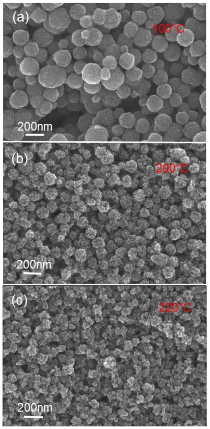
Various powders were prepared using the alkali hydrothermal synthesis method and varying synthesis temperatures and reaction times. To prepare these powders, TiO2 aggregates of spherical shape and different sizes were prepared and used as precursors. The FEG-SEM characterization of precursor powders are shown in Figure 1, and it can be observed that the sizes of spherical aggregates are ranging from 50 to 200 nm.
Figure 1.
FEGSEM images of TiO2 aggregates obtained at different synthesis temperatures: (a) 100, (b) 200, and (c) 220 °C.
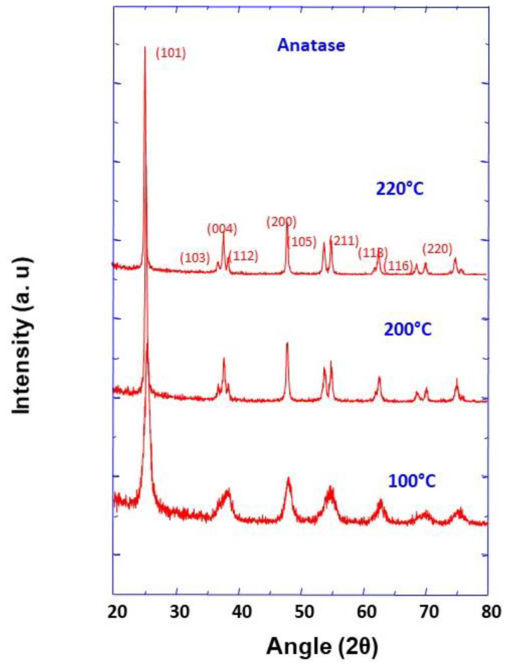
The XRD method was used to characterize the crystalline phase of TiO2 aggregate precursors, and the obtained results are depicted in Figure 2. Several well-resolved peaks were observed and are all assigned to TiO2 anatase phase (JCPDS No. 21-1272), which is proof of the high purity of the prepared precursor powders. Additionally, Scherer analysis was used to calculate the average crystallite sizes at the half-maximum width of the intense peak corresponding to (101) crystallographic plane, and were found to be 9.8, 24.7, and 30.4 nm, for the synthesis temperatures of 100, 200, and 220 °C, respectively.
Figure 2.
XRD pattern of TiO2 nanoparticle aggregates prepared at different synthesis temperatures as indicated.
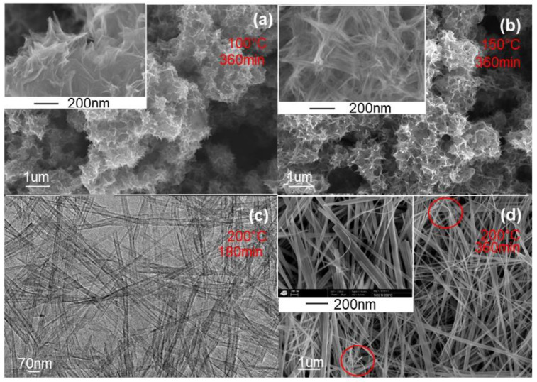
White powders were obtained using TiO2 aggregate precursors whatever the preparation conditions, and their corresponding morphologies are depicted in Figure 3. As it can be observed, at the synthesis temperature of 100 °C, the morphology of the prepared powder is nanourchin-like with a stretched sheet-like network (Figure 3a), whereas at a temperature of 150 °C, the morphology is still nanourchin-like but with a more rolled nanosheet-like network (Figure 3b). From these experiments, it is clear that the temperature increase favors the nanosheet scrolling. This could be explained by the fact that the crystallization enhanced by the temperature increase tends to induce the microstructure to change into rolled nanosheet structure. In fact, to reduce the surface energy of rolled structure, nanosheets reduce the defects and the distortion energy [31]. At a higher temperature of about 200 °C, the FEGSEM characterization of prepared white powder is depicted in Figure 3c,d. It can be observed that TiO2 powder is of nanobelt-like and nanotube morphologies, with monodisperse size. The insert of Figure 3d shows a sticking of several distinguishable nanobelts along their axis direction, forming bundles of nanobelts as a building unit. It can also be observed that their thickness is homogeneous and it is of about 10 nm, their diameter is ranging from 50 to 100 nm with length of around 10 mm. In addition, the nanobelt surface is smooth at the magnification scale, and no contamination was observed. As indicated in Figure 3d, some curved nanobelts were observed, which gives an indication about their high elasticity. From the described experiments, it is clear that the synthesis temperature is an important parameter in the morphology control of TiO2 nanomaterials.
Figure 3.
FEGSEM images of TiO2 nanoparticles with different morphologies obtained at different synthesis times and temperatures: (a) Nanourchin prepared at conditions of 100 °C and 360 min, (b) Nanourchin prepared at conditions of 150 °C and 360 min, (c) TEM image of Nanotube prepared at conditions of 200 °C and 180 min, and (d) Nanobelts prepared at conditions of 200 °C and 360 min.
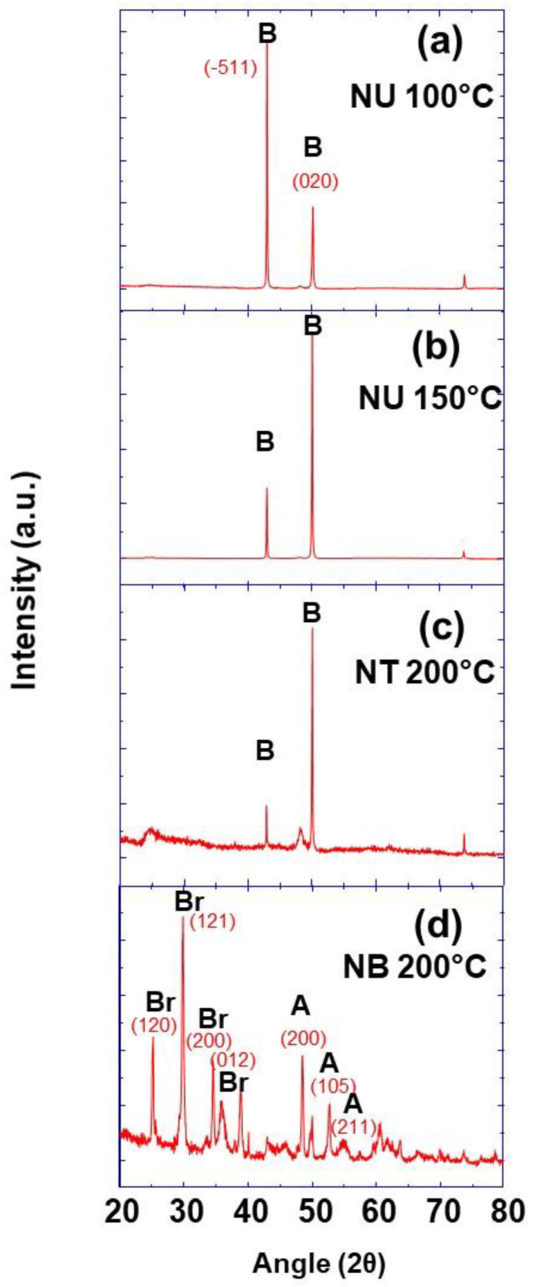
The crystalline structure and phase of prepared TiO2 nanobelt, nanotube, and nanourchin-like powders were studied by the X-ray diffraction method. The obtained XRD patterns are presented in Figure 4, and they show well-resolved peaks in the case of nanourchin and nanotube mophologies attributed to (-511) and (020) crystallographic planes of pure TiO2(B) phase (JCPDS No. 35-0088) (Figure 4a–c). In the case of TiO2 with nanobelt morphology, the observed XRD peaks indicates that the prepared powder is a mixture of anatase (JCPDS 21-1272) and brookite (JCPDS 29-1360) phases (Figure 4d).
Figure 4.
XRD pattern of TiO2 nanoparticles with different morphologies prepared at different synthesis temperatures as indicated, (a) nanoursin 100 °C, (b) nanoursin 150 °C, (c) nanotube 200 °C and (d) nanobelt 200 °C (Br: Brookite; A: Anatase; B: TiO2-B).
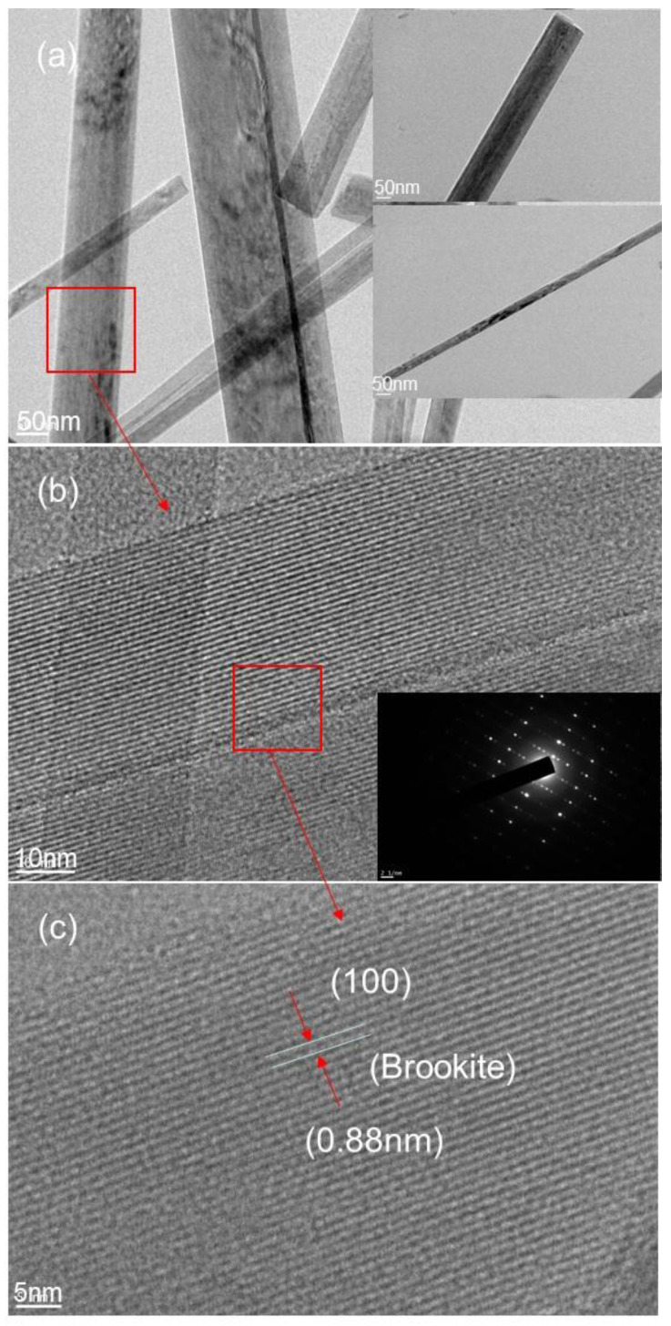
Additionally, among all the peaks, the most intense is the one corresponding to (121) crystallographic plane of brookite. Further details of crystallinity are provided by HRTEM depicted in Figure 5, clearly well resolved lattice planes are shown, and the insert electron diffraction shows well resolved spots (Figure 5b). These spots are the signature that the individual nanobelt is a single crystal. The interplanar distance of about 0.88 nm measured from HRTEM image is assigned to (100) crystallographic plane of brookite, indicating that the growth takes place along the (100) crystallographic plane, which is in good agreement with the result from XRD experiments in terms of brookite formation.
Figure 5.
(a) TEM images of TiO2 nanobelt obtained at synthesis temperature of 200 °C a synthesis time of 6 h at different magnifications; (b) the corresponding HRTEM showing inter atomic crystallographic planes and the insert show the corresponding electron diffraction; (c) another magnification of TiO2 nanobelt.
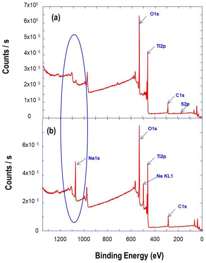
Furthermore, the chemical composition of the powder was provided by XPS analysis, and the obtained spectra are depicted in Figure 6. The XPS survey spectrum in Figure 6a of TiO2 aggregates precursor shows intense peaks corresponding to O1s and Ti2p core levels, and the very weak intensity of the peak corresponding to Na1s. However, the XPS survey spectrum corresponding to TiO2 nanobelt-like and nanourchin-like powders (Figure 6b) shows intense and well resolved peak, corresponding to the core level of Na1s, which is a signature of the formation of sodium titanate (Na2Ti3O7), in addition to those of O1s and Ti2p. It was reported that Na2Ti3O7 is constituted by corrugated strips of edge-sharing TiO6 octahedra [29]. The width of each strips is about three-octahedra, and they are connected through their corner to form stepped layers. Within the sticking layers, sodium cations are located at the positions between the layers.
Figure 6.
XPS survey spectra of prepared TiO2 (a) aggregate precursor (b) nanobelts after synthesis.
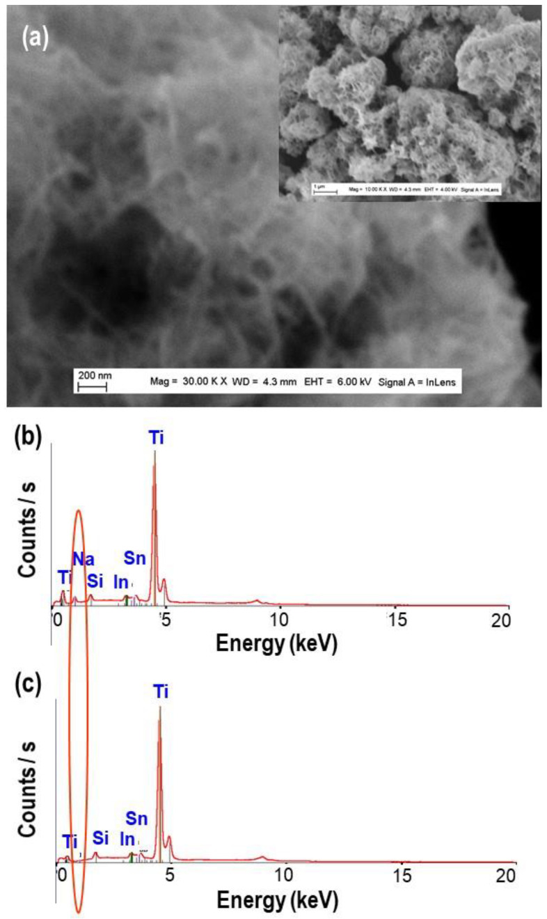
In Figure 7a, it is important to note that nanourchin-like nanoparticles show more enrolled nanosheet with more dense structure, as a consequence of the annealing process. The EDS analyses have been performed to determine the chemical composition of TiO2 nanoparticles, after just synthesis, or after washing and annealing processes. In Figure 7 the obtained EDS spectra are depicted; it should be noted that, on the EDS spectrum of TiO2 nanoparticles, after synthesis shows the presence of Na peak Figure 7b, whereas it is absent in the spectrum after the washing and annealing processes in Figure 7c. In fact, during the washing processes of Na2Ti3O7 by HCl, Na+ ions were exchanged by H+ ions. These results are a clear evidence of the important role played by Na+ ions in the formation of TiO2 nanobelts, nanotube, and nanourchin morphologies.
Figure 7.
(a) FEGSEM images of TiO2 nanourchin obtained at synthesis temperatures of 150 °C, after washing and annealing; (b,c) the corresponding EDS spectrum obtained just after synthesis and after washing and annealing processes, respectively.
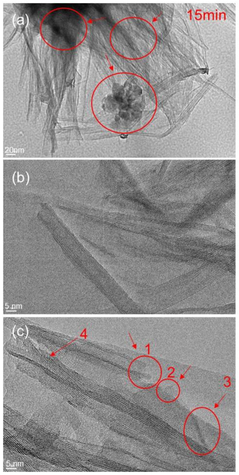
The details of TiO2 nanobelt and nanotube formation mechanisms are further investigated by using high resolution transmission electron microscopy (HRTEM). The influence of hydrothermal reaction time on the morphology of prepared TiO2 nanomaterials is studied at 15, 180, and 360 min. At short reaction time of about 15 min, the morphology of prepared powder is mainly stretched nanosheet-like, with some minor rolled sheet. Closer analysis of prepared powder (Figure 8) shows different stages of the same formation mechanism.
Figure 8.
HRTEM images with different magnifications (a–c) of TiO2 nanotube prepared at synthesis temperature of 200 °C and synthesis time of 15 min.
In fact, the observed nucleation stage can be considered as an integrated growth process of nanobelt structure, from aggregates made of nanoparticles of about 20 nm diameter to nanobelt of several micrometers in length. Similar evolution was observed by other authors [4,29]. Thus, we may assume that the morphologies shown in Figure 8 and Figure 9 represent different stages of the nanobelt growth process.
Figure 9.
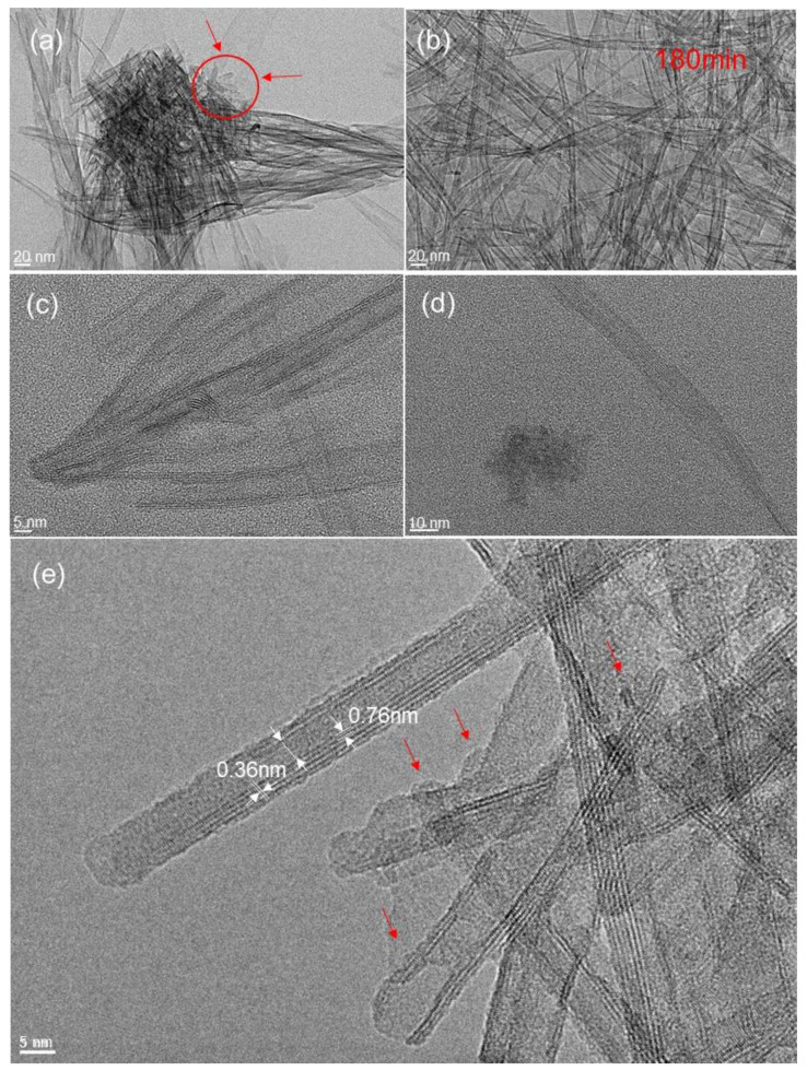
HRTEM images with different magnifications (a–e) of TiO2 nanotube prepared at synthesis temperature of 200 °C and synthesis time of 180 min.
It can be observed that at the earlier stage (reaction time of 15 min) of the nanobelt growth process, coalesced nanoparticles coexist with nanosheet like particles, indicated by zones in Figure 8a,c. Nanoparticles were located at the nanotube edges (region 1 and 2 in Figure 8c), and beside this simple attachment, an alignment of coalesced nanoparticles takes place (region 3 in Figure 8c). In addition, the nanosheet shows both stretched and rolled structures. The indicated region 4 in Figure 8c shows the starting process of nanosheet rolling. However, all these steps are a consequence of different nanobelt growth stages, which will evolve in a later stage to a nanotube structure observed in Figure 9 and nanobelt structure shown in Figure 10. However, at closer inspection of the nanosheet structure at an earlier stage, with a synthesis time of 15 min, we find that it presents an assembly of nanoparticles, whose sizes range from 5 to 20 nm, as indicated in selected region of Figure 8a,c. This proves that these nanoparticles and aggregates are the primary building units for the nanosheet formation process. Furthermore, it is well accepted in the literature that the key point for the formation of nanobelt-like structure is the formation of sodium titanate nanobelt intermediate, in which the sodium ion (Na+) is inserted into space between TiO6 octahedra layers, balancing their negative charges [4,29]. From the present experiments, it can be inferred that the aggregate of TiO2 nanoparticles split up into nanosheets as a consequence of Na+ insertion and their rolling in a second stage to form nanotube in an intermediate stage. Typical TEM and HRTEM patterns of TiO2 nanotube are depicted in Figure 9, with similar structure of nanotube obtained using TiO2 nanoparticles in terms of asymmetrical walls. It can be seen that the nanotube exhibits four layers on one side and two layers on the other (Figure 9e), which indicates that the nanotubes are formed by the scrolling of several layers of nanosheet, as previously observed by other authors. The interplane on both sides is of 0.36 nm, which corresponds to the (010) crystallographic plane, and is the characteristic of monoclinic H2Ti3O7. It was reported for the same materials that the nanotube growth takes place along the (010) direction. Additionally, the interlayer distance between rolled nanosheets is about 0.76 nm closer to different reported values [29].
Figure 10.
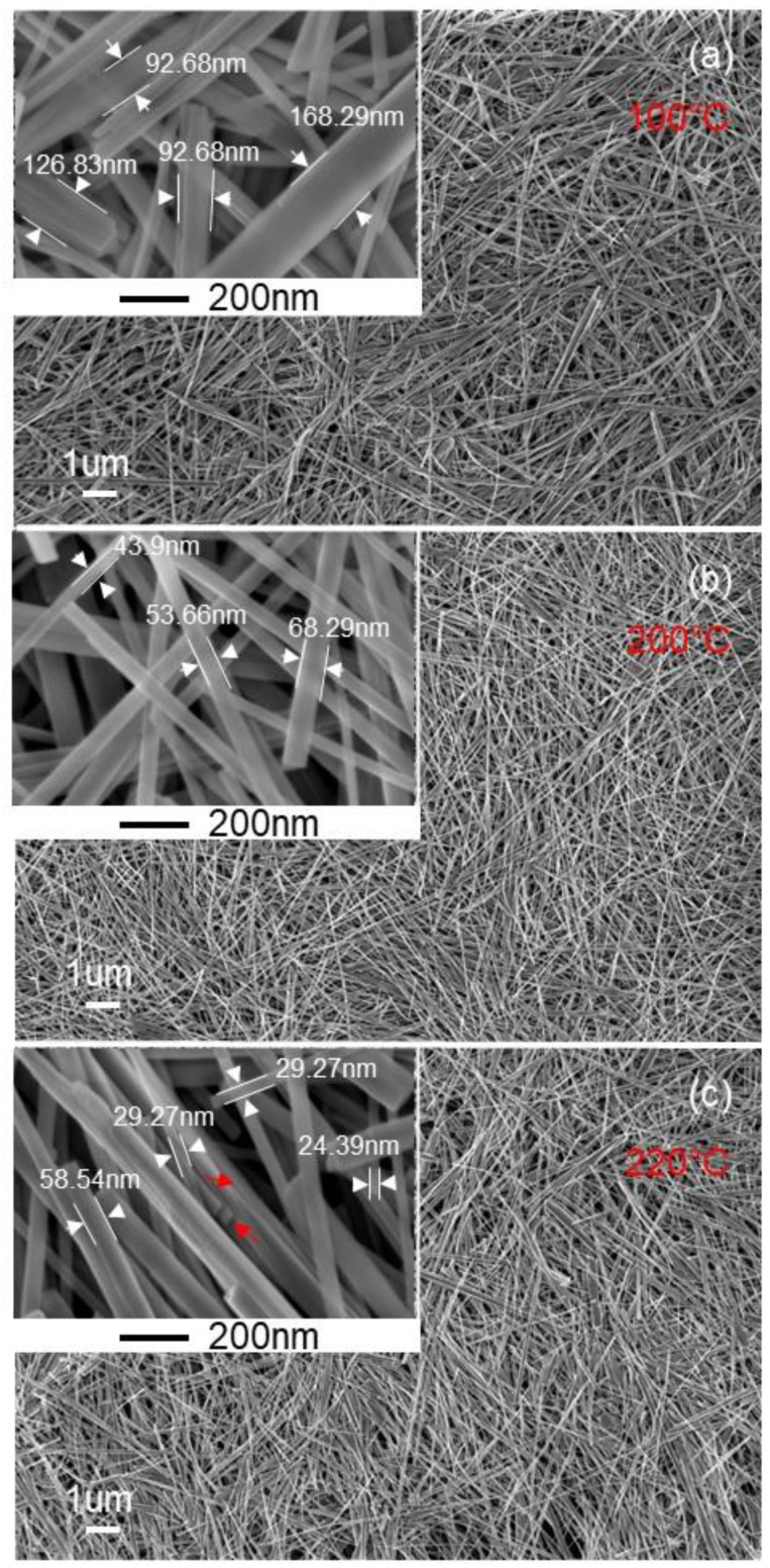
FEGSEM images of TiO2 nanobelts prepared at synthesis temperature of 200 °C and using TiO2 aggregate precursors of different sizes prepared at temperatures of (a) 100 °C, (b) 200 °C, and (c) 220 °C, respectively.
From XPS and EDX analysis in Figure 6 and Figure 7, it is clearly demonstrated that the sodium ions are incorporated in the TiO2 nanobelt, nanotube, and nanourchin, which suggest that it plays a role in their formation mechanisms. These observations indicate that nanobelts are formed by an orderly sticking of nanosheet and their coalescence in later stage; whereas nanourchins are formed by random assembly of the nanosheets.
The size dependence of the TiO2 nanobelt on the size of TiO2 aggregate precursor was demonstrated. Different sizes of TiO2 aggregate precursors were used to prepare TiO2 nanobelt, and the obtained results are depicted on Figure 10. It can be observed that the nanobelt length tends to increase with the increasing of the TiO2 aggregate precursor size. Additionally, the TiO2 nanobelt width increases from 50 to 200 nm (Figure 10), when the TiO2 aggregate precursor size increases from 50 to 200 nm (Figure 1). This confirms that TiO2 nanoparticles play a role in the formation of different observed morphologies. In fact, if we assume that the formation of observed morphologies goes through the TiO2 dissolution and precipitation, the TiO2 nanoparticles size will not have any effects on the final nanoparticle size. Additionally, the observation of TiO2 nanoparticles during the nanotube formation supports the mechanism through which sodium ions (Na+) induce exfoliation of TiO2 aggregates by insertion into the space between TiO6 octahedra layers and their coalescence to form nanosheets at later stage. Furthermore, the present results provide additional arguments to support some reported works in the literature and contradict others [30,32], in which it was claimed that during the hydrothermal synthesis process, TiO2 is dissolved through Ti–O–Ti bonds breaking and formation of sodium titanate nanosheet [29], which is converted to hydrogen titanate during the washing step and at a later stage to TiO2 nanobelt after the annealing process.
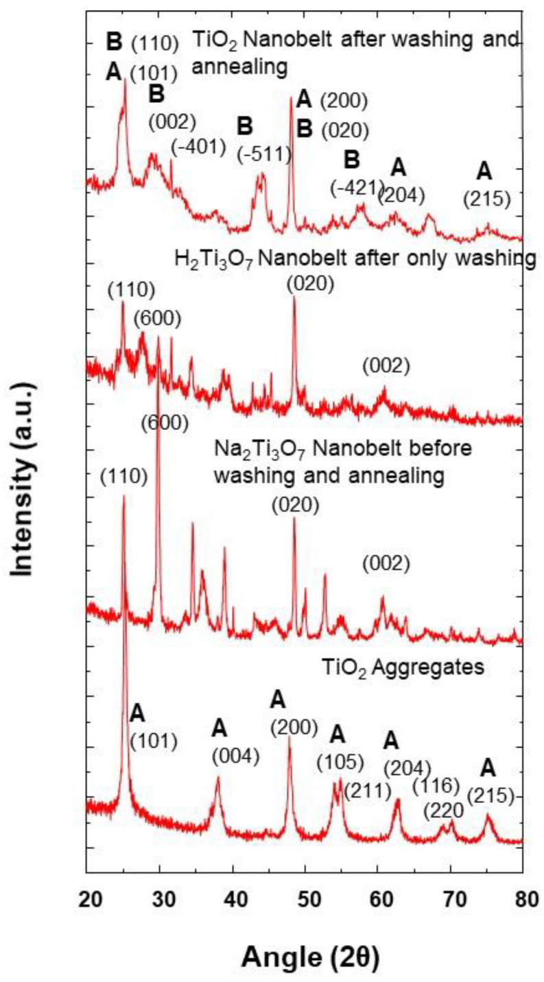
It can be seen from the XRD results that the nanobelt powder, at different synthesis stages (Figure 11), shows a changing of crystalline structure. The TiO2 aggregates precursor is of anatase phase, with tetragonal structure, in which TiO6 octahedra are sharing their face and get stacked in a one-dimensional zigzag chain. During the synthesis of Na2Ti3O7 nanobelts, a crystalline transition takes place, and TiO2 anatase phase is transformed into an orthorhombic structure. In fact, the formation of sodium titanate nanobelt intermediate is obtained through the insertion of sodium ion (Na+) into the space between TiO6 octahedra layers, inducing the distortion of the initial structure.
Figure 11.
XRD pattern of TiO2 nanobelt at different synthesis stages as indicated and at the synthesis temperature of 100 °C (A: Anatase; B: TiO2-B).
From these XRD results obtained at the synthesis temperature of 100 °C, it can be inferred that the anatase TiO2 aggregate structure changes are a consequence of Na+ insertion and a strong repulsion between Na+ ions, which induces a distortion of the anatase crystalline structure. Similar behavior is observed with the insertion of Na+ ion in the case of Na ion batteries charging/discharging cycles [33]. However, after the washing step with hydrochloric acid solution, the H2Ti3O7 nanobelts are obtained as a consequence of proton exchange processes of sodium trititanate. From Figure 11, it can be seen that this exchanging of steps and the resulting orthorhombic structure of H2Ti3O7 (JCPDS Card No. 47-0124) are accompanied by some XRD peak modifications, in terms of the intensity enhancement of some peaks, and their decrease for some others [34,35]. These modifications indicate the distortion of the initial structure after ion exchanges. Additionally, after the annealing process and the removal of protons, a mixture of anatase (JCPDS 21-1272) and TiO2-B (JCPDS 35-0088) phases is obtained at the synthesis temperature of 100 °C. The obtained XRD pattern is similar to that obtained for the same mixture by Beuvier et al. [36]. A phase transition was observed when the morphology changed from nanotube to nanobelt, but with different compositions than those obtained at the synthesis temperature of 200 °C. It was reported by Zhang et al. that the TiO2 nanoparticle size has a strong impact on the phase transformation during the growth of coalesced nanoparticles [37]. In addition, the temperature also plays an important role in the phase transformation of TiO2 nanoparticles [26]. However, as when the temperature is changed the coalescence and/or growth of TiO2 nanoparticles take place, both the temperature and the size contribute to the phase transformation and a formation of different phase mixtures depending on the used synthesis temperature 100 and 200 °C. Furthermore, as it can be observed from Figure 4 and Figure 10, the peaks corresponding to the anatase phase are of lower intensity, which indicates that both of the latter synthesis temperatures produce a lower proportion of anatase, in agreement with different reported works in the literature [36]. During the synthesis process at a given temperature, the phase is also changed due to the insertion of different ions, and it is not necessary to dissolve and precipitate TiO2 octahedra. Furthermore, from these results, it is worth noting that the synthesis temperature plays a crucial role in the phase control of prepared nanobelt powders.
4. Conclusions
Different morphologies of TiO2 nanoparticles have been synthesized, in a large scale using hydrothermal synthesis technique and TiO2 aggregate as a precursor. Both nanotube, nanourchin-like, and nanobelt-like nanoparticles were obtained at low temperatures and over short times. Furthermore, it is demonstrated that a morphology control of prepared TiO2 powders could be achieved through the tuning of the synthesis temperature and time. The mechanisms formation of TiO2 nanobelt-like, nanourchin-like, and nanotube nanoparticles are illustrated to involve TiO2 nanoparticles coalescence and nanosheet intermediate, formed thanks to Na+ ions exfoliation. Furthermore, it was found that the prepared TiO2 nanomaterials properties were governed by those of TiO2 nanoparticles aggregate. It has been demonstrated that it is possible to tune the nanobelt size by using different TiO2 aggregates precursor sizes. Additionally, it was shown that the synthesis temperature enables the tuning of the phase’s composition of the nanobelt powders. The investigation of prepared powders performance, as anode material for Li-ion batteries, is under progress in our group.
Acknowledgments
The authors would like to thank Pierre Dubot from Université Paris Est Créteil, CNRS, ICMPE, UMR 7182, 2-8 rue Dunant F-94320 Thiais, France; for the XPS experiments. The authors would like also to thank the Chinese Scholar Council for supporting W.L. with a scholarship.
Author Contributions
A.T. conceived and designed the experiment and also wrote the paper; W.L. performed experiments and also analyzed the corresponding data. All authors have read and agreed to the published version of the manuscript.
Funding
This research was funded by the European Union′s Horizon 2020 research and innovation program under the Marie Sklodowska-Curie grant agreement No. 734276.
Informed Consent Statement
Not applicable.
Conflicts of Interest
The authors declare no conflict of interest. The funders had no role in the design of the study; in the collection, analyses, or interpretation of data; in the writing of the manuscript; or in the decision to publish the results.
Footnotes
Publisher’s Note: MDPI stays neutral with regard to jurisdictional claims in published maps and institutional affiliations.
References
- 1.Fisher A.C., Peter L.M., Ponomarev E.A., Walker A.B., Wijiayantha K.G.U. Intensity Dependence of the Back Reaction and Transport of Electrons in Dye-Sensitized Nanocrystalline TiO2 Solar Cells. J. Phys. Chem. B. 2000;104:949–958. doi: 10.1021/jp993220b. [DOI] [Google Scholar]
- 2.Tan B., Wu Y. Dye-sensitized solar cells based on anatase TiO2 nanoparticle/nanowire composites. J. Phys. Chem. B. 2006;110:15932–15938. doi: 10.1021/jp063972n. [DOI] [PubMed] [Google Scholar]
- 3.Schneider J., Matsuoka M., Takeuchi M., Zhang J., Horiuchi Y., Anpo M., Bahnemann D.W. Understanding TiO2 Photocatalysis: Mechanisms and Materials. Chem. Rev. 2014;114:9919–9986. doi: 10.1021/cr5001892. [DOI] [PubMed] [Google Scholar]
- 4.Zhao Z., Tian J., Sang Y., Cabot A., Liu H. Structure, Synthesis, and Applications of TiO2 Nanobelts. Adv. Mater. 2015;27:2557–2582. doi: 10.1002/adma.201405589. [DOI] [PubMed] [Google Scholar]
- 5.Sun B., Shi T., Peng Z., Sheng W., Jiang T., Liao G. Controlled fabrication of Sn/TiO2 nanorods for photoelectrochemical water splitting. Nanoscale Res. Lett. 2013;8:1–8. doi: 10.1186/1556-276X-8-462. [DOI] [PMC free article] [PubMed] [Google Scholar]
- 6.Trentler T.J., Hickman K.M., Goel S.C., Viano A.M., Gibbons P.C., Buhro W.E. Solution-liquid-solid growth of crystalline III-V semiconductors: An analogy to vapor-liquid-solid growth. Sci. 1995;270:1791. doi: 10.1126/science.270.5243.1791. [DOI] [Google Scholar]
- 7.Wang F., Dong A., Buhro W.E. Solution–Liquid–Solid Synthesis, Properties, and Applications of One-Dimensional Colloidal Semiconductor Nanorods and Nanowires. Chem. Rev. 2016;116:10888–10933. doi: 10.1021/acs.chemrev.5b00701. [DOI] [PubMed] [Google Scholar]
- 8.Martin C.R. Nanomaterials: A membrane-based synthetic approach. Sci. 1994;266:1961–1966. doi: 10.1126/science.266.5193.1961. [DOI] [PubMed] [Google Scholar]
- 9.Xu G.R., Wang J.N., Li C.J. Template direct preparation of TiO2 nanomaterials with tunable morphologies and their photocatalytic activity research. Appl. Surf. Sci. 2013;279:103–108. doi: 10.1016/j.apsusc.2013.04.043. [DOI] [Google Scholar]
- 10.Choi Y.C., Kim W.S., Park Y.S., Lee S.M., Bae D.J., Lee Y.H., Kim J.M. Catalytic Growth of β-Ga2O3 Nanowires by Arc Discharge. Adv. Mater. 2000;12:746–750. doi: 10.1002/(SICI)1521-4095(200005)12:10<746::AID-ADMA746>3.0.CO;2-N. [DOI] [Google Scholar]
- 11.Morales A.M., Lieber C.M. A laser ablation method for the synthesis of crystalline semiconductor nanowires. Science. 1998;279:208. doi: 10.1126/science.279.5348.208. [DOI] [PubMed] [Google Scholar]
- 12.Duan X., Lieber C.M. General synthesis of compound semiconductor nanowires. Adv. Mater. 2000;12:298–302. doi: 10.1002/(SICI)1521-4095(200002)12:4<298::AID-ADMA298>3.0.CO;2-Y. [DOI] [Google Scholar]
- 13.Li L., Qin X., Wang G., Qi L., Du G., Hu Z. Synthesis of anatase TiO2 nanowire by modifying TiO2 nanoparticles using the microwave heating method. Appl. Surf. Sci. 2011;257:8006–8012. doi: 10.1016/j.apsusc.2011.04.073. [DOI] [Google Scholar]
- 14.Wang H.E., Zheng L.X., Liu C.P., Liu Y.K., Luan C.Y., Cheng H., Bello I. Rapidmicrowave synthesis of porous TiO2 spheres and their applications in dye sensitized solar cells. J. Phys. Chem. C. 2011;115:10419–10425. doi: 10.1021/jp2011588. [DOI] [Google Scholar]
- 15.Crippa M., Callone E., D’Arienzo M., Müller K., Polizzi S., Wahba L., Scotti R. TiO2 nanocrystal grafted on macroporous silica: A novel hybrid organic-inorganic sol-gel approach for the synthesis of highly photoactive composite material. Appl. Catal. B. 2011;104:282–290. doi: 10.1016/j.apcatb.2011.03.018. [DOI] [Google Scholar]
- 16.Poudel B., Wang W.Z., Dames C., Huang J.Y., Kunwar S., Wang D.Z., Ren Z.F. Formation of crystallized titania nanotubes and their transformation into nanowires. Nanotechnology. 2005;16:1935–1940. doi: 10.1088/0957-4484/16/9/086. [DOI] [Google Scholar]
- 17.Asiah M.N., Mamat M.H., Khusaimi Z., Abdullah S., Rusop M., Qurashi A. Surfactant-free seed-mediated large-scale synthesis of mesoporous TiO2 nanowires. Ceram. Int. 2015;41:4266. [Google Scholar]
- 18.Cui L., Hui K.N., Hui K.S., Lee S.K., Zhou W., Wan Z.P., Thuc C.N.H. Facile microwave-assisted hydrothermal synthesis of TiO2 nanotubes. Mater. Lett. 2012;75:175–178. doi: 10.1016/j.matlet.2012.02.004. [DOI] [Google Scholar]
- 19.Byrappa K., Adschiri T. Hydrothermal technology for nanotechnology. Prog. Cryst. Growth Charact. Mater. 2007;53:117–166. doi: 10.1016/j.pcrysgrow.2007.04.001. [DOI] [Google Scholar]
- 20.Yu J., Su Y., Cheng B., Zhou M. Effects of pH on the microstructures and photocatalytic activity of mesoporous nanocrystalline titania powders prepared via hydrothermal method. J. Mol. Catal. A Chem. 2006;258:104–112. doi: 10.1016/j.molcata.2006.05.036. [DOI] [Google Scholar]
- 21.Mamaghani A.H., Haghighat F., Lee C.S. Role of titanium dioxide (TiO2) structural design/morphology in photocatalytic air purification. Appl. Catal. B Environ. 2020;269:118735. doi: 10.1016/j.apcatb.2020.118735. [DOI] [Google Scholar]
- 22.Hoang S., Guo S., Hahn N.T., Bard A.J., Mullins C.B. Visible light driven photoelectrochemical water oxidation on nitrogen modified TiO2 nanowires. Nano Lett. 2012;12:26–32. doi: 10.1021/nl2028188. [DOI] [PubMed] [Google Scholar]
- 23.Pan X., Zhao Y., Liu S., Korzeniewski C.L., Wang S., Fan Z. Comparing graphene-TiO2 nanowire and graphene-TiO2 nanoparticle composite photocatalysts. ACS Appl. Mater. Interface. 2012;4:2944–3950. doi: 10.1021/am300772t. [DOI] [PubMed] [Google Scholar]
- 24.Chen J.Z., Ko W.Y., Yen Y.C., Chen P.H., Lin K.J. Hydrothermally processed TiO2 nanowire electrodes with antireflective and electrochromic properties. ACS Nano. 2012;6:6633–6639. doi: 10.1021/nn300787r. [DOI] [PubMed] [Google Scholar]
- 25.Kasuga T., Hiramatsu M., Hoson A., Sekino T., Niihara K. Formation of titanium oxide nanotube. Langmuir. 1998;14:3160–3163. [Google Scholar]
- 26.Taleb A., Mesguich F., Hérissan A., Colbeau-Justin C., Yanpeng X., Dubot P. Optimized TiO2 nanoparticle packing for DSSC photovoltaic applications. Sol. Energy Mater. Sol. Cells. 2016;148:52–59. doi: 10.1016/j.solmat.2015.09.010. [DOI] [Google Scholar]
- 27.Wang Y.D., Zhang S., Ma C.L., Li H.D. Synthesis and room temperature photoluminescence of ZnO/CTAB ordered layered nanocomposite with flake-like architecture. J. Lumin. 2007;26:661. doi: 10.1016/j.jlumin.2006.10.018. [DOI] [Google Scholar]
- 28.Wang H.X., Li X.X., Tang L. Effect of surfactants on the morphology and properties of TiO2. Appl. Phys. A. 2020;126:448. doi: 10.1007/s00339-020-03638-8. [DOI] [Google Scholar]
- 29.Wu D., Liu J., Zhao X., Li A., Chen Y., Ming N. Sequence of events for the formation of titanate nanotubes, nanowire and nanobelts. Chem. Mater. 2006;18:547–553. doi: 10.1021/cm0519075. [DOI] [Google Scholar]
- 30.Mamaghani A.H., Haghighat F., Lee C.S. Photocatalytic oxidation technology for indoor environment air purification: The state-of-the-art. Appl. Catal. B Environ. 2017;203:247–269. doi: 10.1016/j.apcatb.2016.10.037. [DOI] [Google Scholar]
- 31.Ding L., Chen J., Dong B., Xi Y., Shi L., Liu W., Cao L. Organic macromolecule assisted synthesis of ultralong carbon&TiO2 nanotubes for high performance lithium-ion batteries. Electrochem. Acta. 2016;200:97–105. [Google Scholar]
- 32.Diebold U., Ruzycki N., Herman G.S., Selloni A. One Step towards Bridging the Materials Gap: Surface Studies of TiO2 Anatase. Catal. Today. 2003;85:93–100. doi: 10.1016/S0920-5861(03)00378-X. [DOI] [Google Scholar]
- 33.Massaro A., Munoz-Garc A.B., Maddalena P., Bella F., Meligrana G., Gerbaldi C., Pavone M. First-principles study of Na insertion at TiO2 anatase surfaces: New hints for Na-ion battery design. Nanoscale Adv. 2020;2:274. doi: 10.1039/D0NA00230E. [DOI] [PMC free article] [PubMed] [Google Scholar]
- 34.Yao B.D., Chan Y.F., Zhang X.Y., Zhang W.F., Yang Z.Y., Wang N. Formation mechanism of TiO2 nanotubes. Appl. Phys. Lett. 2003;82:281. doi: 10.1063/1.1537518. [DOI] [Google Scholar]
- 35.Wang W., Varghese O.K., Paulose M., Grimes C.A., Wang Q., Dickey E.C. A study on the growth and structure of titania nanotubes. J. Mater. Res. 2004;19:417. doi: 10.1557/jmr.2004.19.2.417. [DOI] [Google Scholar]
- 36.Beuvier T., Richard-Plouet M., Mancini-Le Granvalet M., Brousse T., Crosnier O., Brohan L. TiO2(B) Nanoribbons as negative electrode material for lithium ion batteries with high rate performance. Inorg. Chem. 2010;49:8457–8464. doi: 10.1021/ic1010192. [DOI] [PubMed] [Google Scholar]
- 37.Zhang H., Banfield J. Understanding polymetric phase transformation behavior during growth of nanocrystalline aggregates: Insghts from TiO2. J. Phys. Chem. B. 2000;104:3481–3487. doi: 10.1021/jp000499j. [DOI] [Google Scholar]


