Abstract
Plants can be regenerated from various explants/tissues via de novo shoot meristem formation. Most of these regeneration pathways are indirect and involve callus formation. Besides plant hormones, the role of polyamines (PAs) has been implicated in these processes. Interestingly, the lateral root primordia (LRPs) of Arabidopsis can be directly converted to shoot meristems by exogenous cytokinin application. In this system, no callus formation takes place. We report that the level of PAs, especially that of spermidine (Spd), increased during meristem conversion and the application of exogenous Spd improved its efficiency. The high endogenous Spd level could be due to enhanced synthesis as indicated by the augmented relative expression of PA synthesis genes (AtADC1,2, AtSAMDC2,4, AtSPDS1,2) during the process. However, the effect of PAs on shoot meristem formation might also be dependent on their catabolism. The expression of Arabidopsis POLYAMINE OXIDASE 5 (AtPAO5) was shown to be specifically high during the process and its ectopic overexpression increased the LRP-to-shoot conversion efficiency. This was correlated with Spd accumulation in the roots and ROS accumulation in the converting LRPs. The potential ways how PAO5 may influence direct shoot organogenesis from Arabidopsis LRPs are discussed.
Keywords: Arabidopsis thaliana, hydrogen peroxide, polyamines, polyamine oxidase, reactive oxygen species, direct shoot regeneration, spermidine
1. Introduction
De novo organogenesis from somatic plant tissues occurs both in nature or in vitro, either directly or indirectly through callus formation [1]. During these processes, explants or calli first form ectopic apical meristems, which subsequently develop into shoots or roots, respectively. Among the plant hormones, auxin and cytokinin are the most important to regulate plant morphogenesis including organ formation. Based on the recognition that high auxin to cytokinin ratios promote root while high cytokinin to auxin ratios shoot formation, the establishment of in vitro plant regeneration systems were elaborated for hundreds of plant species. In the model plant, Arabidopsis thaliana, shoot regeneration is usually achieved via indirect organogenesis from root explants [2,3,4]. In recent years, it has been recognized that callus formation is not required for de novo shoot formation from root tissues and the direct conversion of lateral root primordia (LRP) to shoot meristem can take place in response to cytokinin application [5,6,7]. Three successive phases of this process could be distinguished [7]. First, exogenous cytokinin transiently pauses cell division in the LRPs (mitotic pause) and the regulators of shoot development start to be expressed. This is followed by the conversion phase in which the root meristem is converted into a shoot promeristem with a characteristic cell division pattern. Finally, the promeristem matures and develops into a fully functional shoot meristem.
Auxin and cytokinin interact with other plant growth regulators, including polyamines (PAs) during several physiological and developmental processes [8,9,10]. PAs are highly reactive polycations. The three most common PAs in plants are the diamine putrescine (put), the triamine spermidine (Spd), and the tetramine spermine (spm), although other PAs, such as cadaverine and thermospermine (t-spm), are also present in plants [11]. Several studies indicated that PA metabolic and catabolic pathways are linked to plant growth and development [12]. Among others, polyamines have been reported to be involved in a variety of morphogenic processes such as organogenesis and somatic embryogenesis [13,14,15,16].
In plants, Put is synthesized by two pathways: either directly from ornithine via ORNITHINE DECARBOXYLASE (ODC; EC 4.1.1.17) or indirectly from arginine via ARGININE DECARBOXYLASE (ADC; EC 4.1.1.19). Interestingly, in Arabidospsis thaliana there is no ODC and only the ADC-catalyzed pathway contributes to Put biosynthesis [17]. S-ADENOSYLMETHIONINE DECARBOXYLASEs (SAMDCs) catalyze the formation of an important intermediate of spermidine and spermine biosynthesis, the decarboxylated S-adenosylmethionine (dSAM). The Arabidopsis double mutant samdc1 samdc4/bud2 not expressing two SAMDC isoenzymes is embryo lethal, while the single samdc4/bud2 mutant has a bushy dwarf phenotype demonstrating that SAMDCs are essential for plant development and morphogenesis [18]. It was found that the bud2 mutation results in auxin and cytokinin hypersensitivity suggesting that polyamines may interfere with these plant hormones regulating plant growth [19]. Moreover, t-Spm has recently been identified as a novel plant growth regulator (PGR) which represses xylem differentiation and promotes stem elongation [8,20,21,22]. T-Spm is synthesized by thermospermine synthase ACAULIS5 (ACL5) and acl5 is allelic to thickvein (tkv), which displays defective auxin transport [23,24]. Furthermore, t-Spm potently suppresses the expression of genes involved in auxin signaling, transport, and synthesis [25].
Besides PA synthesis, PA catabolism is also connected to cellular signaling during developmental processes via the generation of H2O2 [26]. In cotton, PAs and their catabolic product H2O2 were found to be essential for the conversion of embryogenic calli into somatic embryos [27]. Polyamine catabolism is mediated mainly by two classes of amine oxidases. One of them is DIAMINE OXIDASE (DAO) which oxidizes mainly Put and the other is the apoplastic POLYAMINE OXIDASE (PAO), which preferentially oxidizes Spd and Spm but not Put yielding aminoaldehydes and H2O2 [28,29]. In cotton somatic embryogenesis, the H2O2 concentration augmented during the formation of embryogenic calli in parallel with the increased expression of GhPAO1 and GhPAO4 and the overall PAO activity [27].
In addition to their terminal oxidation, PAO enzymes catalyze the backconversion of tetraamine to triamine and/or triamine to diamine [9,30]. In the Arabidopsis genome, there are five PAO genes (AtPAO1–AtPAO5) [30]. Two of them (AtPAO1 and AtPAO5) are localized in the cytoplasm and involved in the catabolism/backconversion of Spd, Spm and t-Spm. AtPAO1 and AtPAO5 both catalyze the backconversion reaction of Spm and t-Spm to Spd. However, AtPAO5 unlike the other Arabidopsis PAOs including AtPAO1 has a dehydrogenase rather than oxidase activity [9,31]. Therefore, it was hypothesized that AtPAO5 exerts its regulatory role during plant growth and xylem differentiation maintaining polyamine homeostasis rather than increasing hydrogen peroxide production [8,9]. AtPAO5 has a high affinity for t-Spm and it was hypothesized to contribute to the tightly regulated interplay of auxin and cytokinin during xylem differentiation and plant growth via controlling the t-Spm level [8,9]. The remaining three AtPAOs of Arabidopsis (AtPAO2, AtPAO3, and AtPAO4) have a peroxisomal localization and can oxidize/backconvert both Spd and spm, but not t-Spm [30,32,33,34,35].
Although, the involvement of polyamines in indirect organogenesis was investigated in several studies, the impact of their regulatory role on direct organogenesis has not been shown yet. In this study, our aim was to investigate the involvement of PAs and polyamine metabolism in the direct conversion of lateral root primordia into shoot meristems.
2. Materials and Methods
2.1. Plant Materials and Culture Conditions
Arabidopsis thaliana wild type (WT) plants of the ecotype Columbia (Col-0) were used along with AtPAO2 (35S:PAO2) and AtPAO5 transgenic plants (35S::PAO5) previously described [8,9,30,31]. To induce direct organogenesis the method of Rosspopoff et al. [7] was applied with minor modifications. All plants were grown in a climate-controlled cabinet using 8/16 (light/dark) photoperiod and a constant temperature of 21 °C with an irradiance of 50 µmolm−2s−1 provided by white fluorescent tubes (Sylvania Luxline Plus; Feilo Sylvania Europe Limited, London, UK). Surface-sterilized seeds (70% ethanol for 60 s followed by immersion in 4% commercial sodium hypochlorite solution (having 4.5% active chlorine) for 10 min) were sown and grown for 6 days on solid medium containing full-strength MS (Murashige and Skoog Medium including B5 vitamins, Duchefa Biochemie, Haarlem, The Netherlands), 1% sucrose (Molar Chemicals, Halásztelek, Hungary), 0.6 % agarose (Electran DNA pure grade for electrophoresis: VWR International LLC, Radnor, PA, USA), 0.5 g/L 2-(N-morpholino)ethanesulfonic acid (MES) (Duchefa Biochemie). To induce lateral root (LR) initiation 3.3 µM naphthaleneacetic acid (NAA) (Duchefa Biochemie) priming was applied for 43 h. For synchronization of LR initiation, 1.25 µM 2,3,5-triiodobenzoic acid (TIBA) (Fluka, Chemie GmbH, Buchs, Switzerland) was added to the germination medium before the NAA treatment. To induce the conversion of lateral root primordia (LRP) into a functional shoot meristem (SM), seedlings were transferred onto and cultured on a medium (SM medium) containing full-strength MS salts (Duchefa Biochemie), 2% D(+)-glucose (Molar Chemicals), 0.6 % agarose (Electran DNA pure grade for electrophoresis: VWR International LLC, Radnor, PA, USA) and 8.16 µM isopentenyl-adenine (IPA) (Sigma-Aldrich, St. Louis, MO, USA). Stock solution of spermidine and putrescine, respectively, was prepared in Milli-Q® H2O, filter sterilized with Millex® GV syringe filter (0.22 µm), (Merck Millipore, Burlington, MA, USA) and added to the SM medium at 100 µM final concentration. For sample collection, roots of seedlings were used after 48, 72 and 96 h cytokinin induction (at the 96th hour of induction the seedlings were ≈12 days old). As absolute control, roots of 6-days-old, nontreated seedlings were used. For gene expression analysis, samples were harvested and snap-frozen in liquid N2 and they were stored at −80 °C until usage.
2.2. Gene Expression Analysis
For total RNA extraction Quick-RNA Miniprep Kit (Zymo Research, Irvine, CA, USA) was used which includes the removal of contaminating genomic DNA. NanoDrop™ 2000/2000c spectrophotometer (Thermo Fisher Scientific, Waltham, MA, USA) was used to evaluate the quality and quantity of total isolated RNA, considering the ideal absorbance ratio (1.8 ≤ A260/280 ≤ 2.0). In total, 250–300 ng of total RNA was reverse-transcribed for 60 min at 42 °C and for 10 min at 75 °C in a 20 μL reaction volume using RevertAid First Strand cDNA Synthesis Kit (Thermo Fisher Scientific) according to the manufacturer’s instructions. cDNA products were diluted 1:10 in AccuGENE® water (Lonza, Verviers, Belgium). Primers were designed using Primer 3 software [36] and synthesized by Biocenter Ltd. (Szeged, Hungary). Primer sequences are shown in Supplementary Table S1. Primer sequences were analyzed using OligoAnalyzerTM Tool (Integrated DNA Technologies, Inc., Coralville, IA, USA) and National Center for Biotechnology Information (NCBI) programs (Bethesda (MD): National Library of Medicine (US), National Center for Biotechnology, 1982). Relative mRNA levels were determined by real-time quantitative PCR (RT-qPCR). As reference genes, UBIQUITIN 1 (At3G52590) and PP2A3 (At1G13320)) were used. These genes were selected using the Arabidopsis Regeneration eFP browser at The Bio-Analytic Resource for Plant Biology (bar.u-toronto.ca; [37]) allowing the in silico analysis of transcriptomic data sets of root-to-shoot regeneration experiments. According to these data, the At3G52590 and At1G13320.1 genes have constitutive expression during the process. The RT-qPCR reactions were carried out by the qTOWER 2.0 (Analytic Jena AG, Life Science, Jena, Germany) and CFX384 Touch Real-Time PCR Detection System (Bio-Rad Laboratories Inc., Hercules, CA, USA). Depending on the detection system, the PCR mixture contained (in a total volume of 7 or 14 μL) 1 or 2 μL cDNA, 0.21 or 0.42 μL forward primer, 0.21 or 0.42 μL reverse primer, 3.5 or 7 μL Maxima SYBR Green/ROX qPCR Master Mix (2×) (Thermo Fisher Scientific). Reaction mixture was aliquoted to 96-well plates (non-skirted, white; Thermo Fisher Scientific, Cat no: AB-0600/W) or Hard-Shell® 384-well plates (thin-wall, skirted, clear/white; Bio-Rad, Cat. no: HSP3805). For amplification, a standard two-step thermal cycling profile was used (10 s at 95 °C and 1 min at 60 °C) during 40 cycles, after a 15 min preheating step at 95 °C. Finally, a dissociation stage was added with 95 °C for 15 s, 60 °C for 15 s and 95 °C for 15 s. Data analysis was performed using qPCRsoft (Analytic Jena, AG), Bio-Rad CFX Maestro (Bio-Rad) software and Microsoft Excel 2010. The relative mRNA levels normalized to the average of AT3G52590 and AT1G13320.1 mRNAs were calculated using the (2)−ΔΔCt method. The mRNA level of the initial root tissue was used as control (relative mRNA level: 1). All tested amplification efficiencies were in a narrow range and were not used in the data normalization. Data were averaged from three independent biological experiments with three technical replicates for each gene/sample combination.
2.3. Light Microscopy
The number of regenerated plantlets on seedling roots were determined using an Olympus SZX12 stereo dissection microscope (Olympus Corporation, Sindzsuku, Tokyo, Japan). For the bright field images, white LED light source (Photonic Optics, Vienna, Austria) was used. Photos were captured using an Olympus Camedia C7070 digital camera and the DScaler software (version 4.1.15).
2.4. In Situ Detection of Reactive Oxygen Species
For in situ detection of ROS, 2,7-dichlorodihydrofluorescein diacetate (H2DC-FDA, Sigma-Aldrich, St. Louis, MO, USA) was applied [38,39]. Seedling roots were incubated in 10 μM H2DC-FDA solubilized in 2-Nmorpholine-ethansulphonic acid/potassium chloride (MES/KCl) pH 6.5 for 15 min at room temperature in darkness. After staining, seedling roots were washed once with a dye-free buffer and the fluorescence of the oxidized product of H2DC-FDA, dichlorofluorescein (DCF), was visualized by a fluorescent microscope. To detect fluorescence intensity, a Zeiss Axiowert 200 M-type fluorescent microscope (Carl Zeiss, Germany) equipped with a high-resolution digital camera (Axiocam HR) was used. The fluorescence intensity was estimated by the Axiovision Rel. 4.8 software using a filter set 10 (excitation occurred at 450−490 nm and emission was detected at 515−565 nm). The same camera settings were used for each digital image. Means of pixel intensities were calculated within each image within a 40 μm diameter circle of the converting organ (after 48 h cytokinin induction), early and late shoot promeristem (72 and 96 h after cytokinin induction) (d = 40 μm). The relative fluorescence intensities of at least 20 converting organs/shoot promeristems in each of three replicates were measured using Image J, and mean relative fluorescence intensities were calculated.
2.5. Free Polyamine Determination
Free PA contents were determined as described by [40]. In brief, 100 mg of whole root samples were homogenized in 5% perchloric acid. After centrifugation, 0.5 mL of the supernatant was neutralized with 0.4 mL of 2 M NaOH, then the PAs were derivatized with 10 µL of benzoyl chloride. The benzoylated polyamines were separated by HPLC system (JASCO, HPLC system, Japan) equipped with reverse-phase C18 column (Apex octadecyl, 5 µm; 4.6 mm × 250 mm), eluent was 45% (v/v) acetonitrile/water, with flow rate at 0.5 mL/min monitored with UV detector at 254 nm. Then, 20 µL injection of standards and samples were analyzed. The applied standards were Put, Spd, and Spm in the form of hydrochlorides (Sigma-Aldrich, Germany). The results are the means of three independent biological samples expressed in nmol g−1 fresh weight−1.
2.6. Statistical Analysis and Data Representation
Statistical analysis was performed using SIGMAPLOT12.0 statistical software. Quantitative data are presented as the mean ± SE and the significance of difference between sets of data was determined by one-way analysis of variance (ANOVA) following Duncan’s multiple range tests; p-values of less than 0.05 were considered significant. In some cases, Student’s t-test was used as indicated (* p ≤ 0.05, ** p ≤ 0.01, *** p ≤ 0.001).
3. Results and Discussion
3.1. PA Accumulation Correlates with and Promotes the Formation of Shoot Meristems from Lateral Root Primordia
To investigate the potential involvement of polyamines in the process of direct shoot regeneration, the PA content was determined in cytokinin-treated Arabidopsis roots at 0, 48, 72, and 96 h. These time points mark three main stages of the cytokinin-induced conversion of lateral root primordia (LRP) into a shoot meristem [7]. Following a cytokinin induced mitotic pause of ≈24 h, the LRPs go through a conversion phase (sampled at 48 h) and gradually develop to early (72 h) and late (96 h) shoot promeristems [7].
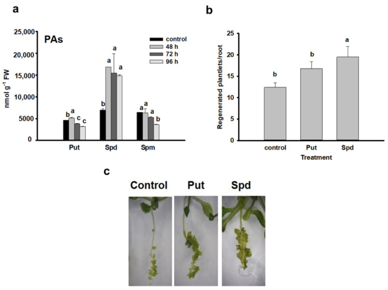
The level of the three main polyamines changed differently during the cytokinin-induced meristem conversion and development process (Figure 1a). The level of putrescine exhibited a slight but significant transient increase at 48 h, followed by decrease at later time points. The amount of spermine slightly but gradually decreased during the process. In contrast, the spermidine concentration exhibited a considerable (≈3-fold) increase in all three time points as compared to the control.
Figure 1.
Polyamines are involved in the conversion of lateral root primordia into shoot meristem. (a) Changes in the polyamine content after 48, 72 and 96 h of cytokinin induction of Arabidopsis Scheme 48 h), and the early (72 h) and late (96 h) shoot promeristem phases [7]. (b) Effect of exogenously applied 100 µM spermidine and putrescine, respectively, on the efficiency of shoot regeneration of wild-type Col seedling roots determined 10 days after cytokinin induction. Images of representative seedlings are shown in (c) Data are means ± SE of three biological replicates with 20 technical replicates each. Different letters indicate significant differences of Duncan’s multiple comparisons (p < 0.05).
Exogenous application of Put and especially Spd enhanced the shoot regeneration efficiency of Arabidopsis seedling roots which further verified the positive effect of these polyamines on direct organogenesis (Figure 1b,c).
The levels of endogenous polyamines have been often found to correlate with the in vitro regeneration efficiency of explants and exogenous polyamines were successfully used in many cases to enhance indirect somatic embryogenesis or shoot organogenesis in various species in a concentration and genotype-dependent manner [14,15,41,42]. In most of these experiments, Put and Spd were used.
Since ethylene synthesis is metabolically linked to the accumulation of PAs as both utilize the same intermediate, S-adenosylmethionine (SAM), it was hypothesized that PA-dependent morphogenic responses might be associated with changes in ethylene levels [16]. Ethylene is a potent inhibitor of in vitro plant regeneration and the enhancement of shoot regeneration by ethylene inhibitors was attributed to accumulation of PAs in Brassica alboglabra explants [43]. Conversion of Put to Spd or Spm involves the addition of aminopropyl moieties from decarboxylated S-adenosylmethionine (dSAM). When Spd and Spm synthesis was blocked inhibiting the SAM DECARBOXYLASE (SAMDC) enzyme, shoot regeneration was prevented in correlation with elevated ethylene production (Cheng, 2002 in Pua, 2007). However, shoot regeneration could be restored to normal level using exogenous PAs without diminishing the ethylene level indicating the direct role of PAs in shoot regeneration. In agreement, inhibition of SAMDC expression by RNA interference in transgenic Arabidopsis strongly promoted shoot regeneration increasing the Spd+spm/Put ratio but not affecting the ethylene level [44].
In accordance with previous observations [44,45], we found that a high endogenous Spd/Put ratio correlates with shoot regeneration and both PAs promoted shoot organogenesis if exogenously applied. To have a better insight into the regulation of endogenous PA levels during the direct shoot organogenesis process, the expression of several genes coding for enzymes implicated in PA metabolism has been investigated in cytokinin-treated roots during the process of LRP-to-SM conversion.
3.2. Put and Spd Synthesis Is Augmented during the Meristem Conversion Process
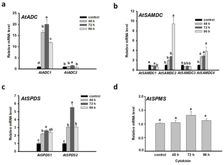
In Arabidopsis, the first step of polyamine biosynthesis, which results in putrescine from arginine or ornithine, is exclusively catalyzed by ARGININE DECARBOXYLASE. This enzyme exists as two isoforms (AtADC1 and AtADC2). AtADC1 is constitutively expressed while AtADC2 is responsive to stress stimuli [46,47,48]. Interestingly, during developmental processes both AtADC1 and AtADC2 are essential [17,49]. In our system, expression of AtADC1 increased ≈12-fold after 48 h cytokinin induction (Figure 2a). This is the time when the LRPs gradually convert to shoot promeristems (“converting organ” state) [7]. During the appearance of early and late shoot promeristems, the mRNA level of AtADC1 remained high, but the expression of AtADC2 gene was also enhanced at a lower degree (Figure 2a). Since the Put content was only slightly enhanced after 48 h cytokinin induction in contrast to Spd (Figure 1a), it is reasonable to assume that the Put produced by ADC1 (or ADC2) was quickly metabolized by DAO or converted to spermidine by SPERMIDINE SYNTHASE (SPDS).
Figure 2.
Expression of genes coding for enzymes involved in polyamine synthesis (AtADC1-2 (a), AtSAMDC1-4 (b), AtSPDS1-2 (c), and AtSPMS (d)) during the direct formation of shoot meristem from lateral root primordia (at the organ conversion (48 h), the early (72 h) and late (96 h) shoot promeristem phases [7]). The mRNA levels of the UBIQUITIN 1 (At3G52590) and PP2A3 (At1G13320.1) genes were used for gene expression normalization. The mRNA level of the untreated root tissue was used as control (relative mRNA level: 1). Data were averaged from three independent biological experiments with three technical replicates each. Standard errors are shown on the columns. The significance of difference between sets of data was determined by one-way analysis of variance (ANOVA) following Duncan’s multiple range tests; a p-value of less than 0.05 was considered significant as indicated by different letters.
Conversion of Put to Spd or Spm by the repetitive addition of the aminopropyl moieties from decarboxylated S-adenosylmethionine (dSAM) is mediated by SPDS and SPERMINE SYNTHASE (SPMS), respectively.
Formation of dSAM is regulated from SAM by the action of SAMDC. Among the five paralogs of AtSAMDC genes, AtSAMDC4 gene was shown to have auxin-regulated promoter elements together with high expression in seedlings, not like the other AtSAMDC genes [50]. The expression of AtSAMDC2 and AtSAMDC4 was enhanced during the root-to-shoot meristem conversion and shoot meristem establishment process while the mRNA levels of AtSAMDC1 and AtSAMDC3 were reduced compared to the control (Figure 2b). The AtSAMDC2 gene exhibited an especially high expression at the latest investigated state (96 h). Downregulation of the AtSAMDC2 gene expression by RNA interference was reported to decrease the shoot regeneration response [44] indicating the importance of PA synthesis during the process. Increased SAMDC activity and Spd level correlated with higher embryogenic response in alfalfa [51].
In Arabidopsis, SPDSs are encoded by two genes, AtSPDS1 and AtSPDS2, which are essential for several developmental processes, such as embryo development and plant survival [52]. After 48 h cytokinin induction, but mainly during the formation of shoot promeristem, the mRNA level of AtSPDS1, but mainly that of AtSPDS2, were elevated (Figure 2c). These results are in accordance with the 3-fold increase of Spd levels in the cytokinin-induced samples in comparison to the control (Figure 1a).
SPMS expression did not change during the converting organ state but slightly increased after 72 h of cytokinin induction (Figure 2d). In agreement, the Spm content did not increase during the shoot promeristem establishment process (Figure 1a).
Altogether, these data suggest that the augmented synthesis and conversion of Put to Spd correlates with direct shoot organogenesis from Arabidopsis roots.
3.3. Ectopically Expressed AtPAO5 Enhances the Direct Conversion of LRPs to SMs
Besides the interference of PA synthesis with ethylene production [16], polyamines might influence morphogenesis via their products of degradation [35,45] or conversion [9]. The PA catabolic enzymes, POLYAMINE OXIDASEs (PAOs) catabolize Spd and Spm and produce 1,3-diaminopropane (DAP) and H2O2 [26]. PAO enzymes also function to convert tetraamine to triamine and/or triamine to diamine (backconversion reactions) [9,30].
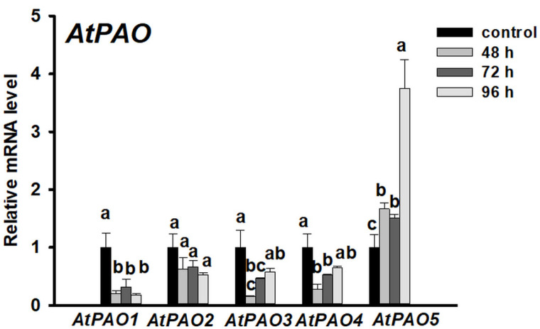
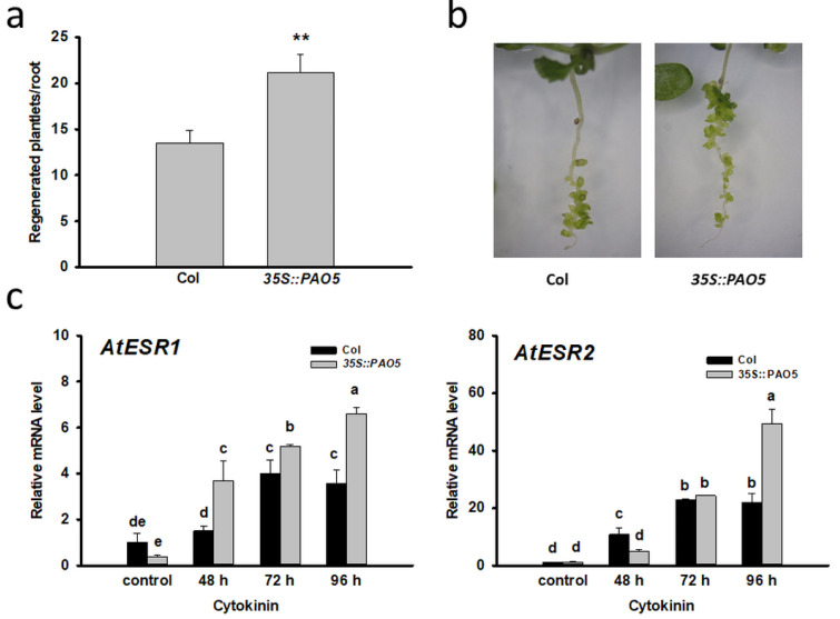
In Arabidopsis five AtPAOs (AtPAO1, AtPAO2, AtPAO3, AtPAO4, AtPAO5) are involved in polyamine catabolism among which only the expression of AtPAO5 increased after 48 h of cytokinin induction and further augmented during the formation of early and late shoot promeristem (Figure 3). To verify the involvement of AtPAO5 in direct shoot organogenesis, AtPAO5-overexpressing transgenic plants (35S:PAO5) were investigated for shoot regeneration efficiency. We found that 35S:PAO5-expressing seedlings had more regenerates/root (Figure 4a,b) and expressed the ENHANCER OF SHOOT REGENERATION 1 and 2 (ESR1 and 2) genes at higher level than the WT ones (Figure 4c). The expression of the genes coding for the ESR1,2 transcription factors was shown to be strongly associated with in vitro shoot organogenesis [53,54] and thus can serve as marker for the conversion process. These data indicate that AtPAO5 expression promotes the direct conversion of LRPs to SMs. AtPAO5 is considered to be unique in comparison to the other Arabidopsis PAOs having a dehydrogenase rather than an oxidase activity [9,30]. Therefore, in order to test whether the effect of 35S:PAO5 expression on direct shoot regeneration is specific, transgenic seedlings overexpressing the 35S:PAO2 gene [55] were also tested for the shoot regeneration rate of cytokinin-treated roots. No difference in the LRP-to-SM conversion frequency of WT and 35S:PAO2 seedlings was detected (Supplementary Figure S1).
Figure 3.
Expression of polyamine oxidase (PAO) genes during direct shoot organogenesis on Arabidopsis roots. Relative mRNA levels of AtPAO1, AtPAO2, AtPAO3, AtPAO4, and AtPAO5 genes after 48, 72 and 96 h of cytokinin induction representing the organ conversion (48 h), the early (72 h) and late (96 h) shoot promeristem phases [7]. The mRNA levels of the UBIQUITIN 1 (At3G52590) and PP2A3 (At1G13320.1) genes were used for gene expression normalization. The mRNA level of the untreated root tissue was used as control (relative mRNA level: 1). Data were averaged from three independent biological experiments with three technical replicates each. Standard errors are shown on the columns. The significance of difference between sets of data was determined by one-way analysis of variance (ANOVA) following Duncan’s multiple range tests; a p-value of less than 0.05 was considered significant as indicated by different letters.
Figure 4.
Ectopic overexpression of AtPAO5 enhances the conversion of lateral root primordia into shoots. (a) Shoot regeneration of seedling roots of WT (Col) and AtPAO5 overexpressing transgenic (35S::PAO5) plants 10 days after cytokinin induction. Data are means ± SE of three biological replicates with 20 seedlings each. For significance analysis Student’s t-test was used (** p ≤ 0.01). (b) Representative images of regenerating seedlings. (c) Relative mRNA levels of AtESR1 and AtESR2 genes in wild type (Col) and AtPAO5-overexpressing (35S::PAO5) seedling roots after 48, 72 and 96 h of cytokinin induction representing the organ conversion (48 h), the early (72 h) and late (96 h) shoot promeristem phases [7]. The mRNA levels of the UBIQUITIN 1 (At3G52590) and PP2A3 (At1G13320.1) genes were used for gene expression normalization. The mRNA level of the untreated root tissue was used as control (relative mRNA level: 1). Data were averaged from three independent biological experiments with three technical replicates each. Standard errors are shown on the columns. The significance of differences between sets of data was determined by one-way analysis of variance (ANOVA) following Duncan’s multiple range tests; a p-value of less than 0.05 was considered significant as indicated by different letters.
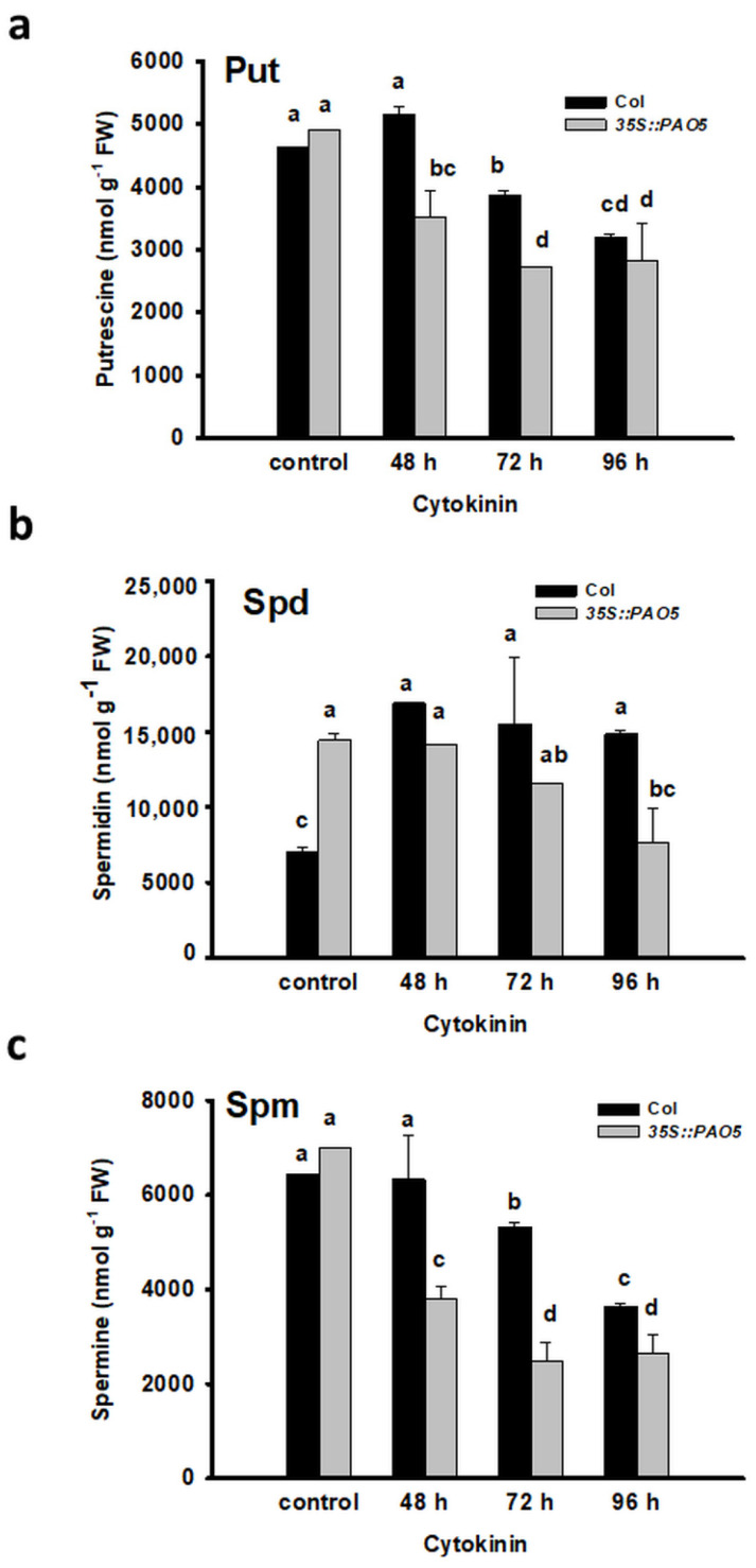
Measuring the polyamine contents in the 35S:PAO5 roots before and during the direct shoot regeneration process revealed that the initial level of Spd, but not that of Put or Spm, was significantly higher in the transgenic roots than in the control (Figure 5). Following the cytokinin-treatment, the level of all three plyamines gradually decreased at a higher rate in the 35S:PAO5 than in the WT roots (Figure 5) indicating that AtPAO5 overexpression influenced the polyamine homeostasis of seedling roots.
Figure 5.
AtPAO5 overexpression influences the PA homeostasis in seedling roots before and during the conversion of lateral root primordia into shoots. Putrescine (Put) (a), spermidine (Spd) (b) and spermine (Spm) (c) contents were determined in 100 mg of root samples of wild type (Col) and transgenic (35S:PAO5) seedlings after 48, 72 and 96 h cytokinin treatment repre-senting the organ conversion (48 h), the early (72 h) and late (96 h) shoot promeristem phases [7]. Data are means ± SE of three biological replicates with 20 technical replicates each. Different let-ters indicate significant differences of Duncan’s multiple comparisons (p < 0.05).
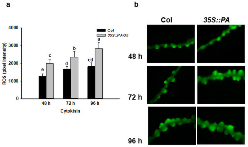
In addition to the altered polyamine content, the ROS level was found to be different in the LRPs of transgenic 35S::PAO5 seedlings compared to the WT ones (Figure 6). Lower ROS accumulation in the atpao5 seedlings has been reported [56] supporting the view that AtPAO5 modify the ROS homeostasis. H2O2 as the product of PA catabolism was found to be essential for the indirect formation of somatic embryos in cotton [27] as well as for lateral root formation in soybean [57]. H2O2 is linked to various plant morphogenic processes [58,59,60], including de novo shoot regeneration [61,62]. H2O2 exerts its effect on shoot regeneration in a context and concentration dependent way: although it inhibits shoot formation at higher doses, it promotes the initial phase of de novo organogenesis at low concentrations [44,55]. It was hypothesized that PAs may influence plant regeneration mediated by H2O2 formed because of their oxidation e.g., by polyamine oxidases [34,44]. Why the overexpresion of AtPAO5 but not that of AtPAO2, the activity of which is also associated with ROS production, enhanced direct shoot regeneration is not known.
Figure 6.
ROS level influenced by 35S::PAO5 expression correlates with the process of direct shoot meri-stem formation. Representative diagram (a) and images (b) of 2,7-dichlorodihydro-fluorescein diacetate (H2DC-FDA)-derived DCF fluorescence in WT (Col) and transgenic (35S::PAO5) seed-ling roots after 48, 72 and 96 h cytokinin treatment representing the organ conversion (48 h), the early (72 h) and late (96 h) shoot promeristem phases [7]. Scale bars = 100 µm. Data are means ± SE of three biological replicates with twenty technical replicates each. Different letters indicate significant differences of Duncan’s multiple comparisons (p < 0.05).
It has to be mentioned, however, that the two enzymes have different cellular localization (AtPAO5 cytoplasmic [9], AtPAO2 peroxisomal [30]) and AtPAO5 is unique among the five Arabidopsis PAOs considering its high affinity for t-Spm as a substrate [9]. AtPAO5, was shown to be involved in the maintenance of t-Spm homeostasis that is required for normal plant development: the atpao5 mutant lacking t-Spm oxidation and the acl5 mutant lacking t-Spm synthesis both exhibit growth defects [8,9]. Therefore, maintaining t-Spm homeostasis was attributed as the primary function of AtPAO5 [9]. Nevertheless, AtPAO5 was also reported to serve as a spermine oxidase/dehydrogenase in 35S:PAO5-expressing transgenic plants [31]. It was hypothesized that although the preferential in vivo substrate of AtPAO5 is t-Spm, if ectopically overexpressed it can efficiently access and metabolize Spm [9].
Transgenic Arabidopsis plants with up- or downregulated ARABIDOPSIS FLAVIN-CONTAINING AMINE OXIDASE 1 (AtFAO1) (AtFAO1 is an earlier designation of AtPAO5) expression were shown to exhibit strongly altered indirect shoot regeneration abilities from root explants [45]. While the regeneration efficiency of root explants with downregulated AtFAO1/AtPAO5 activity was elevated, those ones with upregulated activity were only poorly regenerative [45]. This latter observation seems to be contradictory to our results, since we found increased conversion of LRPs to shoots in the line overexpressing AtPAO5. This contradiction might be due to the excision of the seedling roots followed by auxin-induced callus formation during the indirect process studied by Lim et al. (2006) [45], while we used intact seedlings treated by high concentration of cytokinin in the direct shoot regeneration pathway. AtPAO5 was shown to influence the interplay of auxin and cytokinin during xylem differentiation [8]. Although shoot meristem formation may follow the same steps during the direct and indirect pathways, the difference in the hormonal and redox environment of surrounding tissues (callus or LRP) might result in altered sensitivity of the initial events towards the products/consequences of ectopic AtPAO5 activity.
4. Conclusions
Polyamines have already been shown to control plant morphogenesis and in vitro plant regeneration in several experimental systems. Here we provide evidence that they are also involved in the cytokinin-induced direct (without intervening callus formation) conversion of lateral root primordia (LRPs) of intact seedlings into shoot meristems. This process that includes the correct temporal and spatial organization of cell divisions and cell fate decisions was found to correlate with increased accumulation of spermidine (Spd). In agreement, the expression of genes related to Spd synthesis (AtADC1,2, AtSAMDC2,4, AtSPDS1,2) and backconversion of t-Spm/Spm to Spd (AtPAO5 but not AtPAO1-4) was increased following the cytokinin treatment. In agreement, the ectopic overexpression of AtPAO5 in transgenic seedlings increased the Spd level and the shoot regeneration rate of roots. Furthermore, exogenous Spd treatment could also promote the conversion of lateral root primordia to shoot meristems. Interestingly, this observation is in contradiction with reports about indirect shoot organogenesis from excised Arabidopsis root segments where the overexpression of AtPAO5 reduced the spermidine level and the regeneration potential. This indicates that PAs and PA metabolism affect the shoot regeneration process in Arabidopsis in a context-dependent manner.
Ectopic expression of AtPAO5 also resulted in elevated ROS level in the LRPs undergoing conversion to shoot meristems and the contribution of this ROS to regeneration enhancement cannot be excluded. It is to be noted that although AtPAO1-4 are also capable of Spm backconversion and associated ROS production, only AtPAO5 was found to show elevated expression in the investigated shoot regeneration pathway, and the ectopic expression of AtPAO5 but not that of AtPAO2 could increase the regeneration efficiency. These observations implicate a specific role for AtPAO5 in the process. Since AtPAO5 was shown to have specifically high affinity for t-Spm, one can suppose that it may also influence shoot regeneration controlling t-Spm homeostasis. This possibility, however, still needs to be experimentally addressed.
Supplementary Materials
The following are available online at https://www.mdpi.com/2223-7747/10/2/305/s1, Supplementary Figure S1 AtPAO2 overexpression didn’t influence the conversion rate of lateral root primordia into shoots. Supplementary Table S1. Sequences of the oligonucleotide primers used in the qPCR experiments.
Author Contributions
Conceptualization, A.F. and K.G.; methodology, D.B. and Á.S.; formal analysis, K.G.; investigation, N.K., P.B., and Á.S.; data curation, K.G.; writing—original draft preparation, K.G. and A.F.; writing—review and editing, A.F., K.G. and N.K.; visualization, K.G.; supervision, K.G. and A.F.; project administration, K.G.; funding acquisition, K.G. All authors have read and agreed to the published version of the manuscript.
Funding
This work was supported by grants from National Research, Development, and Innovation Fund (Grant no. FK 128997 and FK 129061). Katalin Gémes was supported by the János Bolyai Research Scholarship of the Hungarian Academy of Sciences (Grant no. 00580/19/8) and the New National Excellence Program of the Ministry for Innovation and Technology (ÚNKP-20-5-SZTE-648).
Institutional Review Board Statement
Not applicable.
Informed Consent Statement
Not applicable.
Data Availability Statement
Data sharing is not applicable to this article.
Conflicts of Interest
The authors declare no conflict of interest. The funders had no role in the design of the study; in the collection, analyses, or interpretation of data; in the writing of the manuscript, or in the decision to publish the results.
Footnotes
Publisher’s Note: MDPI stays neutral with regard to jurisdictional claims in published maps and institutional affiliations.
References
- 1.Ikeuchi M., Ogawa Y., Iwase A., Sugimoto K. Plant Regeneration: Cellular Origins and Molecular Mechanisms. Development. 2016;143:1442–1451. doi: 10.1242/dev.134668. [DOI] [PubMed] [Google Scholar]
- 2.Atta R., Laurens L., Boucheron-Dubuisson E., Guivarc’h A., Carnero E., Giraudat-Pautot V., Rech P., Chriqui D. Pluripotency of Arabidopsis Xylem Pericycle Underlies Shoot Regeneration from Root and Hypocotyl Explants Grown in Vitro. Plant J. 2009;57:626–644. doi: 10.1111/j.1365-313X.2008.03715.x. [DOI] [PubMed] [Google Scholar]
- 3.Bernula D., Benkő P., Kaszler N., Domonkos I., Valkai I., Szőllősi R., Ferenc G., Ayaydin F., Fehér A., Gémes K. Timely Removal of Exogenous Cytokinin and the Prevention of Auxin Transport from the Shoot to the Root Affect the Regeneration Potential of Arabidopsis Roots. Plant Cell Tissue Organ Cult. 2020;140:327–339. doi: 10.1007/s11240-019-01730-3. [DOI] [Google Scholar]
- 4.Che P., Lall S., Howell S.H. Developmental Steps in Acquiring Competence for Shoot Development in Arabidopsis Tissue Culture. Planta. 2007;226:1183–1194. doi: 10.1007/s00425-007-0565-4. [DOI] [PubMed] [Google Scholar]
- 5.Chatfield S.P., Capron R., Severino A., Penttila P.-A., Alfred S., Nahal H., Provart N.J. Incipient Stem Cell Niche Conversion in Tissue Culture: Using a Systems Approach to Probe Early Events in WUSCHEL-Dependent Conversion of Lateral Root Primordia into Shoot Meristems. Plant J. 2013;73:798–813. doi: 10.1111/tpj.12085. [DOI] [PubMed] [Google Scholar]
- 6.Kareem A., Radhakrishnan D., Wang X., Bagavathiappan S., Trivedi Z.B., Sugimoto K., Xu J., Mähönen A.P., Prasad K. Protocol: A Method to Study the Direct Reprogramming of Lateral Root Primordia to Fertile Shoots. Plant Methods. 2016;12 doi: 10.1186/s13007-016-0127-5. [DOI] [PMC free article] [PubMed] [Google Scholar]
- 7.Rosspopoff O., Chelysheva L., Saffar J., Lecorgne L., Gey D., Caillieux E., Colot V., Roudier F., Hilson P., Berthomé R., et al. Direct Conversion of Root Primordium into Shoot Meristem Relies on Timing of Stem Cell Niche Development. Development. 2017;144:1187–1200. doi: 10.1242/dev.142570. [DOI] [PubMed] [Google Scholar]
- 8.Alabdallah O., Ahou A., Mancuso N., Pompili V., Macone A., Pashkoulov D., Stano P., Cona A., Angelini R., Tavladoraki P. The Arabidopsis Polyamine Oxidase/Dehydrogenase 5 Interferes with Cytokinin and Auxin Signaling Pathways to Control Xylem Differentiation. J. Exp. Bot. 2017;68:997–1012. doi: 10.1093/jxb/erw510. [DOI] [PubMed] [Google Scholar]
- 9.Kim D.W., Watanabe K., Murayama C., Izawa S., Niitsu M., Michael A.J., Berberich T., Kusano T. Polyamine Oxidase5 Regulates Arabidopsis Growth through Thermospermine Oxidase Activity. Plant Physiol. 2014;165:1575–1590. doi: 10.1104/pp.114.242610. [DOI] [PMC free article] [PubMed] [Google Scholar]
- 10.Thiruvengadam M., Ill-Min C., Se-Chul C. Influence of Polyamines on in Vitro Organogenesis in Bitter Melon (Momordica Charantia, L.) J. Med. Plants Res. 2012;6:3579–3585. doi: 10.5897/JMPR12.246. [DOI] [Google Scholar]
- 11.Smith T.A. Polyamines. Annu. Rev. Plant Physiol. 1985;36:117–143. doi: 10.1146/annurev.pp.36.060185.001001. [DOI] [Google Scholar]
- 12.Chen D., Shao Q., Yin L., Younis A., Zheng B. Polyamine Function in Plants: Metabolism, Regulation on Development, and Roles in Abiotic Stress Responses. Front. Plant Sci. 2019;9 doi: 10.3389/fpls.2018.01945. [DOI] [PMC free article] [PubMed] [Google Scholar]
- 13.Roustan J.-P., Chraibi K.M., Latché A., Fallot J. Relationship between Ethylene and Polyamine Synthesis in Plant Regeneration. In: Pech J.C., Latché A., Balagué C., editors. Cellular and Molecular Aspects of the Plant Hormone Ethylene, Proceedings of the International Symposium on Cellular and Molecular Aspects of Biosynthesis and Action of the Plant Hormone Ethylene, Agen, France, 31 August–4 September 1992. Springer; Dordrecht, The Netherlands: 1993. pp. 365–366. Current Plant Science and Biotechnology in Agriculture. [Google Scholar]
- 14.Tiburcio A.F., Alcázar R. Potential Applications of Polyamines in Agriculture and Plant Biotechnology. In: Alcázar R., Tiburcio A.F., editors. Polyamines: Methods and Protocols. Springer; New York, NY, USA: 2018. pp. 489–508. Methods in Molecular Biology. [DOI] [PubMed] [Google Scholar]
- 15.Kakkar R.K., Nagar P.K., Ahuja P.S., Rai V.K. Polyamines and Plant Morphogenesis. Biol. Plant. 2000;43:1–11. doi: 10.1023/A:1026582308902. [DOI] [Google Scholar]
- 16.Pua E.-C. Regulation of Plant Morphogenesis In Vitro: Role of Ethylene and Polyamines. In: Xu Z., Li J., Xue Y., Yang W., editors. Biotechnology and Sustainable Agriculture 2006 and Beyond. Springer; Dordrecht, The Netherlands: 2007. pp. 89–95. [Google Scholar]
- 17.Urano K., Hobo T., Shinozaki K. Arabidopsis ADC Genes Involved in Polyamine Biosynthesis Are Essential for Seed Development. FEBS Lett. 2005;579:1557–1564. doi: 10.1016/j.febslet.2005.01.048. [DOI] [PubMed] [Google Scholar]
- 18.Ge C., Cui X., Wang Y., Hu Y., Fu Z., Zhang D., Cheng Z., Li J. BUD2, Encoding an S-Adenosylmethionine Decarboxylase, Is Required for Arabidopsis Growth and Development. Cell Res. 2006;16:446–456. doi: 10.1038/sj.cr.7310056. [DOI] [PubMed] [Google Scholar]
- 19.Cui X., Ge C., Wang R., Wang H., Chen W., Fu Z., Jiang X., Li J., Wang Y. The BUD2 Mutation Affects Plant Architecture through Altering Cytokinin and Auxin Responses in Arabidopsis. Cell Res. 2010;20:576–586. doi: 10.1038/cr.2010.51. [DOI] [PubMed] [Google Scholar]
- 20.Kakehi J., Kuwashiro Y., Niitsu M., Takahashi T. Thermospermine Is Required for Stem Elongation in Arabidopsis thaliana. Plant Cell Physiol. 2008;49:1342–1349. doi: 10.1093/pcp/pcn109. [DOI] [PubMed] [Google Scholar]
- 21.Miyamoto M., Shimao S., Tong W., Motose H., Takahashi T. Effect of Thermospermine on the Growth and Expression of Polyamine-Related Genes in Rice Seedlings. Plants. 2019;8:269. doi: 10.3390/plants8080269. [DOI] [PMC free article] [PubMed] [Google Scholar]
- 22.Takano A., Kakehi J.-I., Takahashi T. Thermospermine Is Not a Minor Polyamine in the Plant Kingdom. Plant Cell Physiol. 2012;53:606–616. doi: 10.1093/pcp/pcs019. [DOI] [PubMed] [Google Scholar]
- 23.Alcázar Hernández R., Bitrian M., Zarza X., Fernández Tiburcio A. Recent Advances in Pharmaceutical Sciences. Volume II. Transworld Research Network; Kerala, India: 2012. Polyamine metabolism and signaling in plant abiotic stress protection; pp. 29–47. [Google Scholar]
- 24.Clay N.K., Nelson T. Arabidopsis Thickvein Mutation Affects Vein Thickness and Organ Vascularization, and Resides in a Provascular Cell-Specific Spermine Synthase Involved in Vein Definition and in Polar Auxin Transport. Plant Physiol. 2005;138:767–777. doi: 10.1104/pp.104.055756. [DOI] [PMC free article] [PubMed] [Google Scholar]
- 25.Yoshimoto K., Takamura H., Kadota I., Motose H., Takahashi T. Chemical Control of Xylem Differentiation by Thermospermine, Xylemin, and Auxin. Sci. Rep. 2016;6:21487. doi: 10.1038/srep21487. [DOI] [PMC free article] [PubMed] [Google Scholar]
- 26.Wang W., Paschalidis K., Feng J.-C., Song J., Liu J.-H. Polyamine Catabolism in Plants: A Universal Process With Diverse Functions. Front. Plant Sci. 2019;10 doi: 10.3389/fpls.2019.00561. [DOI] [PMC free article] [PubMed] [Google Scholar]
- 27.Cheng W.-H., Wang F.-L., Cheng X.-Q., Zhu Q.-H., Sun Y.-Q., Zhu H.-G., Sun J. Polyamine and Its Metabolite H2O2 Play a Key Role in the Conversion of Embryogenic Callus into Somatic Embryos in Upland Cotton (Gossypium Hirsutum, L.) Front. Plant Sci. 2015;6 doi: 10.3389/fpls.2015.01063. [DOI] [PMC free article] [PubMed] [Google Scholar]
- 28.Angelini R., Cona A., Federico R., Fincato P., Tavladoraki P., Tisi A. Plant Amine Oxidases “on the Move”: An Update. Plant Physiol. Biochem. 2010;48:560–564. doi: 10.1016/j.plaphy.2010.02.001. [DOI] [PubMed] [Google Scholar]
- 29.Pottosin I., Velarde-Buendía A.M., Bose J., Zepeda-Jazo I., Shabala S., Dobrovinskaya O. Cross-Talk between Reactive Oxygen Species and Polyamines in Regulation of Ion Transport across the Plasma Membrane: Implications for Plant Adaptive Responses. J. Exp. Bot. 2014;65:1271–1283. doi: 10.1093/jxb/ert423. [DOI] [PubMed] [Google Scholar]
- 30.Fincato P., Moschou P.N., Spedaletti V., Tavazza R., Angelini R., Federico R., Roubelakis-Angelakis K.A., Tavladoraki P. Functional Diversity inside the Arabidopsis Polyamine Oxidase Gene Family. J. Exp. Bot. 2011;62:1155–1168. doi: 10.1093/jxb/erq341. [DOI] [PubMed] [Google Scholar]
- 31.Ahou A., Martignago D., Alabdallah O., Tavazza R., Stano P., Macone A., Pivato M., Masi A., Rambla J.L., Vera-Sirera F., et al. A Plant Spermine Oxidase/Dehydrogenase Regulated by the Proteasome and Polyamines. J. Exp. Bot. 2014;65:1585–1603. doi: 10.1093/jxb/eru016. [DOI] [PubMed] [Google Scholar]
- 32.Kamada-Nobusada T., Hayashi M., Fukazawa M., Sakakibara H., Nishimura M. A Putative Peroxisomal Polyamine Oxidase, AtPAO4, Is Involved in Polyamine Catabolism in Arabidopsis thaliana. Plant Cell Physiol. 2008;49:1272–1282. doi: 10.1093/pcp/pcn114. [DOI] [PubMed] [Google Scholar]
- 33.Moschou P.N., Delis I.D., Paschalidis K.A., Roubelakis-Angelakis K.A. Transgenic Tobacco Plants Overexpressing Polyamine Oxidase Are Not Able to Cope with Oxidative Burst Generated by Abiotic Factors. Physiol. Plant. 2008;133:140–156. doi: 10.1111/j.1399-3054.2008.01049.x. [DOI] [PubMed] [Google Scholar]
- 34.Ono Y., Kim D.W., Watanabe K., Sasaki A., Niitsu M., Berberich T., Kusano T., Takahashi Y. Constitutively and Highly Expressed Oryza Sativa Polyamine Oxidases Localize in Peroxisomes and Catalyze Polyamine Back Conversion. Amino Acids. 2012;42:867–876. doi: 10.1007/s00726-011-1002-3. [DOI] [PubMed] [Google Scholar]
- 35.Tavladoraki P., Cona A., Angelini R. Copper-Containing Amine Oxidases and FAD-Dependent Polyamine Oxidases Are Key Players in Plant Tissue Differentiation and Organ Development. Front. Plant Sci. 2016;7 doi: 10.3389/fpls.2016.00824. [DOI] [PMC free article] [PubMed] [Google Scholar]
- 36.Rozen S., Skaletsky H. Primer3 on the WWW for General Users and for Biologist Programmers. Methods Mol. Biol. 2000;132:365–386. doi: 10.1385/1-59259-192-2:365. [DOI] [PubMed] [Google Scholar]
- 37.Winter D., Vinegar B., Nahal H., Ammar R., Wilson G.V., Provart N.J. An “Electronic Fluorescent Pictograph” Browser for Exploring and Analyzing Large-Scale Biological Data Sets. PLoS ONE. 2007;2:e718. doi: 10.1371/journal.pone.0000718. [DOI] [PMC free article] [PubMed] [Google Scholar]
- 38.Benkő P., Jee S., Kaszler N., Fehér A., Gémes K. Polyamines Treatment during Pollen Germination and Pollen Tube Elongation in Tobacco Modulate Reactive Oxygen Species and Nitric Oxide Homeostasis. J. Plant Physiol. 2020;244:153085. doi: 10.1016/j.jplph.2019.153085. [DOI] [PubMed] [Google Scholar]
- 39.Gémes K., Poór P., Horváth E., Kolbert Z., Szopkó D., Szepesi Á., Tari I. Cross-Talk between Salicylic Acid and NaCl-Generated Reactive Oxygen Species and Nitric Oxide in Tomato during Acclimation to High Salinity. Physiol. Plant. 2011;142:179–192. doi: 10.1111/j.1399-3054.2011.01461.x. [DOI] [PubMed] [Google Scholar]
- 40.Takács Z., Poór P., Tari I. Comparison of Polyamine Metabolism in Tomato Plants Exposed to Different Concentrations of Salicylic Acid under Light or Dark Conditions. Plant Physiol. Biochem. 2016;108:266–278. doi: 10.1016/j.plaphy.2016.07.020. [DOI] [PubMed] [Google Scholar]
- 41.Minocha S.C., Minocha R. Role of Polyamines in Somatic Embryogenesis. In: Bajaj Y.P.S., editor. Somatic Embryogenesis and Synthetic Seed I. Springer; Berlin/Heidelberg, Germany: 1995. pp. 53–70. Biotechnology in Agriculture and Forestry. [Google Scholar]
- 42.Pua E.-C. Morphogenesis in Cell and Tissue Cultures. In: Soh W.-Y., Bhojwani S.S., editors. Morphogenesis in Plant Tissue Cultures. Springer; Dordrecht, The Netherlands: 1999. pp. 255–303. [Google Scholar]
- 43.Pua E.-C., Teo S.-H., Loh C.-S. Interactive Role of Ethylene and Polyamines on Shoot Regenerabillty of Chinese Kale (Brassica Alboglabra Bailey) in vitro. J. Plant Physiol. 1996;149:138–148. doi: 10.1016/S0176-1617(96)80186-6. [DOI] [Google Scholar]
- 44.Hu W.-W., Gong H., Pua E.-C. Modulation of SAMDC Expression in Arabidopsis thaliana Alters in Vitro Shoot Organogenesis. Physiol. Plant. 2006;128:740–750. doi: 10.1111/j.1399-3054.2006.00799.x. [DOI] [Google Scholar]
- 45.Lim T.S., Chitra T.R., Han P., Pua E.-C., Yu H. Cloning and Characterization of Arabidopsis and Brassica Juncea Flavin-Containing Amine Oxidases. J. Exp. Bot. 2006;57:4155–4169. doi: 10.1093/jxb/erl193. [DOI] [PubMed] [Google Scholar]
- 46.Rossi F.R., Marina M., Pieckenstain F.L. Role of Arginine Decarboxylase (ADC) in Arabidopsis thaliana Defence against the Pathogenic Bacterium Pseudomonas Viridiflava. Plant Biol. 2015;17:831–839. doi: 10.1111/plb.12289. [DOI] [PubMed] [Google Scholar]
- 47.Takahashi T., Tong W. Regulation and Diversity of Polyamine Biosynthesis in Plants. In: Kusano T., Suzuki H., editors. Polyamines: A Universal Molecular Nexus for Growth, Survival, and Specialized Metabolism. Springer; Tokyo, Japan: 2015. pp. 27–44. [Google Scholar]
- 48.Urano K., Yoshiba Y., Nanjo T., Ito T., Yamaguchi-Shinozaki K., Shinozaki K. Arabidopsis Stress-Inducible Gene for Arginine Decarboxylase AtADC2 Is Required for Accumulation of Putrescine in Salt Tolerance. Biochem. Biophys. Res. Commun. 2004;313:369–375. doi: 10.1016/j.bbrc.2003.11.119. [DOI] [PubMed] [Google Scholar]
- 49.Sánchez-Rangel D., Chávez-Martínez A.I., Rodríguez-Hernández A.A., Maruri-López I., Urano K., Shinozaki K., Jiménez-Bremont J.F. Simultaneous Silencing of Two Arginine Decarboxylase Genes Alters Development in Arabidopsis. Front. Plant Sci. 2016;7 doi: 10.3389/fpls.2016.00300. [DOI] [PMC free article] [PubMed] [Google Scholar]
- 50.Majumdar R., Shao L., Turlapati S.A., Minocha S.C. Polyamines in the Life of Arabidopsis: Profiling the Expression of S-Adenosylmethionine Decarboxylase (SAMDC) Gene Family during Its Life Cycle. BMC Plant Biol. 2017;17:264. doi: 10.1186/s12870-017-1208-y. [DOI] [PMC free article] [PubMed] [Google Scholar]
- 51.Huang X.-L., Li X.-J., Li Y., Huang L.-Z. The Effect of AOA on Ethylene and Polyamine Metabolism during Early Phases of Somatic Embryogenesis in Medicago Sativa. Physiol. Plant. 2001;113:424–429. doi: 10.1034/j.1399-3054.2001.1130317.x. [DOI] [PubMed] [Google Scholar]
- 52.Imai A., Matsuyama T., Hanzawa Y., Akiyama T., Tamaoki M., Saji H., Shirano Y., Kato T., Hayashi H., Shibata D., et al. Spermidine Synthase Genes Are Essential for Survival of Arabidopsis. Plant Physiol. 2004;135:1565–1573. doi: 10.1104/pp.104.041699. [DOI] [PMC free article] [PubMed] [Google Scholar]
- 53.Banno H., Ikeda Y., Niu Q.-W., Chua N.-H. Overexpression of Arabidopsis ESR1 Induces Initiation of Shoot Regeneration. Plant Cell. 2001;13:2609–2618. doi: 10.1105/tpc.010234. [DOI] [PMC free article] [PubMed] [Google Scholar]
- 54.Matsuo N., Makino M., Banno H. Arabidopsis ENHANCER OF SHOOT REGENERATION (ESR)1 and ESR2 Regulate in Vitro Shoot Regeneration and Their Expressions Are Differentially Regulated. Plant Sci. 2011;181:39–46. doi: 10.1016/j.plantsci.2011.03.007. [DOI] [PubMed] [Google Scholar]
- 55.Wimalasekera R., Schaarschmidt F., Angelini R., Cona A., Tavladoraki P., Scherer G.F.E. POLYAMINE OXIDASE2 of Arabidopsis Contributes to ABA Mediated Plant Developmental Processes. Plant Physiol. Biochem. 2015;96:231–240. doi: 10.1016/j.plaphy.2015.08.003. [DOI] [PubMed] [Google Scholar]
- 56.Ferdousy N., Arif T., Sagor G. Polyamine Oxidase 5 (PAO5) Mediated Antioxidant Response to Promote Salt Tolerance in Arabidopsis thaliana. J. Bangladesh Agric. Univ. 2020;1 doi: 10.5455/JBAU.103110. [DOI] [Google Scholar]
- 57.Su G.-X., Zhang W.-H., Liu Y.-L. Involvement of Hydrogen Peroxide Generated by Polyamine Oxidative Degradation in the Development of Lateral Roots in Soybean. J. Integr. Plant Biol. 2006;48:426–432. doi: 10.1111/j.1744-7909.2006.00236.x. [DOI] [Google Scholar]
- 58.Considine M.J., Foyer C.H. Redox Regulation of Plant Development. Antioxid Redox Signal. 2013;21:1305–1326. doi: 10.1089/ars.2013.5665. [DOI] [PMC free article] [PubMed] [Google Scholar]
- 59.Schmidt R., Schippers J.H.M. ROS-Mediated Redox Signaling during Cell Differentiation in Plants. Biochim. Biophys. Acta (BBA) Gen. Subj. 2015;1850:1497–1508. doi: 10.1016/j.bbagen.2014.12.020. [DOI] [PubMed] [Google Scholar]
- 60.Zeng J., Dong Z., Wu H., Tian Z., Zhao Z. Redox Regulation of Plant Stem Cell Fate. EMBO J. 2017;36:2844–2855. doi: 10.15252/embj.201695955. [DOI] [PMC free article] [PubMed] [Google Scholar]
- 61.Shin J., Bae S., Seo P.J. De Novo Shoot Organogenesis during Plant Regeneration. J. Exp. Bot. 2020;71:63–72. doi: 10.1093/jxb/erz395. [DOI] [PubMed] [Google Scholar]
- 62.Zhang H., Zhang T.T., Liu H., Shi D.Y., Wang M., Bie X.M., Li X.G., Zhang X.S. Thioredoxin-Mediated ROS Homeostasis Explains Natural Variation in Plant Regeneration. Plant Physiol. 2018;176:2231–2250. doi: 10.1104/pp.17.00633. [DOI] [PMC free article] [PubMed] [Google Scholar]
Associated Data
This section collects any data citations, data availability statements, or supplementary materials included in this article.
Supplementary Materials
Data Availability Statement
Data sharing is not applicable to this article.


