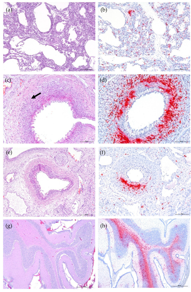
Figure 1.
Histology (H&E stain, a,c,e,g) and PCV-3 ISH (hematoxylin counterstain, b,d,f,h) results. (a) Fetal lung from case No. 3 with no apparent histopathologic lesions. (b) PCV-3 genome in macrophage-like cells and in a perivascular area in lung fetus from case No. 3. (c) Moderate tunica media swelling in a splenic artery with a mild lymphocytic infiltration at perivascular and arteriolar locations (arrow) of a stillborn piglet from case No. 52. (d) PCV-3 detection in smooth muscle of spleen artery and perivascular inflammation in the same fetus. (e) Kidney–pelvis artery of a stillborn piglet from case No. 52. (f) PCV-3 detection in smooth muscle and in lamina propria of a kidney artery of the same fetus. (g) Cerebellum from a stillborn piglet with no apparent histopathologic lesions of fetus from case No. 52. (h) High amount of PCV-3 nucleic acid in cerebellum white matter and mild-to-moderate in grey matter of the same animal.

