Abstract
The expanded CAG repeat number in HTT gene causes Huntington disease (HD), which is a severe, dominant neurodegenerative illness. The accurate determination of the expanded allele size is crucial to confirm the genetic status in symptomatic and presymptomatic at-risk subjects and avoid genetic polymorphism-related false-negative diagnoses. Precise CAG repeat number determination is critical to discriminate the cutoff between unexpanded and intermediate mutable alleles (IAs, 27–35 CAG) as well as between IAs and pathological, low-penetrance alleles (i.e., 36–39 CAG repeats), and it is also critical to detect large repeat expansions causing pediatric HD variants. We analyzed the HTT-CAG repeat number of 14 DNA reference materials and of a DNA collection of 43 additional samples carrying unexpanded, IAs, low and complete penetrance alleles, including large (>60 repeats) and very large (>100 repeats) expansions using a novel triplet-primed PCR-based assay, the AmplideX PCR/CE HTT Kit. The results demonstrate that the method accurately genotypes both normal and expanded HTT-CAG repeat numbers and reveals previously undisclosed and very large CAG expansions >200 repeats. We also show that this technique can improve genetic test reliability and accuracy by detecting CAG expansions in samples with sequence variations within or adjacent to the repeat tract that cause allele drop-outs or inaccuracies using other PCR methods.
Keywords: Huntington disease, HTT-CAG repeats, novel diagnostic test, TP-PCR
1. Introduction
Huntington disease (HD; OMIM 143100) is an autosomal, dominantly inherited, progressive, neurodegenerative disorder caused by an expansion of a coding CAG trinucleotide repeat in the exon 1 of huntingtin (HTT) gene [1]. It generally manifests in adulthood, after a presymptomatic life stage of years, depending on the penetrance of the CAG mutation [2], which affects cortical–striatal connections with brain cell dysfunction and loss [3]. The main clinical manifestations appear with the unpredictable and concurrent appearance of neurological motor abnormalities (e.g., coexistence of hyper- and hypo-kinetic movement disorders, incoordination), cognitive decline (e.g., executive function abnormalities, dementia) and behavioral changes (e.g., uncontrolled emotional state, apathy, depression with suicide propensity, obsessions and perseveration, psychosis) that progressively and severely affect, altogether, the individual functional capacity until cachexia and death [4], after 15 years on average [5]. The abnormal gene product, the Huntingtin protein, is ubiquitously expressed and contains an elongated polyglutamine stretch encoded by the CAG trinucleotide repeats. Normal alleles contain up to 35 CAG repeats, whereas HD patients carry expansions of 36 or more repeats [6]. Although complete penetrance of HD is observed for CAG sizes of ≥40, only a proportion of those with a CAG repeat length of 36–39 (i.e., low penetrance alleles) shows signs or symptoms of HD within a normal life span [2,7]. Expansions of the mutation length are associated with earlier age at onset and death [5], with the first clinical manifestations occurring in young people, i.e., juvenile-onset HD (joHD), when the disease starts ≤20 years of age [8]. CAG repeat expansions beyond 60–80 trinucleotides are rare and affect children who manifest pediatric HD (PHD), with more severe disease course and an atypical phenotype compared to adults, along with reduced life span and specific neuropathological patterns [9]. However, the expanded CAG mutation penetrance, intergenerational parent–child change (i.e., CAG instability) [10], and mosaicism (i.e., cellular CAG length variability within and among the single individual tissues) [9,11,12], as well as the age at onset anticipation of offspring and the severity of disease progression, also depend, altogether, on additional factors, in addition to the CAG mutation length, including gene modifiers [13,14], loss of interruptions (LOI) in the expanded HTT-CAG sequence [13,15,16,17], and yet unknown environmental factors [18].
Predictive and diagnostic testing of HD require accurate sizing of the CAG repeat to distinguish the edge between normal repeats and intermediate mutable CAG alleles (IAs, 27–35 CAG) that are potentially responsible for new HTT mutations in offspring [2], and between the IAs and expanded mutations (i.e., beyond 35 repeats) [19]. PCR-based assays for sizing the HTT-CAG repeat typically involve amplification using primers flanking the CAG repeat region, followed by capillary electrophoresis (CE) [20,21]. Whenever only a single peak is detected, additional tests such as the PCR amplification of the adjacent CCG region is usually performed to exclude errors and PCR amplification failure or, in the past, Southern blot analysis to detect large expanded alleles [22,23]. The negative correlation between repeat length and amplification efficiency represents a significant deficiency of repeat-flanking PCR. Flanking sequence polymorphisms may also cause allele-specific PCR failure (allele drop-out) and lead to misdiagnosis [24,25]. In marked contrast, the more recent triplet-primed PCR (TP-PCR), a strategy that pairs a flanking primer with one that anneals randomly within the repeat to generate different-sized amplicons, produces robust amplification and reliable detection of all expanded alleles regardless of size. This is because TP-PCR products of expanded alleles generate a characteristic CE pattern that can be easily distinguished from the pattern of unexpanded alleles [26], which eliminates the need to perform the labor-intensive Southern blot methodology. In HD, the TP-PCR strategy has been used to successfully detect an expanded allele of >150 CAG repeats [27], and to detect and size an expanded allele of approximatively 180 CAG repeats [28]. Therefore, the American College of Medical Genetics and Genomics (ACMG) committee has indicated the TP-PCR as the preferred method for genetic HD testing and recommends the use of “appropriate controls that include a range of HTT CAG sizes”, which may be accomplished through the “use of external or internal standards” [29].
In this study, we characterized a novel TP-PCR method, the AmplideX PCR/CE HTT Kit, to determine assay performance and robustness. We retrospectively analyzed genetic reference materials for HD genetic testing obtained from the National Institute of General Medical Sciences Human Genetic Cell Repository at the Coriell Cell Repositories and compared them with the DNA collected from our cohort, including samples of subjects with unexpanded and expanded alleles of variable genotype and size. We also assessed the effect of sequence polymorphisms within and adjacent to the repeat tract to determine the impact, if any, on accurate genotyping using this method.
2. Results
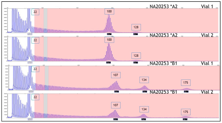
HD reference materials (n = 14) were previously determined by PCR measurement agreement across 10 laboratories and DNA sequence analysis. Allele lengths ranged from 15 to 100 CAG repeats, having the appropriate alleles to span the HD state from normal to joHD [30]. The allele(s), as identified by CAG repeat length value, and genotype were determined for each sample using the AmplideX PCR/CE HTT Kit. As shown in Table 1, all genotypes (as defined by categorical bin) obtained using the kit were in agreement with published genotypes across all cell line samples examined, with only one cell line DNA showing a single CAG repeat difference from the expected repeat length. This lone deviation, sample NA20210, has a reference heterozygous compound genotype of 17 and 74 CAG repeats, although we observed 17 and 75 repeats (see Table 1 for details). However, this difference lies well within error limits recommended by both ACMG [29] and EMQN [31] and, furthermore, the sample was reported as 17 and 75 repeats by DNA sequencing [30]. Moreover, in sample NA20253, a 3rd CAG repeat peak at 128 CAGs was repeatedly detected in the electropherogram in addition to the expected 22 and 100 CAG repeats, which were both correctly identified. These results are in accordance with an external investigation of this cell line that revealed the existence of this 3rd peak and possibly a 4th peak across two Coriell cell-line lots (Figure 1, compare A2 & B1). The 3rd peak of 128 repeats in lot A2 shifted to 134 repeats in lot B1, while also becoming more prominent; a 4th peak of 175 repeats appeared in lot B1 only. The primary expanded peak of 100 CAGs also shifted in B1 to 107 repeats, whereas the unexpanded peak remained unchanged at 22 repeats. Lot B1 was a subsequent passage of the parent cell line, and thus, the shift in the large expanded alleles likely reflects mutations that increased in length with passage of the cell line in this unstable region of the HTT gene. Similarly, in sample NA20252, a 3rd CAG repeat peak was identified at 63 CAGs in the electropherogram that was not reported in the Kalman study [30] (Figure 2).
Table 1.
Huntington disease (HD) reference samples with published allele sizes [30] compared to those determined using the AmplideX PCR/CE HTT Kit.
| Vendor ID | Input (ng) | Categorical Bin | Published (Kalman et al., 2007) |
Measured AmplideX PCR/CE HTT Kit |
||
|---|---|---|---|---|---|---|
| Allele 1 | Allele 2 | Allele 1 | Allele 2 | |||
| NA20245 | 20 | Normal | 15 | 15 | 15 | 15 |
| NA20206 | 20 | Normal | 17 | 18 | 17 | 18 |
| NA20207 | 20 | Normal | 19 | 21 | 19 | 21 |
| NA20246 | 20 | Normal | 15 | 24 | 15 | 24 |
| NA20247 | 20 | Intermediate | 15 | 29 | 15 | 29 |
| NA20248 | 20 | Reduced Penetrance | 17 | 36 | 17 | 36 |
| NA20249 | 20 | Reduced Penetrance | 22 | 39 | 22 | 39 |
| NA20208 | 20 | Expanded | 35 | 45 | 35 | 45 |
| NA20209 | 20 | Expanded | 45 | 47 | 45 | 47 |
| NA20210 | 20 | Expanded | 17 | 74 | 17 | 75 |
| NA20250 | 20 | Expanded | 15 | 40 | 15 | 40 |
| NA20251 | 20 | Expanded | 39 | 50 | 39 | 50 |
| NA20252 | 20 | Expanded | 22 | 65 & 66 | 22 | 66 * |
| NA20253 | 20 | Expanded | 22 | 100 | 22 | 100 ** |
Allele genotypes for Coriell samples shown from Kalman et al. [30] are the mode; error limits were set at ±1 for alleles ≤42 repeats and ±3 repeats for alleles >42 CAG repeats, according to the European Molecular Genetics Quality Network best practice guidelines for the molecular genetic testing of Huntington disease [31]. * A 63 CAG mosaic allele was also detected. ** A 128 CAG mosaic allele was also detected. Underlined and in bold, the difference between the two methodologies.
Figure 1.
HTT PCR/capillary electrophoresis (CE) analysis of two lots (A2, B1) of NA20253 shows additional peaks beyond the first expanded CAG repeat peak.
Figure 2.
HTT PCR/CE analysis of NA20252 shows an additional peak (63 CAG) beyond the reference genotype of 22/66.
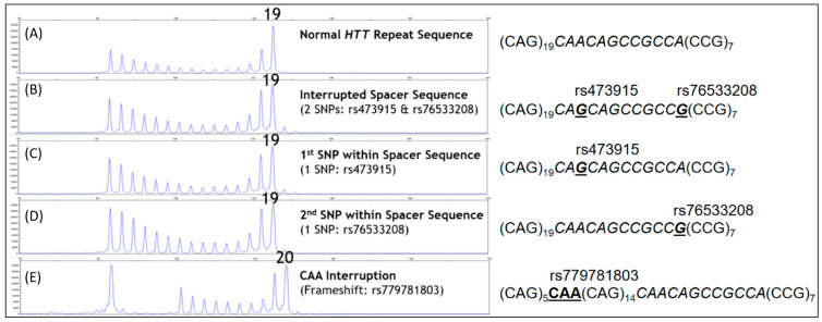
Having demonstrated the assay accuracy with HD reference materials, we next assessed the robustness of the kit to polymorphisms adjacent to and within the CAG repeat tract. As a first step, synthetic templates with 19 CAG repeats and three well-established polymorphisms (rs473915, rs76533208, and rs779781803) were amplified either alone or in combination at copy-number inputs comparable to that of the HTT gene when using genomic DNA. Both rs473915 and rs76533208 are loss-of-interruption A-to-G SNPs with profound impacts on HD age of onset [15,16,17]. They are also known to cause allele drop-outs in PCR [24,25]. By comparison, the rs779781803 variant interrupts the CAG tract with a CAA sequence. As shown in Figure 3, templates bearing all three polymorphisms, including one with rs473915 and rs76533208 on the same molecule, were robustly amplified and genotyped at or within one repeat of the correct value. We further evaluated 10 other sequence variations immediately downstream of the repeat segment that were most prevalent among HTT alleles sequenced elsewhere (Tables S1 and S2) [13]. Templates with each of these variations were also readily amplified in the PCR/CE kit assay, resulting in genotypes that were within a single repeat number of the correct value except for three samples that had deletions within the canonical CAACAGCCGCCA spacer sequence and shifted the amplicon CE mobility accordingly (Tables S1 and S2 and Figure S1).
Figure 3.
Known polymorphisms adjacent to or within the CAG repeat tract. The AmplideX PCR/CE HTT Kit accurately genotypes templates with known polymorphisms adjacent to or within the CAG repeat tract. (A) Template with 19 repeats showing the expected CE electropherogram and corresponding genotype for the relevant portion of the HTT reference sequence. (B) DNA with two SNPs (rs473915 A>G and rs76533208 A>G) interrupting the 12-bp spacer region (shown in italics) is successfully amplified to produce the expected 19 CAG result. (C,D) PCR/CE genotyping of each SNP (rs473915 or rs76533208, respectively) results in the correct 19 CAG call. (E) A CAA frameshift (rs779781803) within the repeat sequence shows a repeat-primed amplification pattern and a terminal peak corresponding to 20 CAG, rather than 19 CAG, due to the mobility shift caused by the triplet interruption. Note that the CAA interruption causes a signal “dip” in the repeat-primed pattern; this dip flags result as suspicious for unexpected sequence variation.
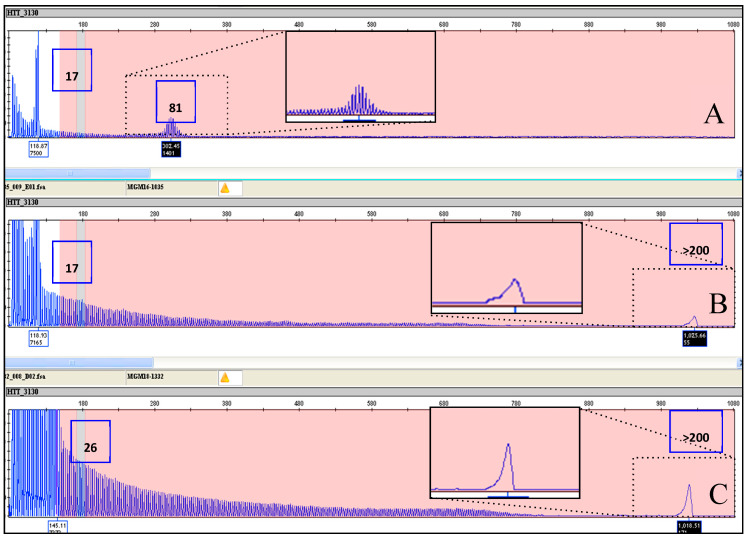
Our next step was to analyze a collection of 43 DNA samples, provided by LIRH Foundation to the Huntington and Rare Diseases Unit of CSS-Mendel Institute, section in Rome, Italy, of IRCCS Casa Sollievo della Sofferenza Research Hospital. These included 2 samples with alleles in the normal range (6–26 CAGs), 2 samples with IAs (27–35 CAGs), 6 samples with incomplete penetrance alleles (36–39 CAGs), and 33 samples carrying alleles with full penetrance (>40 CAGs). Of the alleles with full penetrance, 20 were from joHD/PHD patients carrying CAG expansions larger than 60 repeats. Of these, 41 samples had been previously tested in our laboratory using either fluorescent repeat-flanking PCR or an alternative triplet-primed PCR method, or both, whereas 4 samples had been tested in other HD reference laboratories. Primer sequences and reaction conditions of the fluorescent repeat-flanking PCR method and of the tripled-primed PCR method are described in the methods section. Similar to Coriell cell lines, the genotypes obtained using the AmplideX PCR/CE HTT Kit were identical, or with discrepancies comprised within the acceptable error limits established by the HD genetic testing guidelines [29,31], also in this series (see Table 2 for details). The assay accurately sized 18 samples with very large alleles comprised between 60 and 100 CAG repeats, and it detected two exceptionally large alleles with more than 200 CAG repeats (Table 2 and Figure 4A). In particular, samples MGM16-1035 and MGM18-1332, which could not be detected by standard fluorescent PCR, were clearly identified by an AmplideX PCR/CE HTT Kit and determined to have an expanded allele of >200 CAG repeats (Figure 4B,C). Nevertheless, only a stuttering pattern with a continuous repeat peak pattern and a pile-up peak at the end of the run was seen, since the upper limit for accurately size-expanded alleles is less of 200 CAG repeats.
Table 2.
HD samples used for this validation and corresponding genotypes obtained using fluorescent repeat-flanking PCR.
| LAB ID | Categorical Bin | “In House” PCR | AmplideX PCR/CE HTT Kit | ||
|---|---|---|---|---|---|
| Allele 1 | Allele 2 | Allele 1 | Allele 2 | ||
| MGM17-1760 | Normal | 17 | 17 | 17 | 17 |
| MGM18-1250 | Normal | 17 | 17 | 17 | 17 |
| MGM17-0052 * | Intermediate | 15 | 29 | 15 | 29 |
| MGM17-0089 | Intermediate | 19 | 32 | 19 | 32 |
| MGM16-0689 | Reduced penetrance | 17 | 37 | 17 | 37 |
| HD157-02 | Reduced penetrance | 17 | 37 | 17 | 37 |
| MGM16-0828 | Reduced penetrance | 17 | 37 | 17 | 37 |
| MGM17-1738 | Reduced penetrance | 17 | 39 | 17 | 39 |
| MGM15-1651 | Reduced penetrance | 24 | 39 | 24 | 39 |
| HD23-05 | Reduced penetrance | 33 | 39 | 33 | 39 |
| MGM16-0607 | Full penetrance | 20 | 40 | 20 | 40 |
| MGM18-0177 | Full penetrance | 14 | 41 | 14 | 41 |
| CTRL-DNA_S0162119 * | Full penetrance | 17 | 41 | 17 | 41 |
| MGM16-0196 | Full penetrance | 17 | 41 | 17 | 41 |
| MGM18-0239 | Full penetrance | 40 | 40 | 40 | 40 |
| CTRL-DNA_S0162120 * | Full penetrance | 15 | 42 | 15 | 42 |
| MGM16-0086 | Full penetrance | 18 | 42 | 18 | 42 |
| MGM17-1838 | Full penetrance | 17 | 43 | 17 | 43 |
| HD672-01 | Full penetrance | 17 | 44 | 17 | 44 |
| MGM16-0250 | Full penetrance | 17 | 46 | 16 | 46 |
| MGM18-0181 | Full penetrance | 40 | 47 | 40 | 47 |
| HD423-02 | Full penetrance | 22 | 50 | 22 | 50 |
| HD09-08 | Full penetrance | 18 | 59 | 18 | 59 |
| HD87-07 | Full penetrance | 17 | 62 | 17 | 62 |
| MGM17-1716 | Full penetrance | 17 | 63 | 17 | 63 |
| MGM17-0054 * | Full penetrance | 22 | 63 | 22 | 63 |
| MGM18-0054 | Full penetrance | 22 | 63 | 22 | 63 |
| MGM18_0054 | Full penetrance | 22 | 63 | 22 | 63 |
| HD438-01 | Full penetrance | 17 | 66 | 17 | 66 |
| MGM17-1387 | Full penetrance | 20 | 74 | 20 | 74 |
| HD09-11 | Full penetrance | 20 | 74 | 20 | 74 |
| MGM16-1030 | Full penetrance | 26 | 76 | 26 | 76 |
| HD315-01 | Full penetrance | 26 | 78 | 26 | 78 |
| MGM16-1033 | Full penetrance | 17 | 80 | 17 | 81 |
| MGM16-1032 | Full penetrance | 33 | 86 | 33 | 87 |
| HD636-01 | Full penetrance | 17 | 86 | 17 | 86 |
| HD09-12 | Full penetrance | 18 | 86 | 18 | 86 |
| MGM16-1031 | Full penetrance | 19 | 88 | 19 | 89 |
| HD379-05 | Full penetrance | 18 | 89 | 18 | 89 |
| MGM16-1034 | Full penetrance | 18 | 96 | 18 | 96 |
| HD130-05 | Full penetrance | 25 | 98 | 25 | 98 |
| MGM16-1035 | Full penetrance | 17 | 84 | 17 | >200 |
| MGM18-1332 | Full penetrance | 26 | 95 | 26 | >200 |
Acceptable error limits are ±1 repeat for alleles ≤42 and ±3 repeats for alleles >42 [31]. * Samples tested in other HD reference laboratories. Underlined and in bold is the difference between the two methodologies.
Figure 4.
Electropherogram results for samples with unusually large CAG expansions. (A) Electable 16. showing alleles of 17/81 CAG repeats and a continuous stuttering after the first prominent peak. (B,C) show sample MGM16-1035 and sample MGM18-1332, respectively, with the presence of a pile-up peak at the end of the run representing the high CAG expansion of 200 repeats (zoom of pile-up in B,C).
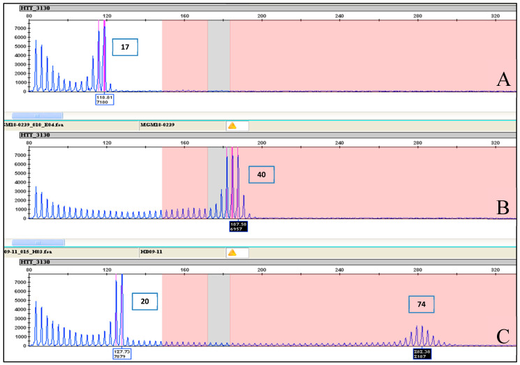
Samples with true homozygous normal genotypes were distinguished from heterozygote samples with expanded alleles by the absence of a stuttering pattern denoting an expanded allele (Figure 5A,B). As expected, an electrophoretic pattern overlapping that of normal homozygotes was observed in the case of extremely rare genotypes with double expanded alleles (Figure 5C).
Figure 5.
Difference between homo- and heterozygous alleles electropherogram results. (A) Electropherogram results of sample MGM17-1760 showing a homozygous allele of 17 CAG repeats. (B) Electropherogram results of sample MGM18-0239 showing a homozygous allele of 40 CAG repeats. (C) Electropherogram results of sample HD09-11 showing a heterozygous allele of 20 CAG repeats and an expanded allele of 74 CAG repeats. Note the continuous stuttering after the prominent peak at 20 CAG repeats to peak at 74 CAG repeat in the close-up view, which is an indication of the presence of an expanded allele.
3. Discussion
With the improved clinical and genetic knowledge of neurodegenerative diseases, the accuracy and sensitivity of genetic testing technologies is strongly required to confirm the wide spectrum of heterogeneous clinical manifestations. Reliable genetic tests help to properly stratify patients and possibly address them to clinical research trials with the potentiality of disease modification, which are emerging in recent years.
Concerning HD, the accurate sizing of the CAG repeat number, throughput, efficiency, and inter-laboratory accuracy are cornerstones for genetic testing. In the present study, we evaluated the accuracy of the AmplideX PCR/CE HTT Kit in sizing the number of CAG repeats in the HTT gene. As for the other protocols based on TP-PCR, this assay provides an electrophoretic pattern characterized by a continuous stuttering of peaks representing CAG expansion after the first prominent peak (see Figure 1, Figure 2, Figure 3, Figure 4 and Figure 5 for details). For this reason, this method easily distinguishes between a healthy homozygote (i.e., an individual carrying two unexpanded alleles of identical size) and heterozygotes with large expanded alleles that could be hard to distinguish with other PCR-based methods and confused with healthy homozygotes. In HD laboratory testing, it is extremely important to precisely discriminate the CAG repeat variability falling in the edge between the normal and the IA range, and those falling between the IA range and pathological low penetrance range of triplets. In these cases, the difference of a few CAG repeats may represent a burden for genetic diagnosis, mainly when the genetic test result is addressed to presymptomatic, at-risk people. In this study, all our samples were genotyped precisely, demonstrating the accuracy of this method for diagnosing HD. The very few discrepancies in genotyping we observed with respect to the reference samples were perhaps due to the use of different size standards in CE runs—e.g., Asuragen assay uses ABI ROX1000 and “in house” fluorescent repeat-flanking PCR assay uses ABI ROX500—and were for highly expanded and mosaic alleles and, anyhow, within the tolerated error according to the international EMQN/CMGS [31] and ACMG [29] guidelines for HD genetic testing, allowing us to assign each sample to its correct genotypic class (see Table 1 and Table 2 for details).
Compared to the other published systems, this method has also the advantage of allowing the accurate genotyping of allelic expansions up to hundreds of CAG repeats, therefore permitting the accurate genotyping of challenging cases such as those with large expansions that are typical of the joHD form. Furthermore, as demonstrated for the first time in this study, this method allows the safe identification of allelic expansions with more than 200 repeats, which are present in the rarest PHD form. In these cases, even if it is not possible to calculate the accurate number of triplets, the method clearly identified the expanded allele, which appears as a pile-up peak at the end of the run. As expected, the system was also capable of correctly genotyping expanded homozygous genotypes, which are visible as a single expanded peak in the full mutation range with the absence of a peak in the normal range. Finally, this method also correctly genotyped cell-line reference materials that are used as size references for HD molecular diagnosis and identified previously undetected extended expanded mosaic alleles (see Figure 1 and Figure 2) as additional peaks in the electropherograms of previously published samples [30].
In terms of cost, time, and effort, compared to other apparently less expensive methods, such as fluorescent repeat-flanking PCR, the AmplideX PCR/CE HTT Kit has the advantage of simplifying the testing algorithm by lowering, if not eliminating, the number of samples needing further testing by CGG or CGG+CAG, and thus reducing the time and effort spent in testing. Other HTT screening methods based on TP-PCR have also been developed, such as TP-PCR melting curve analysis [32]. While being cost-effective and amenable to high throughput, TP-PCR melting curve analysis is not able to determine the exact allele sizes in a DNA sample. This aspect represents an important limitation in the clinical setting, because all expansion-positive samples (HD-affected), as well as those samples within the cutoffs between unexpanded and intermediate alleles, or between IAs and pathological, low-penetrance alleles, should be followed up with sizing confirmation.
There is a need to standardize and improve the accuracy, sensitivity, and the specificity of the genetic determination of the precise expanded trinucleotide number within different populations for diagnostic purposes [33]. This is particularly true in light of potential results of new, upcoming, disease-modifying and neuroprotective therapies that could be used to treat people at the very first HD or presymptomatic life stages in the future.
These results also demonstrate the utility of the assay for research purposes. For example, CAG repeat mosaicism, a phenomenon occurring in dynamic expanded mutations such as in HD, represents an important research topic with implication with clinics [12,15,16], mainly in case of large size alleles [9]. Thus, genetic labs committed to research on HD may benefit from careful discrimination of mosaicism detection for research purposes in the future. Another relevant example is related to the clinical trials recently aiming to test experimental antisense oligonucleotide therapies that recommended precise CAG number and patient’s age in a math formula for inclusion criteria determination. Finally, there is strong need to raise awareness among families with at-risk children by the global HD community (e.g., pharma, families, and researchers) after the European Medicines Agency (EMA) removed a class waiver that allowed sponsors to exclude children and adolescents from clinical studies for a number of conditions, including HD, and recommended a Pediatric Investigation Plan (PIP) for future therapeutic trials [34]. Such recommendations aim to extend medicines following future positive trial results to minors, who are still excluded from all clinical experimental therapies. Therefore, the first step is to approach such a young population and their parents through careful counselling [35] by a clinical diagnosis that needs to be confirmed by a reliable genetic test, whenever allowed [36]. For instance, the methodology we describe here contributes to reliably detect highly expanded mutations above 200 CAG repeats thanks to the visible smear between the normal and the pathological alleles.
We are aware that new upcoming technologies aiming to detect intra-CAG repeat patterns [15,16,17] or bioinformatics tools [37] will help improve the genotyping of repeat-expansion alleles in the future. However, a highly qualitative vs. quantitative, PCR-based measurement of the triplet number for a wide delivery by genetic lab services is urgently needed. Therefore, the methodology we describe might help standardize HD diagnosis among genetic laboratories, with implications for genetic pathologies other than HD, e.g., Fragile X (FMR1 gene) [38], ALS/FTD (C9orf72 gene) [39], and Myotonic Dystrophy Type I (DMPK gene) [40], while providing the sensitivity and versatility to detect a broader spectrum of allelic conditions within HD itself. Remarkably, this assay was shown to successfully amplify and accurately genotype DNA harboring rare polymorphisms near the CAG repeat region that can cause allelic drop-outs, thus mitigating, if not eliminating, false-negative results that challenge current HTT PCR technologies [41]. The improved sensitivity is particularly needed in cases of presymptomatic HD, where normal allele homozygosity may rarely mask the expanded allele amplification [24,25]. By reducing such risks, more definitive results are possible from the initial test with the potential for diagnostic labs to shorten turn-around times by minimizing complex follow-on testing, such as allele sequencing.
4. Materials and Methods
4.1. Samples
Study cohorts included (1) DNA samples of a set of 14 HD cell lines with associated genotype data available from Coriell Cell Repositories (Camden, NJ, USA) that are used as reference materials for HTT CAG repeat sizing [30], and (2) DNA samples of a set of 43 additional subjects available from LIRH Foundation and genetically characterized at CSS-Mendel Institute, section in Rome, Italy, of IRCCS Casa Sollievo della Sofferenza Research Hospital (San Giovanni Rorondo, Italy). The Coriell HD reference samples included 4 cases with alleles within the normal range, 1 case with IA, 2 cases with alleles with low penetrance, and 7 samples carrying alleles with complete penetrance. Of the samples with alleles with complete penetrance, 2 harbored alleles larger than 60 CAG repeats, which are generally seen in subjects with joHD and in the rarest PHD variants (Table 1). Our 43 DNA sample collection included 2 cases with alleles within the normal range, 2 cases with IA, 6 with low penetrance, and 33 with complete penetrance. Of the samples with alleles with complete penetrance, 20 harbored alleles larger than 60 CAG repeats, which are generally seen in subjects with joHD and in the rarest PHD variants. Finally, we also included 2 samples from subjects homozygous for CAG repeat expansion (Table 2). These 43 samples had been previously tested in our laboratory using an “in-house” fluorescent repeat-flanking PCR (primer sequences and PCR protocols for this method are available upon request), whereas four had been tested in other HD reference laboratories (Table 2). The HD alleles in the 14 HD cell lines ranged in size from 15 to 100 CAG repeats (refer to [30] for details on cell-line genotypes). The HD alleles from our collection ranged from 37 to >200 CAG repeats.
4.2. AmplideX PCR/CE HTT Kit
The AmplideX PCR/CE HTT Kit (cat#: 49657; Asuragen, Inc., Austin, TX, USA) was used to PCR amplify the HTT trinucleotide CAG fragment starting from 20 ng total of purified genomic DNA, isolated from peripheral blood leukocytes using the Gentra Puregene Blood Kit (Qiagen, Germantown, MD, USA), or the MagCore Genomic DNA Whole Blood Kit (Diatech Lab line, Jesi, Italy). Samples were prepared for the PCR with a master mix from Asuragen containing HTT PCR Mix (5.0 uL), HTT forward, reverse Primer Mix (3.0 uL), and an internal calibrator. Aliquots of the DNA sample, typically 2 uL at 10 ng/uL, and 8 uL master mix were vortex-mixed, centrifuged, and transferred to a thermal cycler. Thermal cycling was performed on Applied Biosystems’ Veriti and 9700 Thermal Cyclers (Applied Biosystems, Foster City, CA, USA) using the following cycle: 95° C for 5 min, 10 cycles of 97° C for 35 s, 64° C for 35 s, 68° C for 4 min, 18 cycles of 97° C for 35 s, 64° C for 35 s, 68° C for 4 min plus 20 sec/cycle, and final extension at 72° C for 10 min, 4° C hold. Then, 2 uL of PCR product was mixed with 11 uL Hi-Di Formamide (Applied Biosystems, Foster City, CA, USA) and 2 uL ROX 1000TM Size Ladder (Asuragen, Inc., Austin, TX, USA). Samples thus prepared were denatured at 95° C for 2 min and then cooled at 4° C and held until ready for analysis by capillary electrophoresis (3130xL Genetic Analyzer, Applied Biosystems, Foster City, CA, USA). The FAM-labeled amplicons were detected using the following fragment analysis protocol: 36cm capillary, 2.5 kV, 20 s injection, and 15 kV run for 2400 s. Capillary electrophoresis run parameters were adjusted to extend POP-7 sizing range beyond 200 repeats. Lower run voltages (2.5 kV, 40 s injection) were used to allow the identification of hyper-expanded allele over to 200 repeats, with an increase in run time and loss in sensitivity due to the spreading of the signal within a “pile-up peak” (>200 repeats). This peak comprises aggregated amplicons too long to be adequately resolved by CE electropherograms, which were processed as .fsa files using GeneMapper v4.1 for analysis and manually annotated for repeat profile initiation and the size of the full-length product. A PCR control admixture sample comprising HTT alleles with 17, 39, 50, and 75 repeats and a no-template control were used in all experiments. Data analysis and interpretation was conducted using the fragment analysis software GeneMapper v4.1 and an Excel-based analysis tool, AmplideX PCR/CE HTT Macro (Asuragen, Inc., Austin, TX, USA). The AmplideX PCR/CE HTT Kit Macro can determine size and mobility correction, repeat size was determined using a linear fit adjustment of the ROX ladder size peaks to the PCR/CE control sample alleles. Alleles are reported as integer CAG repeats. The largest allele size determines genotype category: normal, intermediate, reduced penetrance or expanded. The alleles up to 200 CAG repeats are reported; the alleles >200 repeats are identified as “> 200”.
To determine the effects of polymorphisms in and near the repeat tract, ultramer DNA oligonucleotides (Integrated DNA Technologies, Coralville, IA, USA) were synthesized as controls. Ultramer samples were input at 10,000 copies and amplified on the Veriti Thermal Cycler using the AmplideX PCR/CE HTT Kit following the manufacturer’s protocol guide. Post-PCR allelic detection of the fluorescently-labeled products were resolved by capillary electrophoresis on the 3500xL Genetic Analyzer (Applied Biosystems, Foster City, CA, USA) with a 50cm capillary with the fragment analysis protocol as described above, except that a 19.5 kV run was performed. Genotypes were determined from the mobility of the target amplicon in combination with the repeat-primed peak pattern.
5. Conclusions
In conclusion, the AmplideX PCR/CE HTT Kit provides a rapid, sensitive, and reliable method to accurately genotype the HTT CAG repeat region while extending the detection limit of expanded alleles to over 200 CAG repeats. Thus, this methodology provides a comprehensive molecular diagnostic evaluation to detect the full range of HTT-CAG trinucleotides of all HD subjects, including pediatric forms carrying extremely large, hard-to-detect alleles and avoiding the use of Southern analysis to estimate the size. Finally, the AmplideX PCR technique provides an accurate approach to easily and rapidly detect mutations in those cases where particular nucleotide polymorphisms may erroneously generate false-negative results. A missing genetic diagnosis may have critical implications, both for individuals carrying the risk of HD and their families.
Acknowledgments
We are grateful to all families and patients from the LIRH Foundation Network of Huntington disease patients’ associations (LIRH-Puglia, LIRH-Tuscany, Noi Huntington) for participating in our research initiatives and kindly providing biological samples for research purposes.
Supplementary Materials
Supplementary Materials can be found at https://www.mdpi.com/1422-0067/22/4/1689/s1.
Author Contributions
A.D.L., S.S., J.R.T. and F.S.: designed the experiments; A.M., F.C., S.F. and J.R.T.: performed the experiments; G.J.L., S.S. and A.D.L.: contributed to conceptualization and methodology; F.S.: wrote the first draft. A.D.L., G.J.L. and F.S.: discussed and contributed to the final version. All authors have read and agreed to the published version of the manuscript.
Funding
This study was partially funded by the Italian Ministry of Health (funds from Ricerca Finalizzata, RAREST-JHD project, to FS (RF-2016-02364123) and from Ricerca Corrente 2019-2020 to FS).
Institutional Review Board Statement
The study was conducted according to the guidelines of the Declaration of Helsinki and approved for publication by the Institutional Review Board of LIRH Foundation (protocol code: 1.070121; date of approval: 7 January 2021).
Informed Consent Statement
Informed consent was obtained from all subjects involved in the study.
Data Availability Statement
The data that support the findings of this study are available from the corresponding author upon reasonable request.
Conflicts of Interest
Asuragen markets the AmplideX® PCR/CE HTT kit as a research use only assay for the detection and sizing of CAG repeats in the HTT gene. J.R.T., S.S., and G.J.L. are employees of Asuragen and have or may have stock in the company. The authors declare no conflict of interest.
Footnotes
Publisher’s Note: MDPI stays neutral with regard to jurisdictional claims in published maps and institutional affiliations.
References
- 1.The Huntington’s Disease Collaborative Research Group A novel gene containing a trinucleotide repeat that is expanded and unstable on Huntington’s disease chromosomes. Cell. 1993;72:971–983. doi: 10.1016/0092-8674(93)90585-E. [DOI] [PubMed] [Google Scholar]
- 2.Squitieri F. Neurodegenerative disease: ‘Fifty shades of grey’ in the Huntington disease gene. Nat. Rev. Neurol. 2013;9:421–422. doi: 10.1038/nrneurol.2013.128. [DOI] [PubMed] [Google Scholar]
- 3.Saudou F., Humbert S. The Biology of Huntingtin. Neuron. 2016;89:910–926. doi: 10.1016/j.neuron.2016.02.003. [DOI] [PubMed] [Google Scholar]
- 4.Marder K., Zhao H., Myers R.J., Cudkowics M., Kayson E., Kieburtz K., Orme C., Paulsen J., Penney J.B., Siemers E., et al. Rate of functional decline in Huntington’s disease. Huntington Study Group. Neurology. 2000;54:452–458. doi: 10.1212/WNL.54.2.452. [DOI] [PubMed] [Google Scholar]
- 5.Keum J.W., Shin A., Gillis T., Mysore J.S., Elneel K.A., Lucente D., Hadzi T., Holmans P., Jones L., Orth M., et al. The HTT CAG-Expansion Mutation Determines Age at Death but Not Disease Duration in Huntington Disease. Am. J. Hum. Genet. 2016;98:287–298. doi: 10.1016/j.ajhg.2015.12.018. [DOI] [PMC free article] [PubMed] [Google Scholar]
- 6.Kremer B., Goldberg P., Andrew S.E., Theilmann J., Telenius H., Zeisler J., Squitieri F., Lin B., Bassett A., Almqvist E., et al. A worldwide study of the Huntington’s disease mutation: The sensitivity and specificity of measuring CAG repeats. N. Engl. J. Med. 1994;330:1401–1406. doi: 10.1056/NEJM199405193302001. [DOI] [PubMed] [Google Scholar]
- 7.Rubinsztein D.C., Leggo J., Coles R., Almqvist E., Biancalana V., Cassiman J.J., Chotai K., Connarty M., Craufurd D., Curtis A., et al. Phenotypic characterization of individuals with 30–40 CAG repeats in the Huntington disease (HD) gene reveals HD cases with 36 repeats and apparently normal elderly individuals with 36–39 repeats. Am. J. Hum. Genet. 1996;59:16–22. [PMC free article] [PubMed] [Google Scholar]
- 8.Quarrell O.W.J., Brewer H.M., Squitieri F., Barker R.A., Nance M.A., Landwehrmeyer G.B. In: Juvenile Huntington’s Disease. Quarrell O.W.J., Brewer H.M., Squitieri F., Barker R.A., Nance M.A., Landwehrmeyer G.B., editors. Oxford University Press; Oxford, UK: 2009. [Google Scholar]
- 9.Fusilli C., Migliore S., Mazza T., Consoli F., De Luca A., Barbagallo G., Ciammola A., Gatto E.M., Cesarini M., Etcheverry J.L., et al. Biological and clinical manifestations of juvenile Huntington’s disease: A retrospective analysis. Lancet Neurol. 2018;17:986–993. doi: 10.1016/S1474-4422(18)30294-1. [DOI] [PubMed] [Google Scholar]
- 10.Pearson C.E., Edamura K.N., Cleary J.D. Repeat instability: Mechanisms of dynamic mutations. Nat. Rev. Genet. 2005;6:729–742. doi: 10.1038/nrg1689. [DOI] [PubMed] [Google Scholar]
- 11.Telenius H., Kremer B., Goldberg Y.P., Theilmann J., Andrew S.E., Zeisler J., Adam S., Greenberg C., Ives E.J., Clarke L.A., et al. Somatic and gonadal mosaicism of the Huntington disease gene CAG repeat in brain and sperm. Nat. Genet. 1994;6:409–414. doi: 10.1038/ng0494-409. [DOI] [PubMed] [Google Scholar]
- 12.Kennedy L., Evans E., Chen C.M., Craven L., Detloff P.J., Ennis M., Shelbourne P.F. Dramatic tissue-specific mutation length increases are an early molecular event in Huntington disease pathogenesis. Hum. Mol. Genet. 2003;12:3359–3367. doi: 10.1093/hmg/ddg352. [DOI] [PubMed] [Google Scholar]
- 13.Genetic Modifiers of Huntington’s Disease (GeM-HD) Consortium Identification of Genetic Factors that Modify Clinical Onset of Huntington’s Disease. Cell. 2015;162:516–526. doi: 10.1016/j.cell.2015.07.003. [DOI] [PMC free article] [PubMed] [Google Scholar]
- 14.Hensman Moss D.J., Pardiñas A.F., Langbehn D., Lo K., Leavitt B.R., Roos R.A.C., Durr A., Mead S. Identification of genetic variants associated with Huntington’s disease progression. A genome-wide association study. Lancet Neurol. 2017;16:701–711. doi: 10.1016/S1474-4422(17)30161-8. [DOI] [PubMed] [Google Scholar]
- 15.Ciosi M., Maxwell A., Cumming S.A., Hensman Moss D.J., Alshammari A.M., Flower M.D., Durr A., Leavitt B.R., Roos R.A.C., The Track-HD Team et al. A genetic association study of glutamine-encoding DNA sequence structures, somatic CAG expansion, and DNA repair gene variants, with Huntington disease clinical outcomes. EBioMedicine. 2019;48:568–580. doi: 10.1016/j.ebiom.2019.09.020. [DOI] [PMC free article] [PubMed] [Google Scholar]
- 16.Wright G.E.B., Collins J.A., Kay C., McDonald C., Dolzhenko E., Xia Q., Becanovic K., Drogemoller B.I., Semaka A., Nguyen C.M., et al. Length of Uninterrupted CAG, Independent of Polyglutamine Size, Results in Increased Somatic Instability, Hastening Onset of Huntington Disease. Am. J. Hum. Genet. 2019;104:1116–1126. doi: 10.1016/j.ajhg.2019.04.007. [DOI] [PMC free article] [PubMed] [Google Scholar]
- 17.Black H.F., Wright G.E.B., Collins J.A., Caron N., Kay C., Xia Q., Arning L., Bijlsma E.K., Squitieri F., Nguyen H.P., et al. Frequency of the loss of CAA interruption in the HTT CAG tract and implications for Huntington disease in the reduced penetrance range. Genet. Med. 2020;22:2108–2113. doi: 10.1038/s41436-020-0917-z. [DOI] [PMC free article] [PubMed] [Google Scholar]
- 18.Wexler N.S. The Venezuela Collaborative Research Project: Venezuelan kindreds reveal that genetic and environmental factors modulate Huntington’s disease age of onset. Proc. Natl. Acad. Sci. USA. 2004;101:3498–3503. doi: 10.1073/pnas.0308679101. [DOI] [PMC free article] [PubMed] [Google Scholar]
- 19.Goldberg Y.P., McMurray C.T., Zeisier J., Almqvist E., Sillence D., Richards F., Gacy A.M., Buchanan J., Telenius H., Hayden M.R. Increased instability of intermediate alleles in families with sporadic Huntington disease compared to similar sized intermediate alleles in the general population. Hum. Mol. Genet. 1995;4:1911–1918. doi: 10.1093/hmg/4.10.1911. [DOI] [PubMed] [Google Scholar]
- 20.Blanco S., Suarez A., Gandia-Pla S., Gòmez-Llorente C., Antùnez A., Gòmez-Capilla J.A., Fàrez-Vidal M.E. Use of capillary electrophoresis for accurate determination of CAG repeats causing Huntington disease. An oligonucleotide design avoiding shadow bands. Scand. J. Clin. Lab. Investig. 2009;68:577–584. doi: 10.1080/00365510801915171. [DOI] [PubMed] [Google Scholar]
- 21.Andrew S.E., Goldberg Y.P., Theilmann J., Zeisler J., Hayden M.R. A CCG repeat polymorphism adjacent to the CAG repeat in the Huntington disease gene: Implications for diagnostic accuracy and predictive testing. Hum. Mol. Genet. 1994;3:65–67. doi: 10.1093/hmg/3.1.65. [DOI] [PubMed] [Google Scholar]
- 22.Tóth T., Findlay I., Papp C., Tóth-Pál E., Marton T., Nagy B., Quirke P., Papp Z. Prenatal detection of trisomy 21 and 18 from amniotic fluid by quantitative fluorescent polymerase chain reaction. J. Med. Genet. 1998;35:126–129. doi: 10.1136/jmg.35.2.126. [DOI] [PMC free article] [PubMed] [Google Scholar]
- 23.Guida M., Fenwick R.G., Papp A.C., Snyder P.J., Sedra M., Prior T.W. Southern transfer protocol for confirmation of Huntington disease. Clin. Chem. 1996;42:1711–1712. doi: 10.1093/clinchem/42.10.1711. [DOI] [PubMed] [Google Scholar]
- 24.Gellera C., Meoni C., Castellotti B., Zappacosta B., Girotti F., Taroni F., Di Donato S. Errors in Huntington disease diagnostic test caused by trinucleotide deletion in the IT15 gene. Am. J. Hum. Genet. 1996;59:475–477. [PMC free article] [PubMed] [Google Scholar]
- 25.Magri S., Nanetti L., Mongelli A., Rizzo E., Taroni F., Mariotti C., Gellera C. Missing the pathological expansion in Huntington disease: De novo c.51C > G variant on the expanded allele causing intrafamilial allele dropout. Am. J. Med. Genet. A. 2020 doi: 10.1002/ajmg.a.61973. Online ahead of print. [DOI] [PubMed] [Google Scholar]
- 26.Warner J.P., Barron L.H., Goudie D., Kelly K., Dow D., Fitzpatrick D.R., Brock D.J.H. A general method for the detection of large CAG repeat expansions by fluorescent PCR. J. Med. Genet. 1996;33:1022–1026. doi: 10.1136/jmg.33.12.1022. [DOI] [PMC free article] [PubMed] [Google Scholar]
- 27.Zhao M., Lee C.G., Law H.Y., Chong S.S. Enhanced Detection and Sizing of the HTT CAG Repeat Expansion in Huntington Disease Using an Improved Triplet-Primed PCR Assay. Neurodegener. Dis. 2016;16:348–351. doi: 10.1159/000444714. [DOI] [PubMed] [Google Scholar]
- 28.Jama M., Millson A., Miller C.E., Lyon E. Triplet Repeat Primed PCR Simplifies Testing for Huntington Disease. J. Mol. Diagn. 2013;15:255–262. doi: 10.1016/j.jmoldx.2012.09.005. [DOI] [PubMed] [Google Scholar]
- 29.Bean L., Bayrak-Toydemir P. American College of Medical Genetics and Genomics Standards and Guidelines for Clinical Genetics Laboratories, 2014 edition: Technical standards and guidelines for Huntington disease. Genet. Med. 2014;16:e2. doi: 10.1038/gim.2014.146. [DOI] [PubMed] [Google Scholar]
- 30.Kalman L., Johnson M.A., Beck J., Berry-Kravis E., Buller A., Casey B., Feldman G.L., Handsfield J., Jakupciak J.P., Maragh S., et al. Development of genomic reference materials for Huntington disease genetic testing. Genet. Med. 2007;9:719–723. doi: 10.1097/GIM.0b013e318156e8c1. [DOI] [PubMed] [Google Scholar]
- 31.Losekoot M., Van Belzen M.J., Seneca S., Bauer P., Stenhouse S.A., Barton D.E. European Molecular Genetic Quality Network (EMQN). EMQN/CMGS best practice guidelines for the molecular genetic testing of Huntington disease. Eur. J. Hum. Genet. 2013;21:480–486. doi: 10.1038/ejhg.2012.200. [DOI] [PMC free article] [PubMed] [Google Scholar]
- 32.Zhao M., Cheah F.S.H., Chen M., Lee C.G., Law H.Y., Chong S.S. Improved high sensitivity screen for Huntington disease using a one-step triplet-primed PCR and melting curve assay. PLoS ONE. 2017;12:e0180984. doi: 10.1371/journal.pone.0180984. [DOI] [PMC free article] [PubMed] [Google Scholar]
- 33.Squitieri F., Mazza T., Maffi S., De Luca A., AlSalmi Q., AlHarasi S., Collins J.A., Kay C., Baine-Savanhu F., Landwhermeyer B.G., et al. Tracing the mutated HTT and haplotype of the African ancestor who spread Huntington disease into the Middle East. Genet. Med. 2020;22:1903–1908. doi: 10.1038/s41436-020-0895-1. [DOI] [PubMed] [Google Scholar]
- 34.European Medicines Agency EMA decision CW/0001/2015. [(accessed on 21 December 2018)]; Available online: http://www.ema.europa.eu/docs/en_GB/document_library/Other/2015/07/WC500190385pdf.
- 35.Migliore S., Jankovic J., Squitieri F. Genetic Counseling in Huntington’s Disease: Potential New Challenges on Horizon? Front. Neurol. 2019;30:453. doi: 10.3389/fneur.2019.00453. [DOI] [PMC free article] [PubMed] [Google Scholar]
- 36.Anderson J.A., Hayeems R.Z., Shuman C., Szego M.J., Monfared N., Bowdin S., Zlotnik Shaul R., Mein M.S. Predictive genetic testing for adult-onset disorders in minors: A critical analysis of the arguments for and against the 2013 ACMG guidelines. Clin. Genet. 2015;87:301–310. doi: 10.1111/cge.12460. [DOI] [PubMed] [Google Scholar]
- 37.Dolzhenko E., Deshpande V., Shlesinger F., Krusche P., Petrovski R., Chen S., Emig-Agius D., Gross A., Narzisi G., Bowman B., et al. ExpansionHunter: A sequence-graph-based tool to analyze variation in short tandem repeat regions. Bioinformatics. 2019;35:4754–4756. doi: 10.1093/bioinformatics/btz431. [DOI] [PMC free article] [PubMed] [Google Scholar]
- 38.Filipovic-Sadic S., Sah S., Chen L., Krosting J., Sekinger E., Zhang W., Hagerman P.J., Stenzel T.T., Hadd A.G., Latham G.J., et al. A novel FMR1 PCR method for the routine detection of low abundance expanded alleles and full mutations in fragile X syndrome. Clin. Chem. 2010;56:399–408. doi: 10.1373/clinchem.2009.136101. [DOI] [PMC free article] [PubMed] [Google Scholar]
- 39.Bram E., Javanmardi K., Nicholson K., Culp K., Thibert J.R., Kemppainen J., Le V., Schlageter A., Hadd A., Latham G.J. Comprehensive genotyping of the C9orf72 hexanucleotide repeat region in 2095 ALS samples from the NINDS collection using a two-mode, long-read PCR assay. Amyotroph. Lateral Scler. Front. Degener. 2018;20:107–114. doi: 10.1080/21678421.2018.1522353. [DOI] [PMC free article] [PubMed] [Google Scholar]
- 40.Singh S., Zhang A., Dlouhy S., Bai S. Detection of large expansions in myotonic dystrophy type 1 using triplet primed PCR. Front. Genet. 2014;5:94. doi: 10.3389/fgene.2014.00094. [DOI] [PMC free article] [PubMed] [Google Scholar]
- 41.Thibert J.R., Nicholson K., Wisotsky J., Le V., Latham G.J., Statt S. A Robust PCR/CE Assay Using AmplideX Technology for Rapid and Accurate Genotyping of CAG Repeat Expansions in HTT; Proceedings of the ACMG Annual Clinical Genetics Meeting; Charlotte, NC, USA. 10–14 April 2018. Poster 2018. [Google Scholar]
Associated Data
This section collects any data citations, data availability statements, or supplementary materials included in this article.
Supplementary Materials
Data Availability Statement
The data that support the findings of this study are available from the corresponding author upon reasonable request.


