Abstract
The aim of this systematic review and meta-analysis is to analyze the accuracy of implant placement using computer-assisted dynamic navigation procedures. An electronic literature search was carried out, supplemented by a manual search. The literature search was completed in June 2020. The results of in vitro and clinical studies were recorded separately from each other. For inclusion in the review, the studies had to examine at least the prosthetically relevant parameters for angle deviation, as well as global deviation or lateral deviation at the platform of the implant. Sixteen of 320 articles were included in the investigation: nine in vitro and seven clinical studies. The meta-analysis showed values of 4.1° for the clinical studies (95% CI, 3.12–5.10) and 3.7° for the in vitro studies (95% CI, 2.31–5.10) in terms of the angle deviation. The global deviation at the implant apex of the implant was 1.00 mm for the clinical studies (95% CI, 0.83–1.16) and 0.91 mm for the in vitro studies (95% CI, 0.60–1.12). These values indicate no significant difference between the clinical and in vitro studies. The results of this systematic review show a clinical accuracy of dynamic computer-assisted navigation that is comparable to that of static navigation. However, the dynamic navigation systems show a great heterogeneity that must be taken into account. Moreover, currently there are few clinical data available. Therefore, further investigations into the practicability of dynamic navigation seem necessary.
Keywords: computer-assisted surgery, dynamic navigation, accuracy, prostheses and implants, dental implants, dental implantation, computer-aided surgery
1. Introduction
The objective of an implant prosthetic restoration is the functional and esthetic rehabilitation of the masticatory organ after tooth loss [1]. Prosthetically driven planning has been shown to be suitable for achieving this goal in an optimal and predictable way [2]. When planning implant positions, various aspects must be considered and assessed equally. For example, the bone condition [3], the soft tissue condition [4], the inter-implant distance [5], or the position of the [6] cement space must be taken into account in the planning. Accordingly, the long-term success of an implant restoration is determined by multiple factors [7].
An established process is digital three-dimensional (3D) planning. The actual condition of the alveolar bone is recorded using 3D imaging (computed tomography (CT)) or CBCT (cone beam computed tomography) and merged with the target situation of a digitized prosthetic planning goal [1]. Such digital implant planning can be implemented using computer-assisted procedures [8]. With static computer-assisted procedures, clinically sufficient accuracy can be achieved [9]. The use of drill templates for implants has been sufficiently studied [10], and the use of these templates will achieve predictable results [11]. Various procedures for static guided implant placement have been established. For example, templates are used in which only the pilot drilling is carried out in a guided manner until the implant has been completely prepared and placed using the template [12]. It has been shown, however, that every single step in the digital workflow can lead to inaccuracies [13]. The intraoral positioning and fixation of the templates also have a significant influence on the accuracy [13]. Various authors suspected that implants that are placed using mucosa-supported templates show greater deviations from the planned implant position to the achieved implant position than those placed using tooth-supported templates [10]. Raico Gallardo et al. were able to refute this claim in their meta-analysis [14]. However, major inaccuracies in implantation can usually be traced back to application errors and not to the process per se [15]. In particular, errors in the positioning of the drilling template are to be mentioned here. A disadvantage of full-guided static navigation is listed as the fact that no intraoperative, condition-related changes are possible [11]. The use of closed drilling templates can also lead to bone overheating due to the lack of access for cooling liquid [16].
In addition to static computer-assisted surgical procedures, dynamic procedures are also available [1]. The position of the instruments is recognized in real-time through optical tracking systems using defined markers [17]. The position of the instruments and the three-dimensional planning situation can thus be followed on a screen by the implant surgeon [18]. This method may also be used in other dental issues. For example, endodontic treatments can be performed dynamically [19]. These procedures were first introduced in a variety of mostly preclinical studies [20,21,22], but were not widely used in clinical practice due to their complexity and cost [1]. The further development of computer technology and the associated computer-aided methods have increased the use of dynamic navigation in clinical practice in recent years [1]. The advantages of dynamic navigation are that any implant systems can be used thanks to open-sourced systems and that the disadvantages of storing a static template do not exist, especially with a flapless procedure [1]. Another advantage of dynamic navigation is that the implant placement is carried out with visibility and thus remains controllable, and the plan can be modified intraoperatively during the operation [11]. Another advantage over the use of drill templates is that the procedure is possible even with limited vertical space [23]. However, the complexity of the surgical procedure requires sufficient training of the surgeon and team, and a learning curve for the procedure must be observed [24]. Further development of implant surgical procedures based on technologies of virtual reality and augmented reality is rewarded with an increase in the quality of care [25].
The objective of this review was to determine the accuracy with which the planned implant position can be implemented clinically or in vitro when using dynamic navigation systems.
2. Materials and Methods
2.1. Search Strategy
This systematic review was designed in compliance with the Preferred Reporting Items for Systematic Reviews and Meta-Analyses (PRISMA) guidelines [26]. The accuracy of the implant placement was studied using dynamic computer-assisted surgical procedures. The study included partially edentulous and edentulous patients. The search, presentation, and evaluation were carried out differently according to clinical and in vitro examinations. The question posed was: “How much accuracy can be achieved clinically and on the model with dynamic computer-assisted implant placement?” The protocol of this systematic review was registered in the international database of prospectively registered systematic reviews in health and social care PROSPERO (CRD-No. 42020179128).
2.2. PICO Questions
The selection criteria are shown in Table 1.
Table 1.
Selection criteria.
| Clinical Studies | In Vitro Studies | |
|---|---|---|
| Inclusion criteria |
|
|
| Exclusion criteria |
|
|
The PICO questions when searching for clinical (P in vivo) and in vitro (P in vitro) studies were as follows:
(P) Population in vivo: Edentulous and partially edentulous patients who require an implant-prosthetic restoration;
(P) Population in vitro: Plastic models of edentulous or partially edentulous jaws;
(I) Intervention: Implant placement with a dynamic computer-assisted surgical procedure;
(C) Comparison: Results of the clinical and in vitro investigations; and
(O) Outcome Acccuracy: Deviation between the planned and actual achieved implant position.
2.3. Search Strategy
An electronic search was carried out in the MEDLINE (PubMed), EMBASE via Ovid, and Cochrane Central Register of Controlled Trials via Ovid databases. The electronic search was completed on 30 June 2020.
The search term was: (((((((((((((dental implantation [MeSH Terms]) OR dental implant [MeSH Terms]) AND dental navigation) OR computer aided dental implant) OR three dimensional dental planning) OR 3D dental planning) OR computer assisted dental implant) OR guided dental implant placement) OR dental surgical template) OR dental guided surgery) OR dental surgical guide) OR guided dental implant placement) AND ((dynamic) OR (robot *)).
In addition, a manual search for relevant further literature was carried out on the basis of the bibliographies of the included studies. Only publications in English and German were considered.
2.4. Study Selection
Duplicates were sorted out prior to screening. Two reviewers (C.E. and A.K.) reviewed all titles and abstracts independently. If there were differences of opinion, these were discussed with a third reviewer (S.S.). If a clear decision was made after the discussion, the publication was selected accordingly. If, after the discussion and assessment, a reviewer continued to report doubts, the title was included in the next selection round. No kappa values were calculated. The selection was made according to the criteria described in Table 1.
2.5. Risk of Bias, Quality Assessment, and Interstudy Heterogeneity
A quality assessment and a risk of bias assessment of the included studies were carried out by two independent reviewers (C.E. and S.S.). Since no randomized clinical studies were available for the question of this review article, the quality assessment was carried out in a modified form. The basis for this was the tool for assessing risk of bias in the Cochrane Handbook for Systematic Reviews of Interventions and the matrix for quality assessment by Esposito et al. [27]. The assessment was based on the following points: Allocation concealment, sample size calculation, inclusion and exclusion criteria, incomplete outcome data, blinding of outcome assessment, selective reporting and appropriate statistical analysis, and other bias.
2.6. Data Extraction and Method of Analysis
The data were independently compiled in tables by the investigators. These tables contain the following parameters of the clinical examination: author(s), year of publication, study design (prospective/retrospective), number of patients, number of implants, edentulism, location of the implants, implant system, guide system, and planning software. The following parameters were included in the evaluation of the in vitro examination: author(s), year of publication, number of models, number of implants, edentulism, location of the implants, implant system, guide system, and planning software. The target variables were the deviation between the planned and the actual achieved implant position. The following were recorded in the tables: angle deviation, horizontal coronal and apical deviation, vertical coronal and apical deviation, and coronal and apical global deviation.
The statistical analysis was carried out using the software program R Version 4.0.2 (The R Foundation for Statistical Computing Platform (R Foundation for Statistical Computing, Vienna, Austria.). The meta-analysis was carried out for the parameters of angle deviation and global deviation at the coronal end of the implant. In studies that had not published any information on the confidence interval (CI), the standard deviation (SD) value was used to calculate a CI equivalence. As there was evidence of heterogeneity between the included studies, totals were calculated using random-effects meta-analysis for continuous variables. The models were based on the variances approach of DerSimonian and Laird. The heterogeneity was assessed using Cochran’s Q test (p < 0.001 (CI 95%) and I2 statistic (I2 > 50%)). In addition, a descriptive analysis was carried out between the clinical and in vitro studies using the mean values. For this purpose, the angle deviation and the deviation at the coronal end of the implant were again used. A p-value of <0.05 was considered statistically significant. This descriptive analysis was performed with IBM SPSS® Statistics Version 27.0 (IBM Corp., Armonk, NY, USA).
3. Results
3.1. Study Selection
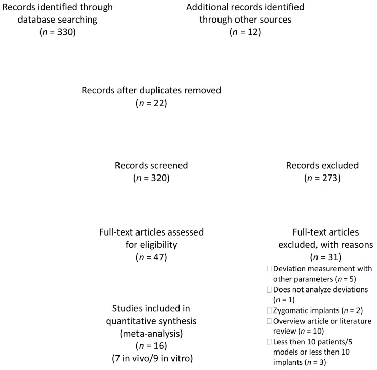
The study selection is described in the flow chart in Figure 1.
Figure 1.
PRISMA flow diagram for the search strategy and selection process for the included studies.
The electronic search yielded 320 hits, which were supplemented by 12 publications from the manual search. After screening the titles and then the abstracts, 47 publications were viewed as full texts. The review of the full texts led to the exclusion of a further 31 publications for the following reasons: deviation measurement with other parameters [22,28,29,30], does not analyze all deviations [31], zygomatic implants [32,33], different research question [17,34,35,36,37,38,39,40,41], overview article or literature review [1,11,24,42,43,44,45,46,47,48], and less then 10 patients/five models or less then 10 implants [23,49,50]. The inclusion criteria were met in nine publications of in vitro studies (Table 2) [20,21,51,52,53,54,55,56,57]. Clinical data could be used from seven publications that met the inclusion criteria (Table 3) [58,59,60,61,62,63,64].
Table 2.
Characteristics of the selected in vitro studies included in the review.
| Study | Number of Models | Number of Implants | Edentulism | Jaw | Implant System | Guide System | Planning Software |
|---|---|---|---|---|---|---|---|
| Brief et al. (2005) [20] (Robo) | 5 | 15 | Partially | Mandible | NR | RoboDent; RoboDent, GmbH, Berlin, Germany | RoboDent; RoboDent, GmbH, Berlin, Germany |
| Brief et al. (2005) (IGI) [20] | 5 | 15 | Partially | Mandible | NR | IGI DenX; Denx Ltd., Moshav Ora, Jerusalem, Israel | IGI DenX; Denx Ltd., Moshav Ora, Jerusalem, Israel |
| Emery et al. (2016) [53] | 27 | 47 | Partially and fully edentulous | Maxilla and mandible | Zimmer/Biomet 3i, Palm Beach, FL, USA | X-Guide; X-Nav Technologies, LLC, Lansdale, PA, USA | X-Guide; X-Nav Technologies, LLC, Lansdale, PA, USA |
| Hoffmann et al. (2005) [21] | 16 | 112 | Fully edentulous | Mandible | NR | Vector-Vision Compact; VVC, BrainLAB, Heimstetten, Germany | NR |
| Kang et al. (2014) (molar) [57] | 10 | 20 | Fully edentulous | Mandible molar region | Dentium implant Fx4314; Dentium, Seoul, Korea | CBYON suite system; CBYON Inc., Mountain View, CA, USA | SimPlant; Materialise Dental, Leuven, Belgium |
| Kang et al. (2014) (canine) [57] | 10 | 20 | Fully edentulous | Mandible canine region | Dentium implant Fx4314; Dentium, Seoul, Korea | CBYON suite system; CBYON Inc., Mountain View, CA, USA | SimPlant; Materialise Dental, Leuven, Belgium |
| Kim et al. (2015) [52] | 10 | 110 | Partially | Maxilla and mandible | Ostem TS; Osstem Implant, Seoul, Korea | Polaris Vicar; Northern Digital Inc., Waterloo, ON, Canada | InVivoDental; Anatomage, San Jose, CA, USA |
| Mediavilla-Guzmán et al. (2019) [54] | 10 | 20 | Partially | Maxilla | BioHorizons, Birmingham, AL, USA | Navident System; ClaroNav Inc., Toronto, ON, Canada | Navident System; ClaroNav Inc., Toronto, ON, Canada |
| Jorba-García et al. (2019) [55] | 6 | 18 | Partially | Mandible | Ticare In-Hex; MG Mozo-Grau, Valladolid, Spain | Navident System; ClaroNav Inc., Toronto, ON, Canada | Navident System; ClaroNav Inc., Toronto, ON, Canada |
| Pellegrino et al. (2020) [56] | 16 | 112 | Edentulous | Maxilla | Southern Implants, Irene, South Africa | ImplaNav; BresMedical, Sydney, Australia | ImplaNav; BresMedical, Sydney, Australia |
| Somogyi-Ganss et al. (2015) [51] | 10 | 80 | Partially | Maxilla and mandible | NR | Prototype Navident; Claron Technology Inc., Toronto, ON, Canada | Prototype Navident; Claron Technology Inc., Toronto, ON, Canada |
NR: not reported.
Table 3.
Characteristics of the selected clinical studies included in the review.
| Study | Design | Number of Patients | Number of Implants | Edentulism | Jaw | Implant System | Guide System | Planning Software |
|---|---|---|---|---|---|---|---|---|
| Aydemir and Arisan (2020) [63] | Prospective | 30 | 43 | Partially | Maxilla | Southern Implants, Irene, South Africa | Navident System; ClaroNav Inc., Toronto, ON, Canada | Navident System; ClaroNav Inc., Toronto, ON, Canada |
| Block et al. (2017a) [59] | Prospective | 80 | 80 | Partially | Maxilla and mandible | NR | X-Guide; X-Nav Technologies, LLC, Lansdale, PA, USA | X-Guide; X-Nav Technologies, LLC, Lansdale, PA, USA |
| Block et al. (2017b) [60] | Prospective | NR (but more than 10) |
219 | Partially | Maxilla and mandible | NR | X-Guide; X-Nav Technologies, LLC, Lansdale, PA, USA | X-Guide; X-Nav Technologies, LLC, Lansdale, PA, USA |
| Kaewsiri et al. (2019) [58] | Prospective | 30 | 30 | Partially | Maxilla and mandible | Straumann Bone level (18), Straumann Bone Level Taper (9), Straumann Tissue level (3) | IRIS-100; EPED Inc., Kaohsiung City, Taiwan | IRIS-100; EPED Inc., Kaohsiung City, Taiwan |
| Pellegrino et al. (2019) [61] | Prospective | 10 | 18 | Partially and fully edentulous | Maxilla and mandible | Southern Implants IBT (16), Co-axis (2), Southern Implants, Irene, South Africa | ImplaNav; BresMedical, Sydney, Australia | ImplaNav; BresMedical, Sydney, Australia |
| Stefanelli et al. (2019) [62] | Retrospective | 89 | 231 | Partially and fully edentulous | Maxilla and mandible | NR | Navident System; ClaroNav Inc., Toronto, ON, Canada | Navident System; ClaroNav Inc., Toronto, ON, Canada |
| Stefanelli et al. (2020) [64] | Retrospective | 59 | 136 | Partially | Maxilla and mandible | Osseotite Tapered, Zimmer/Biomet 3i, Palm Beach, FL, USA | Navident System; ClaroNav Inc., Toronto, ON, Canada | Navident System; ClaroNav Inc., Toronto, ON, Canada |
NR: not reported.
3.2. Quality of the Studies
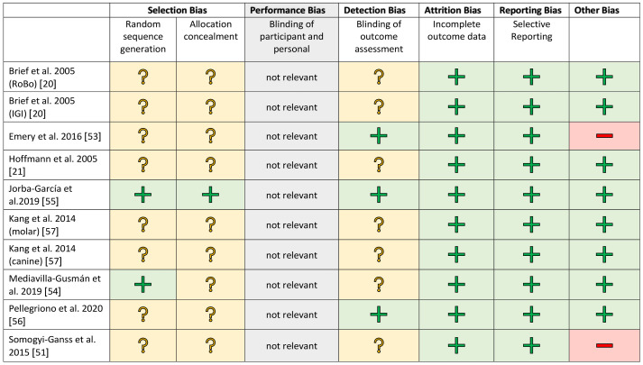
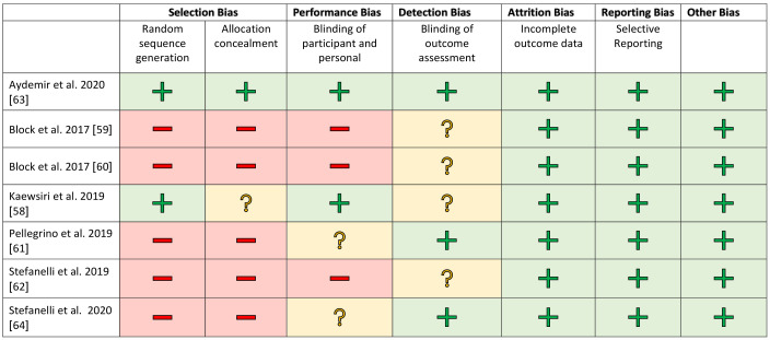
The studies were evaluated separately according to clinical and in vitro investigations in a methodical risk analysis. The risk assessment was carried out using established procedures and adapted to the samples being examined. Selection bias was included for completeness but is negligible in in vitro studies. The risk assessments are shown in Figure 2 and Figure 3.
Figure 2.
Risk of bias graph for the in vitro studies. +/green: low risk of bias; ?/yellow: unclear risk of bias; −/red: high risk of bias.
Figure 3.
Risk of bias graph for the clinical studies. +/green: low risk of bias; ?/yellow: unclear risk of bias; −/red: high risk of bias.
A blinded assessment was clearly described in only a few studies [58,63]. In the other works, this was recorded as an increased risk factor. All studies showed a low risk rating for attrition and reporting bias. The financial participation of an industrial partner was assessed as a further possible risk factor [51,53]. There was no evidence of financial support for the other studies.
3.3. Outcomes
In the nine in vitro studies, seven commercially available navigation systems and one prototype were examined. A total of 125 models were implanted. Five hundred and sixty-nine implants were placed and evaluated. Four commercially available navigation systems were used in the seven clinical studies. A total of 298 patients received implants. Seven hundred and fifty-seven implants were placed and evaluated. The navigation systems all showed a significantly different design, particularly in terms of the structure and the spatial distribution of the markers in the tracking systems for detecting the drill position. Different planning software was also used depending on the system, and various implant systems were used.
In order to compare the planned implant position with the actual achieved implant position, a CBCT was performed postoperatively in all clinical publications. The evaluation was carried out after superimposing the pre- and post-operative CBCT and the planning data. The results of the accuracy tests of the in vitro studies are shown in Table 4 and those of the clinical studies in Table 5.
Table 4.
Summary of the accuracies of the clinical studies.
| Study | Year | Number of Patients | Number of Implants | Angle Deviation | SD | 95% CI | Global Deviation at the Implant Platform | SD | 95% CI | Linear Lateral Deviation at the Implant Platform | SD | 95% CI | Vertical Deviation at the Implant Platform | SD | 95% CI | Global Deviation at the Apex | SD | 95% CI | Linear Lateral Deviation at the Apex | SD | 95% CI | Vertical Deviation at the Apex | SD | 95% CI |
|---|---|---|---|---|---|---|---|---|---|---|---|---|---|---|---|---|---|---|---|---|---|---|---|---|
| Aydemir and Arisan [63] | 2020 | 30 | 43 | 5.59 | 0.39 | 4.87–6.42 | 1.01 | 0.07 | 0.87–1.18 | 1.83 | 0.12 | 1.60–2.10 | ||||||||||||
| Block et al. [59] | 2017a | 80 | 80 | 3.62 | 2.73 | 1.37 | 0.55 | 0.87 | 0.42 | 0.93 | 0.60 | 1.56 | 0.69 | 1.09 | 0.66 | 0.96 | 0.66 | |||||||
| Block et al. [60] | 2017b | NA > 10 | 219 | 2.97 | 2.09 | 2.46–3.44 | 1.16 | 0.59 | 1.03–1.27 | 0.74 | 0.43 | 0.60–0.78 | 0.76 | 0.60 | 0.68–0.94 | 1.29 | 0.65 | 1.14–1.43 | 0.9 | 0.55 | 0.73–0.99 | 0.78 | 0.6 | 0.68–0.94 |
| Kaewsiri et al. [58] | 2019 | 30 | 30 | 3.06 | 1.37 | 2.54–3.57 | 1.05 | 0.44 | 0.89–1.21 | 1.29 | 0.5 | 1.10–1.48 | ||||||||||||
| Pellegrino et al. [61] | 2019 | 10 | 18 | 6.46 | 3.95 | 1.04 | 0.47 | 0.43 | 0.34 | 1.35 | 0.56 | |||||||||||||
| Stefanelli et al. [62] | 2019 | 89 | 231 | 2.26 | 1.62 | 0.71 | 0.40 | 1.00 | 0.49 | |||||||||||||||
| Stefanelli et al. [64] | 2020 | 59 | 136 | 2.50 | 1.04 | 0.67 | 0.29 | 0.99 | 0.33 | 0.55 | 0.25 |
CI: confidence interval; NA > 10: not available, but more than 10 patients; SD: standard deviation.
Table 5.
Summary of the accuracy of the in vitro studies.
| Study | Year | Number of Models | Number of Implants | Angle Deviation | SD | Global Deviation at the Implant Platform | SD | Linear Lateral Deviation at the Implant Platform | SD | Vertical Deviation at the Implant Platform | SD | Global Deviation at the Apex | SD | Linear Lateral Deviation at the Apex | SD | Vertical Deviation at the Apex | SD |
|---|---|---|---|---|---|---|---|---|---|---|---|---|---|---|---|---|---|
| Brief et al. [20] | 2005 (RoBo) | 5 | 15 | 2.12 | 0.78 | 0.35 | 0.17 | 0.60 | 0.20 | 0.47 | 0.18 | 0.32 | 0.21 | ||||
| Brief et al. [20] | 2005 (IGI) | 5 | 15 | 4.21 | 4.76 | 0.65 | 0.58 | 0.94 | 0.40 | 0.68 | 0.31 | 0.61 | 0.36 | ||||
| Emery et al. [53] | 2016 | 27 | 47 | 1.09 | 0.55 | 0.46 | 0.20 | 0.33 | 0.19 | 0.26 | 0.19 | 0.48 | 0.21 | 0.36 | 0.20 | 0.25 | 0.19 |
| Hoffmann et al. [21] | 2005 | 16 | 112 | 4.20 | 1.80 | ||||||||||||
| Jorba-García et al. [55] | 2019 | 6 | 18 | 1.60 | 1.30 | 1.29 | 0.46 | 0.85 | 0.41 | 1.33 | 0.50 | 0.88 | 0.47 | ||||
| Kang et al. [57] | 2014 (molar) | 10 | 20 | 8.97 | 3.83 | 3.03 | 1.81 | 0.76 | 0.84 | 2.76 | 1.03 | 1.96 | 0.93 | ||||
| Kang et al. [57] | 2014 (canine) | 10 | 20 | 12.37 | 4.18 | 2.06 | 1.43 | 1.14 | 1.25 | 3.31 | 2.07 | 1.42 | 1.01 | ||||
| Kim et al. [52] | 2015 | 10 | 110 | 2.64 | 1.31 | 0.41 | 0.12 | 0.56 | 0.14 | ||||||||
| Mediavilla-Guzmán et al. [54] | 2019 | 10 | 20 | 4.00 | 1.41 | 0.85 | 0.48 | 1.18 | 0.60 | ||||||||
| Pelleriono et al. [56] | 2020 | 16 | 112 | 4.24 | 2.52 | 1.58 | 0.80 | 0.75 | 0.74 | 1.61 | 0.75 | 0.70 | 0.67 | ||||
| Somogyi-Ganss et al. [51] | 2015 | 10 | 80 | 2.99 | 1.68 | 1.14 | 0.55 | 1.71 | 0.61 | 1.18 | 0.56 | 1.04 | 0.71 |
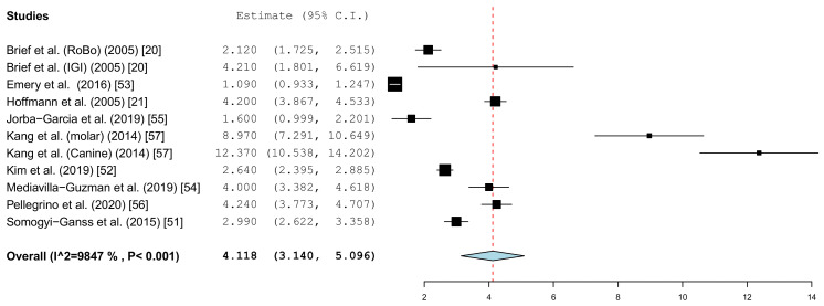
Different parameters were recorded in the publications. With the inclusion criteria, the minimum requirement was defined as the angle deviation and the linear or global deviation at the coronal end of the implant. The representation in the forest plot diagrams and the descriptive comparison between clinical and in vitro examinations were therefore limited to these two prosthetically relevant target values. The angular deviation of the in vitro studies is shown in Figure 4. In the in vitro examinations, the global deviation was given in five papers and the linear coronal deviation of the implant in seven papers. For this reason, two forest plots were calculated.
Figure 4.
Forest plot demonstrating the angular deviation (°) for all of the selected in vitro articles.
Figure 5 summarizes the global deviations at the implant platform. Figure 6 shows the global deviation at the implant apex. The following Figure 7 shows the lateral deviations at the implant platform without looking at the horizontal deviation. In the clinical studies, data on both the angular deviation (Figure 8) and the global deviation at the implant platform were available (Figure 9). Figure 10 summarized the global deviation at the implant apex. All of the forest plots confirmed a substantial heterogeneity in all of the parameters in both groups. The results of these analyses were p < 0.001 and I2 = 97.4%–99.6%.
Figure 5.
Forest plot demonstrating the global deviation (mm) at the implant platform measured for all of the selected in vitro articles.
Figure 6.
Forest plot demonstrating the global deviation (mm) at the implant apex measured for all of the selected in vitro articles.
Figure 7.
Forest plot demonstrating linear lateral deviation (mm) at the implant platform measured for all of the selected in vitro articles.
Figure 8.
Forest plot demonstrating the angular deviation (°) for all of the selected clinical articles.
Figure 9.
Forest plot demonstrating the global deviation (mm) at the implant platform measured for all of the selected clinical articles.
Figure 10.
Forest plot demonstrating the global deviation (mm) at the implant apex measured for all of the selected clinical articles.
3.3.1. Coronal Deviation
The results of the meta-analysis showed comparable mean values. The global deviation in the clinical studies was 1.00 mm (95% CI, 0.83–1.16). In the in vitro studies, the global deviation was 0.91 mm (95% CI, 0.60–1.21) and the lateral deviation was 1.01 mm (95% CI, 0.68–1.34). The forest plots demonstrate that the accuracy was highly system-dependent. Both evaluation groups showed outliers for the mean accuracy. It can also be seen from the graph that the scatter around the mean values in the clinical examinations is higher.
3.3.2. Apical Deviation
The results of the meta-analysis showed comparable mean values. The global deviation in the clinical studies was 1.33 mm (95% CI, 0.98–1.68). In the in vitro studies, this global deviation was 1.04 mm (95% CI, 0.76–1.33). The forest plots demonstrate that the accuracy was highly system-dependent. Both evaluation groups showed outliers for the mean accuracy. It can also be seen from the graph that the scatter around the mean values in the clinical examinations is higher.
3.3.3. Angle Deviation
The results of the meta-analysis also showed comparable mean values (clinical 4.1° (95% CI, 3.14–5.10) and in vitro 3.7° (95% CI, 2.31–5.10)). The forest plots showed little scatter in both the clinical and in vitro studies.
The dynamic navigation also showed comparable accuracies compared to the static guided surgery. The mean values of this meta-analysis for dynamic navigation and the values from two [10,65] meta-analyses for static guided implant placement are shown in Table 6. This table is supplemented with the results of an examination [66] of the accuracy of freehand implants. Thus far, there has been no systematic review, as only a few studies have been published. To classify the in vitro studies on the clinical studies, we carried out a static analysis for their variance. This analysis should have a descriptive value, since there is not yet enough data on an actual one. No statistically significant differences in the achieved accuracy could be detected when comparing in vitro and clinical studies. For the angle deviation, the Mann–Whitney U test indicated a significance level of p = 0.964 and a deviation at the implant shoulder of p = 0.685.
Table 6.
Comparison the data from the present study of the angle deviation and the global deviation at the coronal end of the implants between dynamic and static navigation and a work on freehand implant placement (mean value and standard error (if specified)).
| Study | Angle Deviation | Global Coronal Deviation | Global Apical Deviation |
|---|---|---|---|
| Mean | Mean | Mean | |
| Dynamic navigation in vitro | 4.1° | 1.03 mm | 1.04 mm |
| Dynamic navigation clinical | 3.7° | 1.00 mm | 1.33 mm |
| Static navigation—clinical review 1 [65] | 3.6° | 1.10 mm | 1.40 mm |
| Static navigation—clinical review 2 [10] | 3.5° | 1.2 mm | 1.40 mm |
| Freehand implant placement [66] | 9.9° | 2.77 mm | 2.91 mm |
4. Discussion
In the present meta-analysis, the accuracy between the planned and actually achieved implant positions was evaluated using dynamic computer-assisted navigation. In their study design, all examinations showed clearly different influencing factors. Various navigation systems with fundamental differences in the structure of the optical tracking system and the arrangement of the markers were used. Furthermore, different implant planning programs and different implants were used.
The high degree of heterogeneity of the measured parameters suggests that the evaluated studies examined navigation systems with significantly different qualities. The structure and arrangement of the marker structures seems to lead to different results. For reviews of computer-assisted static guided implants, there was a lower heterogeneity [65]. Moreover, the present study could not make any statement on clinical suitability for practice.
In this meta-analysis, in vitro and clinical studies were handled separately. This was based on clinical experience and the assumption that intraoral implementation in the patient is difficult. The opening of the mouth, movements of the patient, or the restricted view of the operating field can all have an influence [67]. Therefore, in this meta-analysis, it was hypothesized that there would be a significant difference between these two study designs. This was supported by a systematic review of static navigation by Bover-Ramos et al., in which significant differences between in vitro and clinical studies were found [65]. There were differences in the horizontal apical deviation and the angle deviation, and this statement was supported by Schneider’s meta-analysis [68]. Significant differences could not be detected in the evaluation of the dynamic navigation presented here. An evaluation of the subgroups in which in vitro and clinical studies with a similar study design—particularly the use of only one navigation system—are considered could not be carried out due to the as yet low number of studies. Therefore, a final assessment that the in vitro-achieved accuracy can also actually be achieved clinically cannot be made. The included studies showed moderate limitations in methodological quality and were thus in the range of the usual quality requirements [14]. In all clinical studies, the accuracy was determined by overlaying post-operative CBCTs. This should be assessed critically in further studies for considerations of radiation protection, since non-radiological examination methods are also available [15].
In the present evaluation, the focus was on the angle deviation and the deviation at the implant platform. This was justified in the prosthetic importance of these parameters. Inclined implant axes make it difficult to correctly design the approximal contacts. In their meta-analysis, Omori et al. found that after one year of follow-up, implants supporting angulated abutments yielded significantly more marginal bone loss than those supporting straight abutments [69]. The coronal position of the implant in particular has a decisive influence on the esthetic result [70]. Since our working group evaluated computer-assisted implants mainly as a primary prosthetic tool and only secondarily as a surgical tool, the values at the apex of the implant were not evaluated separately. Various authors have argued that this level of precision is necessary when assessing possible damage to relevant anatomical structures, e.g., the inferior alveolar nerves or the maxillary sinus [65]. An assessment of the lateral deviation at the implant tip makes only limited sense, however, since this value is directly dependent on the length of the implant used. However, in order to evaluate the possible risk of injury-sensitive anatomical structures such as the sinus, the mental foramen, and the mandible canal, these values were also evaluated. The dependence of the deviation on length is a purely geometric function and therefore has no clinical justification. Minimum distances to vulnerable anatomical structures must be observed in all procedures for guided implant placement [71]. The safety clearances given in the literature are between 1.0 [72] and 0.5 mm horizontally [73] and 1.7 [65] and 1.2 mm vertically [73]. Maintaining the horizontal position should be more reliable with a dynamic system that can be checked visually than with static systems in which a template covers the operating field.
The present evaluation also showed that the accuracy depends heavily on the navigation system. The considerable differences of the technique used was demonstrated by the work of Kang et al. The mean values in the work by Kang et al. showed significant deviations from the mean values calculated in the meta-analysis in the group of in vitro studies [57]. Overall, the studies showed a high heterogeneity of the data. A similar picture emerged in the clinical studies in the work of Pellegrino et al., as well as that of Aydemir and Arisan [61,63]. There were also considerable differences in the spread of the values in the individual studies. The influence of the planning software cannot be determined from the available data. Static navigation studies have shown that the accuracy of superimposing 3D datasets in software programs is different. For example there are significant differences in the accuracy of the matching of Standard Tessellation Language (STL) data and Digital Imaging and Communication in Medicine (DICOM) data [74]. All of the limitations and co-factors mentioned for static navigation, which influence the accuracy [10], can be transferred to the planning programs used here.
The results of this meta-analysis are in the range of the results obtained from meta-analyses for static navigation [10,65,67]. Further investigations must show whether this also applies to dynamic navigation systems that have not been investigated so far. There are also only studies by one working group of individual systems. Therefore, there are no statements about the robustness of the values when the systems are used by different surgeons. In particular, in addition to the accuracy, the feasibility in everyday clinical practice must also be examined in further studies. The parameters to be considered here are the time required, the surgical difficulty in use, and, last but not least, the costs.
5. Conclusions
With dynamic computer-assisted navigation, mean deviations between the planned and actually achieved implant position of 1.00 mm clinically and 0.91 mm in vitro were calculated at the platform of the implant. The mean global deviation at the implant apex was 1.33 mm clinically and 1.04 mm in vitro. The angle deviation averaged 4.1° in clinical examinations and 3.7° in in vitro examinations. These values were significantly different. These results of dynamic navigation show similar values as described in various systematic reviews for static navigation. The usual surgical safety distances must therefore also be observed with dynamic navigation. With further studies, recommendations can possibly be given for areas of application in which static and dynamic processes have advantages.
Author Contributions
Conceptualization, S.S., A.K., and C.E.; methodology, S.S.; software, S.S.; validation, S.S., C.E., A.K., and R.G.L.; formal analysis and writing—original draft preparation, S.S.; writing—review and editing, A.K., C.E., and R.G.L.; visualization, S.S.; supervision, R.G.L. All authors critically reviewed and edited the manuscript before submission. All authors read and agreed to the published version of the manuscript.
Funding
This research received no external funding.
Institutional Review Board Statement
Not applicable.
Informed Consent Statement
Not applicable.
Data Availability Statement
Not applicable.
Conflicts of Interest
The authors declare no conflict of interest.
Footnotes
Publisher’s Note: MDPI stays neutral with regard to jurisdictional claims in published maps and institutional affiliations.
References
- 1.D’Haese J., Ackhurst J., Wismeijer D., De Bruyn H., Tahmaseb A. Current state of the art of computer-guided implant surgery. Periodontology 2000. 2017;73:121–133. doi: 10.1111/prd.12175. [DOI] [PubMed] [Google Scholar]
- 2.Garber D.A., Belser U.C. Restoration-driven implant placement with restoration-generated site development. Compend. Contin. Educ. Dent. 1995;16:796, 798–802, 804. [PubMed] [Google Scholar]
- 3.Albrektsson T., Chrcanovic B., Ostman P.O., Sennerby L. Initial and long-term crestal bone responses to modern dental implants. Periodontology 2000. 2017;73:41–50. doi: 10.1111/prd.12176. [DOI] [PubMed] [Google Scholar]
- 4.Romanos G.E., Delgado-Ruiz R., Sculean A. Concepts for prevention of complications in implant therapy. Periodontology 2000. 2019;81:7–17. doi: 10.1111/prd.12278. [DOI] [PubMed] [Google Scholar]
- 5.Al Amri M.D. Influence of interimplant distance on the crestal bone height around dental implants: A systematic review and meta-analysis. J. Prosthet. Dent. 2016;115:278–282. doi: 10.1016/j.prosdent.2015.09.001. [DOI] [PubMed] [Google Scholar]
- 6.Staubli N., Walter C., Schmidt J.C., Weiger R., Zitzmann N.U. Excess cement and the risk of peri-implant disease—A systematic review. Clin. Oral. Implant. Res. 2017;28:1278–1290. doi: 10.1111/clr.12954. [DOI] [PubMed] [Google Scholar]
- 7.Bosshardt D.D., Chappuis V., Buser D. Osseointegration of titanium, titanium alloy and zirconia dental implants: Current knowledge and open questions. Periodontology 2000. 2017;73:22–40. doi: 10.1111/prd.12179. [DOI] [PubMed] [Google Scholar]
- 8.Wismeijer D., Joda T., Flugge T., Fokas G., Tahmaseb A., Bechelli D., Bohner L., Bornstein M., Burgoyne A., Caram S., et al. Group 5 ITI consensus report: Digital technologies. Clin. Oral. Implant. Res. 2018;29(Suppl. 16):436–442. doi: 10.1111/clr.13309. [DOI] [PubMed] [Google Scholar]
- 9.Al Yafi F., Camenisch B., Al-Sabbagh M. Is digital guided implant surgery accurate and reliable? Dent. Clin. N. Am. 2019;63:381–397. doi: 10.1016/j.cden.2019.02.006. [DOI] [PubMed] [Google Scholar]
- 10.Tahmaseb A., Wu V., Wismeijer D., Coucke W., Evans C. The accuracy of static computer-aided implant surgery: A systematic review and meta-analysis. Clin. Oral. Implant. Res. 2018;29(Suppl. 16):416–435. doi: 10.1111/clr.13346. [DOI] [PubMed] [Google Scholar]
- 11.Gargallo-Albiol J., Barootchi S., Salomo-Coll O., Wang H.L. Advantages and disadvantages of implant navigation surgery. A systematic review. Ann. Anat. 2019;225:1–10. doi: 10.1016/j.aanat.2019.04.005. [DOI] [PubMed] [Google Scholar]
- 12.Abduo J., Lau D. Accuracy of static computer-assisted implant placement in anterior and posterior sites by clinicians new to implant dentistry: In vitro comparison of fully guided, pilot-guided, and freehand protocols. Int. J. Implant. Dent. 2020;6:10. doi: 10.1186/s40729-020-0205-3. [DOI] [PMC free article] [PubMed] [Google Scholar]
- 13.Tahmaseb A., Wismeijer D., Coucke W., Derksen W. Computer technology applications in surgical implant dentistry: A systematic review. Int. J. Oral. Maxillofac. Implant. 2014;29:25–42. doi: 10.11607/jomi.2014suppl.g1.2. [DOI] [PubMed] [Google Scholar]
- 14.Raico Gallardo Y.N., da Silva-Olivio I.R.T., Mukai E., Morimoto S., Sesma N., Cordaro L. Accuracy comparison of guided surgery for dental implants according to the tissue of support: A systematic review and meta-analysis. Clin. Oral. Implant. Res. 2017;28:602–612. doi: 10.1111/clr.12841. [DOI] [PubMed] [Google Scholar]
- 15.Schnutenhaus S., Edelmann C., Rudolph H., Luthardt R.G. Retrospective study to determine the accuracy of template-guided implant placement using a novel nonradiologic evaluation method. Oral. Surg. Oral. Med. Oral. Pathol. Oral. Radiol. 2016;121:e72–e79. doi: 10.1016/j.oooo.2015.12.012. [DOI] [PubMed] [Google Scholar]
- 16.Liu S.H., Cao C.R., Lin W.C., Shu C.M. Experimental and numerical simulation study of the thermal hazards of four azo compounds. J. Hazard Mater. 2019;365:164–177. doi: 10.1016/j.jhazmat.2018.11.003. [DOI] [PubMed] [Google Scholar]
- 17.Du Y., Wangrao K., Liu L., Liu L., Yao Y. Quantification of image artifacts from navigation markers in dynamic guided implant surgery and the effect on registration performance in different clinical scenarios. Int. J. Oral. Maxillofac. Implant. 2019;34:726–736. doi: 10.11607/jomi.7179. [DOI] [PubMed] [Google Scholar]
- 18.Vercruyssen M., Fortin T., Widmann G., Jacobs R., Quirynen M. Different techniques of static/dynamic guided implant surgery: Modalities and indications. Periodontology 2000. 2014;66:214–227. doi: 10.1111/prd.12056. [DOI] [PubMed] [Google Scholar]
- 19.Gambarini G., Galli M., Stefanelli L.V., Di Nardo D., Morese A., Seracchiani M., De Angelis F., Di Carlo S., Testarelli L. Endodontic microsurgery using dynamic navigation system: A case report. J. Endod. 2019;45:1397–1402. doi: 10.1016/j.joen.2019.07.010. [DOI] [PubMed] [Google Scholar]
- 20.Brief J., Edinger D., Hassfeld S., Eggers G. Accuracy of image-guided implantology. Clin. Oral. Implant. Res. 2005;16:495–501. doi: 10.1111/j.1600-0501.2005.01133.x. [DOI] [PubMed] [Google Scholar]
- 21.Hoffmann J., Westendorff C., Gomez-Roman G., Reinert S. Accuracy of navigation-guided socket drilling before implant installation compared to the conventional free-hand method in a synthetic edentulous lower jaw model. Clin. Oral. Implant. Res. 2005;16:609–614. doi: 10.1111/j.1600-0501.2005.01153.x. [DOI] [PubMed] [Google Scholar]
- 22.Kramer F.J., Baethge C., Swennen G., Rosahl S. Navigated vs. conventional implant insertion for maxillary single tooth replacement. Clin. Oral. Implant. Res. 2005;16:60–68. doi: 10.1111/j.1600-0501.2004.01058.x. [DOI] [PubMed] [Google Scholar]
- 23.Chen J.T. A novel application of dynamic navigation system in socket shield technique. J. Oral. Implantol. 2019;45:409–415. doi: 10.1563/aaid-joi-D-19-00072. [DOI] [PubMed] [Google Scholar]
- 24.Block M.S., Emery R.W. Static or dynamic navigation for implant placement-choosing the method of guidance. J. Oral. Maxillofac. Surg. 2016;74:269–277. doi: 10.1016/j.joms.2015.09.022. [DOI] [PubMed] [Google Scholar]
- 25.Ayoub A., Pulijala Y. The application of virtual reality and augmented reality in oral & maxillofacial surgery. BMC Oral. Health. 2019;19:238. doi: 10.1186/s12903-019-0937-8. [DOI] [PMC free article] [PubMed] [Google Scholar]
- 26.Moher D., Liberati A., Tetzlaff J., Altman D.G., The PRISMA Group Reprint—Preferred reporting items for systematic reviews and meta-analyses: The PRISMA statement. Phys. Ther. 2009;89:873–880. doi: 10.1093/ptj/89.9.873. [DOI] [PubMed] [Google Scholar]
- 27.Esposito M., Coulthard P., Worthington H.V., Jokstad A. Quality assessment of randomized controlled trials of oral implants. Int. J. Oral. Maxillofac. Implant. 2001;16:783–792. [PubMed] [Google Scholar]
- 28.Gaggl A., Schultes G., Karcher H. Navigational precision of drilling tools preventing damage to the mandibular canal. J. Craniomaxillofac. Surg. 2001;29:271–275. doi: 10.1054/jcms.2001.0239. [DOI] [PubMed] [Google Scholar]
- 29.Zheng G., Gu L., Wu Z., Huang Y., Kang L. The implementation of an integrated computer-aided system for dental implantology. Ann. Int. Conf. IEEE Eng. Med. Biol. Soc. 2008;2008:58–61. doi: 10.1109/IEMBS.2008.4649090. [DOI] [PubMed] [Google Scholar]
- 30.Chen Y.T., Chiu Y.W., Peng C.Y. Preservation of inferior alveolar nerve using the dynamic dental implant navigation system. J. Oral. Maxillofac. Surg. 2020;78:678–679. doi: 10.1016/j.joms.2020.01.007. [DOI] [PubMed] [Google Scholar]
- 31.Wittwer G., Adeyemo W.L., Schicho K., Gigovic N., Turhani D., Enislidis G. Computer-guided flapless transmucosal implant placement in the mandible: A new combination of two innovative techniques. Oral. Surg. Oral. Med. Oral. Pathol. Oral. Radiol. Endod. 2006;101:718–723. doi: 10.1016/j.tripleo.2005.10.047. [DOI] [PubMed] [Google Scholar]
- 32.Zhou W., Fan S., Wang F., Huang W., Jamjoom F.Z., Wu Y. A novel extraoral registration method for a dynamic navigation system guiding zygomatic implant placement in patients with maxillectomy defects. Int. J. Oral. Maxillofac. Surg. 2020 doi: 10.1016/j.ijom.2020.03.018. [DOI] [PubMed] [Google Scholar]
- 33.Schramm A., Gellrich N.C., Schimming R., Schmelzeisen R. Computer-assisted insertion of zygomatic implants (Branemark system) after extensive tumor surgery. Mund Kiefer. Gesichtschir. 2000;4:292–295. doi: 10.1007/s100060000211. [DOI] [PubMed] [Google Scholar]
- 34.Wittwer G., Adeyemo W.L., Wagner A., Enislidis G. Computer-guided flapless placement and immediate loading of four conical screw-type implants in the edentulous mandible. Clin. Oral. Implant. Res. 2007;18:534–539. doi: 10.1111/j.1600-0501.2007.01370.x. [DOI] [PubMed] [Google Scholar]
- 35.Berdougo M., Fortin T., Blanchet E., Isidori M., Bosson J.L. Flapless implant surgery using an image-guided system. A 1- to 4-year retrospective multicenter comparative clinical study. Clin. Implant Dent. Relat. Res. 2010;12:142–152. doi: 10.1111/j.1708-8208.2008.00146.x. [DOI] [PubMed] [Google Scholar]
- 36.Wanschitz F., Birkfellner W., Watzinger F., Schopper C., Patruta S., Kainberger F., Figl M., Kettenbach J., Bergmann H., Ewers R. Evaluation of accuracy of computer-aided intraoperative positioning of endosseous oral implants in the edentulous mandible. Clin. Oral. Implant. Res. 2002;13:59–64. doi: 10.1034/j.1600-0501.2002.130107.x. [DOI] [PubMed] [Google Scholar]
- 37.Golob Deeb J., Bencharit S., Carrico C.K., Lukic M., Hawkins D., Rener-Sitar K., Deeb G.R. Exploring training dental implant placement using computer-guided implant navigation system for predoctoral students: A pilot study. Eur. J. Dent. Educ. 2019;23:415–423. doi: 10.1111/eje.12447. [DOI] [PubMed] [Google Scholar]
- 38.Essig H., Rana M., Kokemueller H., Zizelmann C., von See C., Ruecker M., Tavassol F., Gellrich N.C. Referencing of markerless CT data sets with cone beam subvolume including registration markers to ease computer-assisted surgery—A clinical and technical research. Int. J. Med. Robot. 2013;9:e39–e45. doi: 10.1002/rcs.1444. [DOI] [PubMed] [Google Scholar]
- 39.Gordon C.R., Murphy R.J., Coon D., Basafa E., Otake Y., Al Rakan M., Rada E., Susarla S., Swanson E., Fishman E., et al. Preliminary development of a workstation for craniomaxillofacial surgical procedures: Introducing a computer-assisted planning and execution system. J. Craniofac. Surg. 2014;25:273–283. doi: 10.1097/SCS.0000000000000497. [DOI] [PMC free article] [PubMed] [Google Scholar]
- 40.Widmann G., Stoffner R., Keiler M., Zangerl A., Widmann R., Puelacher W., Bale R. A laboratory training and evaluation technique for computer-aided oral implant surgery. Int. J. Med. Robot. 2009;5:276–283. doi: 10.1002/rcs.258. [DOI] [PubMed] [Google Scholar]
- 41.Chen X., Lin Y., Wu Y., Wang C. Real-time motion tracking in image-guided oral implantology. Int. J. Med. Robot. 2008;4:339–347. doi: 10.1002/rcs.215. [DOI] [PubMed] [Google Scholar]
- 42.Hultin M., Svensson K.G., Trulsson M. Clinical advantages of computer-guided implant placement: A systematic review. Clin. Oral. Implant. Res. 2012;23(Suppl. 6):124–135. doi: 10.1111/j.1600-0501.2012.02545.x. [DOI] [PubMed] [Google Scholar]
- 43.Block M.S. Static and dynamic navigation for dental implant placement. J. Oral. Maxillofac. Surg. 2016;74:231–233. doi: 10.1016/j.joms.2015.12.002. [DOI] [PubMed] [Google Scholar]
- 44.Marquardt P., Witkowski S., Strub J. Three-dimensional navigation in implant dentistry. Eur. J. Esthet. Dent. 2007;2:80–98. [PubMed] [Google Scholar]
- 45.Block M.S. Accuracy using static or dynamic navigation. J. Oral. Maxillofac. Surg. 2016;74:2–3. doi: 10.1016/j.joms.2015.11.002. [DOI] [PubMed] [Google Scholar]
- 46.Brignardello-Petersen R. Similar deviation between planned and placed implants when using static and dynamic computer-assisted systems in single-tooth implants. J. Am. Dent. Assoc. 2019;150:e171. doi: 10.1016/j.adaj.2019.06.009. [DOI] [PubMed] [Google Scholar]
- 47.Azari A., Nikzad S. Computer-assisted implantology: Historical background and potential outcomes—A review. Int. J. Med. Robot. 2008;4:95–104. doi: 10.1002/rcs.188. [DOI] [PubMed] [Google Scholar]
- 48.Miller R.J., Bier J. Surgical navigation in oral implantology. Implant Dent. 2006;15:41–47. doi: 10.1097/01.id.0000202637.61180.2b. [DOI] [PubMed] [Google Scholar]
- 49.Wagner A., Wanschitz F., Birkfellner W., Zauza K., Klug C., Schicho K., Kainberger F., Czerny C., Bergmann H., Ewers R. Computer-aided placement of endosseous oral implants in patients after ablative tumour surgery: Assessment of accuracy. Clin. Oral. Implant. Res. 2003;14:340–348. doi: 10.1034/j.1600-0501.2003.110812.x. [DOI] [PubMed] [Google Scholar]
- 50.Herklotz I., Beuer F., Kunz A., Hildebrand D., Happe A. Navigation in implantology. Int. J. Comput. Dent. 2017;20:9–19. [PubMed] [Google Scholar]
- 51.Somogyi-Ganss E., Holmes H.I., Jokstad A. Accuracy of a novel prototype dynamic computer-assisted surgery system. Clin. Oral. Implant. Res. 2015;26:882–890. doi: 10.1111/clr.12414. [DOI] [PubMed] [Google Scholar]
- 52.Kim S.G., Lee W.J., Lee S.S., Heo M.S., Huh K.H., Choi S.C., Kim T.I., Yi W.J. An advanced navigational surgery system for dental implants completed in a single visit: An in vitro study. J. Craniomaxillofac. Surg. 2015;43:117–125. doi: 10.1016/j.jcms.2014.10.022. [DOI] [PubMed] [Google Scholar]
- 53.Emery R.W., Merritt S.A., Lank K., Gibbs J.D. Accuracy of dynamic navigation for dental implant placement-model-based evaluation. J. Oral. Implantol. 2016;42:399–405. doi: 10.1563/aaid-joi-D-16-00025. [DOI] [PubMed] [Google Scholar]
- 54.Mediavilla Guzman A., Riad Deglow E., Zubizarreta-Macho A., Agustin-Panadero R., Hernandez Montero S. Accuracy of computer-aided dynamic navigation compared to computer-aided static navigation for dental implant placement: An in vitro study. J. Clin. Med. 2019;8:2123. doi: 10.3390/jcm8122123. [DOI] [PMC free article] [PubMed] [Google Scholar]
- 55.Jorba-Garcia A., Figueiredo R., Gonzalez-Barnadas A., Camps-Font O., Valmaseda-Castellon E. Accuracy and the role of experience in dynamic computer guided dental implant surgery: An in-vitro study. Med. Oral. Patol. Oral. Cir. Bucal. 2019;24:e76–e83. doi: 10.4317/medoral.22785. [DOI] [PMC free article] [PubMed] [Google Scholar]
- 56.Pellegrino G., Bellini P., Cavallini P.F., Ferri A., Zacchino A., Taraschi V., Marchetti C., Consolo U. Dynamic navigation in dental implantology: The influence of surgical experience on implant placement accuracy and operating time. An in vitro study. Int. J. Environ. Res. Public. Health. 2020;17:2153. doi: 10.3390/ijerph17062153. [DOI] [PMC free article] [PubMed] [Google Scholar]
- 57.Kang S.H., Lee J.W., Lim S.H., Kim Y.H., Kim M.K. Verification of the usability of a navigation method in dental implant surgery: In vitro comparison with the stereolithographic surgical guide template method. J. Craniomaxillofac. Surg. 2014;42:1530–1535. doi: 10.1016/j.jcms.2014.04.025. [DOI] [PubMed] [Google Scholar]
- 58.Kaewsiri D., Panmekiate S., Subbalekha K., Mattheos N., Pimkhaokham A. The accuracy of static vs. dynamic computer-assisted implant surgery in single tooth space: A randomized controlled trial. Clin. Oral. Implant. Res. 2019;30:505–514. doi: 10.1111/clr.13435. [DOI] [PubMed] [Google Scholar]
- 59.Block M.S., Emery R.W., Lank K., Ryan J. Implant placement accuracy using dynamic navigation. Int. J. Oral. Maxillofac. Implant. 2017;32:92–99. doi: 10.11607/jomi.5004. [DOI] [PubMed] [Google Scholar]
- 60.Block M.S., Emery R.W., Cullum D.R., Sheikh A. Implant placement is more accurate using dynamic navigation. J. Oral. Maxillofac. Surg. 2017;75:1377–1386. doi: 10.1016/j.joms.2017.02.026. [DOI] [PubMed] [Google Scholar]
- 61.Pellegrino G., Taraschi V., Andrea Z., Ferri A., Marchetti C. Dynamic navigation: A prospective clinical trial to evaluate the accuracy of implant placement. Int. J. Comput. Dent. 2019;22:139–147. [PubMed] [Google Scholar]
- 62.Stefanelli L.V., DeGroot B.S., Lipton D.I., Mandelaris G.A. Accuracy of a dynamic dental implant navigation system in a private practice. Int. J. Oral. Maxillofac. Implant. 2019;34:205–213. doi: 10.11607/jomi.6966. [DOI] [PubMed] [Google Scholar]
- 63.Aydemir C.A., Arisan V. Accuracy of dental implant placement via dynamic navigation or the freehand method: A split-mouth randomized controlled clinical trial. Clin. Oral. Implant. Res. 2020;31:255–263. doi: 10.1111/clr.13563. [DOI] [PubMed] [Google Scholar]
- 64.Stefanelli L.V., Mandelaris G.A., DeGroot B.S., Gambarini G., De Angelis F., Di Carlo S. Accuracy of a novel trace-registration method for dynamic navigation surgery. Int. J. Periodontics Restor. Dent. 2020;40:427–435. doi: 10.11607/prd.4420. [DOI] [PubMed] [Google Scholar]
- 65.Bover-Ramos F., Vina-Almunia J., Cervera-Ballester J., Penarrocha-Diago M., Garcia-Mira B. Accuracy of Implant placement with computer-guided surgery: A systematic review and meta-analysis comparing cadaver, clinical, and in vitro studies. Int. J. Oral. Maxillofac. Implant. 2018;33:101–115. doi: 10.11607/jomi.5556. [DOI] [PubMed] [Google Scholar]
- 66.Vercruyssen M., Cox C., Coucke W., Naert I., Jacobs R., Quirynen M. A randomized clinical trial comparing guided implant surgery (bone- or mucosa-supported) with mental navigation or the use of a pilot-drill template. J. Clin. Periodontol. 2014;41:717–723. doi: 10.1111/jcpe.12231. [DOI] [PubMed] [Google Scholar]
- 67.Jung R.E., Schneider D., Ganeles J., Wismeijer D., Zwahlen M., Hammerle C.H., Tahmaseb A. Computer technology applications in surgical implant dentistry: A systematic review. Int. J. Oral. Maxillofac. Implant. 2009;24:92–109. [PubMed] [Google Scholar]
- 68.Schneider D., Marquardt P., Zwahlen M., Jung R.E. A systematic review on the accuracy and the clinical outcome of computer-guided template-based implant dentistry. Clin. Oral. Implant. Res. 2009;20(Suppl. 4):73–86. doi: 10.1111/j.1600-0501.2009.01788.x. [DOI] [PubMed] [Google Scholar]
- 69.Omori Y., Lang N.P., Botticelli D., Papageorgiou S.N., Baba S. Biological and mechanical complications of angulated abutments connected to fixed dental prostheses: A systematic review with meta-analysis. J. Oral. Rehabil. 2020;47:101–111. doi: 10.1111/joor.12877. [DOI] [PubMed] [Google Scholar]
- 70.Forna N., Agop-Forna D. Esthetic aspects in implant-prosthetic rehabilitation. Med. Pharm. Rep. 2019;92:S6–S13. doi: 10.15386/mpr-1515. [DOI] [PMC free article] [PubMed] [Google Scholar]
- 71.Nickenig H.J., Eitner S., Rothamel D., Wichmann M., Zoller J.E. Possibilities and limitations of implant placement by virtual planning data and surgical guide templates. Int. J. Comput. Dent. 2012;15:9–21. [PubMed] [Google Scholar]
- 72.Soares M.M., Harari N.D., Cardoso E.S., Manso M.C., Conz M.B., Vidigal G.M., Jr. An in vitro model to evaluate the accuracy of guided surgery systems. Int. J. Oral. Maxillofac. Implant. 2012;27:824–831. [PubMed] [Google Scholar]
- 73.Sicilia A., Botticelli D., Working G. Computer-guided implant therapy and soft- and hard-tissue aspects. The Third EAO Consensus Conference 2012. Clin. Oral. Implant. Res. 2012;23(Suppl. 6):157–161. doi: 10.1111/j.1600-0501.2012.02553.x. [DOI] [PubMed] [Google Scholar]
- 74.Park J.H., Hwang C.J., Choi Y.J., Houschyar K.S., Yu J.H., Bae S.Y., Cha J.Y. Registration of digital dental models and cone-beam computed tomography images using 3-dimensional planning software: Comparison of the accuracy according to scanning methods and software. Am. J. Orthod. Dentofacial. Orthop. 2020;157:843–851. doi: 10.1016/j.ajodo.2019.12.013. [DOI] [PubMed] [Google Scholar]
Associated Data
This section collects any data citations, data availability statements, or supplementary materials included in this article.
Data Availability Statement
Not applicable.


