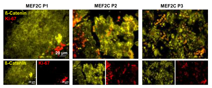
Figure 4.
Ki-67 and ß-catenin (CTNNB1) expression in resected human breast cancer brain metastases. Double-labeling immunofluorescence analysis was performed in tissue sections from breast cancer brain metastases representative of each myocyte enhancer factor 2C (MEF2C) expression phenotype (P1, P2, and P3). Analysis of the proliferation marker Ki-67 (red) in metastases labeled with β-catenin (yellow) revealed the presence of Ki-67 positive cells at the periphery of metastasis in which β-catenin was mainly expressed at the cell membrane (corresponding to MEF2C P1), as well as an increasing number of Ki-67 positive cells and their presence inside metastases as β-catenin dislocated to the cytosol and nucleus (corresponding to MEF2C P2 and P3). Ten fields of one representative section of each MEF2C phenotype were analyzed.

