



Abstract
A 59-year-old male active smoker presented with a 6-month history of cough and breathlessness and was found to have a right upper lobe mass. Histology revealed this to be an adenoid cystic carcinoma (ACC) of the lung, while local lymph node dissection revealed a synchronous diagnosis of chronic lymphocytic leukaemia (CLL). The connection between CLL and solid organ malignancy is well documented, but the reporting of ACC in this context is novel. Mechanisms linking the two processes are revealed with the possibility of causality, and heightened vigilance for the development of primary lung tumours in CLL, and their management, is recommended.
Keywords: lung cancer (oncology), haematology (incl blood transfusion)
Background
Adenoid cystic carcinoma (ACC) is a rare malignancy characterised histologically by nests or cords of epithelial cells surrounding glandular structures. ACC tends to be a slow-growing tumour; however, local and distant recurrence (up to 50%) is common and so the tumour often poses long-term challenges.
The salivary glands are the principal site of origin and therefore this tumour is most often encountered by head and neck oncologists.1 The lung is the most common site of metastatic activity.2
Occasionally, however, primary ACC may evolve in sites such as the lung, under the umbrella term pulmonary salivary gland tumours (PSGT). PSGTs are less likely to have an indolent evolution. Metastatic deposits are difficult to distinguish from primary lesions radiologically or histologically; therefore, careful assessment for a salivary gland primary is required before diagnosis.3 The cornerstone of management is complete surgical resection; however, there is consistent literature to suggest that radiotherapy can yield similar disease control, either in addition to a partial resection or as a definitive treatment.4 For example, Hogerle et al recently reported a similar 5-year and 10-year overall survival between surgery+radiotherapy (92%+82%) versus radiotherapy alone (100%+83%) in a small series.4 There is more anecdotal evidence for chemotherapy.5 Genomic alterations involving the Notch pathway, chromatin remodelling, TERT promoter and MYB/MYBL1 re-arrangements have been described in both head and neck, and pulmonary ACC.6–8 However, none of these oncogenic events is currently targetable via licensed therapeutic agents.
Chronic lymphocytic leukaemia (CLL), a malignant clonal expansion of B or T lymphocytes, is the most common blood malignancy of adulthood, with an incidence of approximately 4 per 100 000 persons per year. Under the microscope, the vastly abundant white cells appear normal but are functionally immature, leading to immunocompromise. The natural history is usually benign and requires no specific treatment; however, for symptomatic disease (including rare cases of high-grade lymphoma transformation), tailored treatment, including chemotherapy and radiation, may be indicated. In the first instance, active surveillance of CLL with serum monitoring is often instituted.9
Case presentation
A 59-year-old male patient presented with a 6-month history of non-productive cough and breathlessness in October 2018. No associated symptoms were reported at the time of diagnosis. The patient was an active lifelong smoker and had a medical history of atrial fibrillation and cardiomyopathy, taking rivaroxaban and ramipril. The remainder of the history was unremarkable.
A screening chest X-ray requested in primary care showed a bulky right hilum with right upper lobe (RUL) consolidation, suspicious for malignancy. Subsequent referral to the chest clinic resulted in CT imaging of the chest which revealed an obstructing 15 mm nodule within the RUL bronchus (figure 1), with associated right hilar and paratracheal lymphadenopathy, the latter of which was noted to have a fatty centre. The ensuing bronchoscopy-guided biopsy of the RUL nodule showed evidence of epithelial tumour cells staining for cytokeratins, smooth muscle actin (SMA) and p63 and negative for TTF1, S100, CK20 and CD56 (table 1). This was felt to be consistent with an ACC. Human epidermal growth factor receptor 2 (HER-2) overexpression was not tested for.
Figure 1.
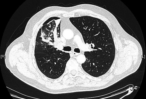
Presentation CT scan. A 15 mm nodule is seen to obstruct the right main bronchus, with distal plugging and partial right upper lobe collapse.
Table 1.
Summary of expected histological markers in adenoid cystic carcinoma and those found in this case
| Markers | Current case | Ravi et al34 | Raman and Sherlin35 | Teixeira et al36 | McHugh et al37 | Argyris et al38 | Chu and Weiss39 |
| Cytokeratins | |||||||
| SMA | |||||||
| p63 | |||||||
| VIM | 38% |
Green =positive; orange = intermediate; red = negative. For example, in the case of Teixeira et al, samples were positive for SMA, p63 and VIM, but negative for cytokeratins. The literature predicted the markers that were found on review of the tumour in this case.
SMA, smooth muscle actin; VIM, vimentin.
There was radiological uncertainty about cysts seen in the liver on CT, but a fluorodeoxyglucose-positron emission tomography CT ruled this out or any other site of metastatic disease, as well as demonstrating no primary malignancy in the salivary glands. The patient underwent right upper lobectomy and perihilar lymph node dissection from stations 2R through 12. The interbronchial tumour, measuring 21×14×12 mm, was pathologically consistent with ACC. The lymph nodes exposed no features of metastatic ACC, but revealed instead a diffuse B-cell infiltrate staining for CD3, CD5, CD19, CD20, CD23 and BCL-2 while negative for cyclin D1, consistent with B-cell CLL. The final staging of the resected ACC was pT1c N0 pL0 R1, R1 on account of infiltration of the endobronchial tumour through seromucinous glands to perihilar fat at the bronchial margin. Representative microscopic images of both lesions are shown in figures 2–4. On the basis of the positive resection margin, the multidisciplinary team consensus was to offer adjuvant radiotherapy to the ACC tumour bed, of which he completed a radical schedule of 55 Gy in 20 fractions via a four-field conformal plan with wedges in May 2019.
Figure 2.

Histopathological microscopic images. (A) Endobronchial polypoid involvement by an adenoid cystic carcinoma (ACC). The tumour shows predominantly (B) tubular and focally (C) cribriform architecture. (D) In other parts of the tumour, the glands were lying within basement membrane-like matrix. (E) Individual glands comprised of either basaloid (small and centrally-placed nuclei with clear cytoplasm) or luminal (enlarged and hyperchromatic nuclei with eosinophilic cytoplasm) cells. (F) Rarely, there is a dual population of cells within the same gland where the luminal cells were surrounded by an outer layer of basaloid cells. This is a classical finding in ACC. (G) The tumour infiltrated the soft tissue plane surrounding the bronchial cartilage. (H) There was perineural invasion, another common feature of ACC. (I–K) Station 2R lymph node showed normal lymphoid follicular architecture largely effaced by a monotypic proliferation of small lymphocytes. (L) A small residual reactive follicle. (M) The lymphocytes were positive for CD3, (N) CD20 and (O) Bcl-2 in keeping with chronic lymphocytic leukaemia/small lymphocytic lymphoma. (P) Ki67 labelling index approximately 5%. Scale bar=100 µm
Figure 3.

Station 2R lymph node lymphocytes demonstrating positive staining for CD5.
Figure 4.

Station 2R lymph node lymphocytes demonstrating positive staining for CD23.
Following his radiotherapy, the patient was reviewed by the haematologists regarding his CLL. A mildly raised white cell count (11.7×10⁹/L) and lymphocyte count (6.66×10⁹/L) was noted prior to surgery, the first result being from October 2018. Interestingly, his counts normalised after radiotherapy (white cell count 7.7×10⁹/L and neutrophils 2.4×10⁹/L). There was no other site of lymphadenopathy noted on the preoperative cross-sectional imaging. Given the normalisation of white cell count, confirmatory flow cytometry was not possible and therefore the patient has entered a period of 6 monthly active surveillance.
Outcome and follow-up
At the time of writing, 6 months postadjuvant radiotherapy, a restaging CT scan has shown postradiotherapy changes in the treatment field and no sign of local or metastatic recurrence. The patient is currently taking dexamethasone for grade 2 radiation-induced pneumonitis.
Discussion
The concurrent diagnosis of CLL with solid organ malignancy is well documented in the literature;10–17 however, to the best of our knowledge (following review of international medical literature), this is the first reported case of concurrent ACC with CLL, at least in the UK. Herein, the connections between CLL and solid organ malignancy are explored.
CLL and solid malignancies: a retrospective
CLL has precedence for diagnosis at surgical lymph node harvesting for primary solid organ malignancies. Farma et al conducted a retrospective review of patients treated for both conditions between 1995 and 2009 at a large centre in Philadelphia, USA, and identified malignant melanoma to be the most common concurrent malignancy alongside CLL, with 52 such patients identified in that period, followed by breast cancer. Approximately two-thirds of these patients had an antecedent diagnosis of solid malignancy, but one-third had the diagnosis of CLL made at sentinel lymph node biopsy during the surgical excision of their tumour.10
Furthermore, a large retrospective study of almost 5000 patients with CLL at the National Cancer Institute, Bethesda, Maryland, by Greene et al in the late 1970s demonstrated an increased incidence of secondary cancers, including of the lung, of which there was a relative risk (RR) of 1.5 (95% CI 1.1 to 2.1) over the expected population rate. Of interest, there were also two incidences of salivary gland tumours, which reflected an RR of 3.4 over the expected population rate for this sample size. The increased incidence of lung cancer persisted for up to 6 years after the diagnosis of CLL, indicating an intrinsic association.11 Adding weight to this is that the association was independent of treatment with chemotherapy or radiotherapy, as the increased RR of lung cancer (1.66× in Amadori and Ronconi’s single-centre study) in active treatment groups persisted in watchful waiting groups,12 meaning that the increased incidence of solid malignancy cannot be attributed to the cellular toxicity of CLL treatment. Rovira Daudi et al and Fukaura et al,13 14 among other groups, have also reported synchronous squamous cell carcinomas of the lung with CLL, with Travis et al showing a twofold increased incidence of lung carcinoma.15
Understanding the link between CLL and solids tumours: the immune hypothesis and tobacco smoking
Interplay between the immune system and malignancy is increasingly understood, both in terms of the immune system’s critical role in managing malignant processes, and the functional limitation that haematological malignancy has on the immune system. Escape from immunoediting, the process by which the immune system recognises and either kills or contains neoplastic growth, has been shown to be a seminal event in the progression of malignancy.18 It follows that a state of immunodeficiency might weaken the body’s ability to eradicate malignancy, and therefore allow earlier and quicker progression of cancer. CLL is a cause of immunodeficiency as lymphocytes are abundant but immature, with limited functionality.
In the context of lung malignancy, overexpression of the cellular enzyme uridine-diphospho glucuronosyltransferase (UGT-2B17; not tested in our case) results in particular interest as it has been documented in patients with poor prognostic CLL,19 variants of which have been associated with the incidence of multiple solid tumour types, including adenocarcinoma of the lung and prostate.20 21 In this particular framework, UGT2B17 has a key role in the breakdown of a major metabolite of tobacco smoke, 4-(methylnitrosamino)-1-(3-pyridyl)-1-butanol (NNAL).22 Deletion of the enzyme leads to increased NNAL levels, which is a selective inducer of lung adenocarcinoma in experimental models.23 24 In view of the patient’s smoking history, we could (with caution) ask ourselves if such a mechanism may have played a role in the development of a similar carcinoma of glandular tissue, ACC, in this case.
Moreover, that the association between the two is independent of previous chemotherapy or radiotherapy, adds further weight to the possibility of a potentially causative factor such as the immune hypothesis. Indeed, Parekh et al demonstrated a number of patients with CLL who went on to develop multiple separate solid malignancies through a review of patients treated for both lung cancer and CLL between the years 1977 and 1998 at the Memorial Sloan-Kettering Cancer Centre (MSKCC) in New York. Of approximately 1400 patients treated for CLL during that period, 2% went on to develop either small cell or non-small cell lung cancer (NSCLC), with an average latency of 10 years. Of these, 85% of patients were tobacco smokers. Of these 26 patients with lung cancer, 10 went on to develop a third primary malignancy, 4 developed a fourth and 1 went on to develop a fifth primary malignancy. The MSKCC study was also able to demonstrate that the best prognoses were held by patients with resectable stage 1 NSCLC, but that significantly worse outcomes compared with those expected on a population basis exist for those with inoperable lung cancer who had concurrent CLL.16 17
HER-2 overexpression: a possible mechanism of CLL related oncogenesis?
It is notable that such lung carcinomas that develop in patients with CLL appear to have an excess of HER-2 overexpression; immunohistochemistry 2+ or 3+ were found in 57%–64% of such patients in small series, compared with a background rate of 16%–36%.25–28 HER-2-positive cells have been demonstrated in vitro to induce a cytotoxic T-cell response,29 and so it follows that immunosuppressive conditions such as CLL could hamper the response to these tumours, potentially explaining their increased prevalence in these patients. HER-2 was not tested for in our patient—this would have been interesting, given that HER-2 overexpression has been demonstrated in salivary gland tumours too and could open up the possibility of directed therapy with monoclonal antibody antagonists.30
Further aspects: is there evidence for reverse causality?
In our case, the concurrence of PSGT and CLL diagnoses makes it difficult to assume a direction of association. Is there a possibility of causality emanating not from CLL, but from the PSGT? While an excess of CLL cases have not been demonstrated among patients initially diagnosed with solid organ malignancies,11 secondary acute myeloid leukaemia (AML) diagnoses have. While chemoradiotherapy for solid organ tumours and resulting cellular toxicity is the most tangible possible causal factor here,31 the mutational landscape of PSGT is also of interest, including a predominance of epigenetic modulators, particularly histone acetyltransferases (HACT), the most commonly mutated being KAT6A, and methyltransferases, including KMT2D.32 HACTs help to loosen winding of DNA around centromeres, making transcription of tumour suppressor genes possible. Chromosomal translocation of the HACT KAT6A has been associated with AML,33 providing a feasible causal link between PSGT and haematological malignancy. Given that the CLL tissue was resected close to the primary ACC, we could ask ourselves whether such a mechanism could have been at play in the presented case.
Conclusion
We describe a novel case of concurrent PSGT and CLL. The literature reveals an excess of solid organ malignancies in patients with CLL, in whom immunodeficiency and tobacco smoking have the potential to be causal factors. The identification of patients with CLL developing multiple separate solid organ tumours strengthens the argument of causality. Certainly, with the increased risk and apparently worse outcomes of secondary lung cancer in CLL, vigilance for secondary cancer should be heightened in this group of patients. Finally, full resection, followed by adjuvant radiotherapy, appears to yield the optimum outcomes in the management of PSGT.
Learning points.
Clinicians should be alert to the increased risk of solid organ malignancy in patients with chronic lymphocytic leukaemia (CLL).
It is possible that immune immaturity in CLL is a causal factor in this association.
In accordance with the medical literature, complete surgical resection±adjuvant radiotherapy remains the primary management strategy for pulmonary and salivary gland tumours.
Tobacco smoking is the single most important modifiable factor implicated in this association, encouraging cessation of which is highly advised.
Human epidermal growth factor receptor 2 testing may be particularly relevant in lung tumours diagnosed in patients with CLL.
Acknowledgments
Our deepest gratitude goes to oncology nurse consultant Mark Foulkes (Royal Berkshire Hospital) and Professor Andrew Nicholson (Royal Brompton & Harefield National Health Service Foundation Trust) for their valuable contributions to image collection and opinion on histopathological data.
Footnotes
Contributors: PSW synthesised the literature, wrote the background and discussion and produced revisions of the case report. YZZ collated and presented relevant histopathological images. KB produced cross-sectional imaging. GS provided the case description and edited revisions of the case report. All authors approved the final submission.
Funding: The authors have not declared a specific grant for this research from any funding agency in the public, commercial or not-for-profit sectors.
Competing interests: None declared.
Patient consent for publication: Obtained.
Provenance and peer review: Not commissioned; externally peer-reviewed.
References
- 1.Garg M, Tudor-Green B, Bisase B. Current thinking in the management of adenoid cystic carcinoma of the head and neck. Br J Oral Maxillofac Surg 2019;57:S0266-4356(19)30308-0:716–21. 10.1016/j.bjoms.2019.07.021 [DOI] [PubMed] [Google Scholar]
- 2.NORD . Rare disease database. Available: https://rarediseases.org/rare-diseases/adenoid-cystic-carcinoma/ [Accessed 08 Oct 2019].
- 3.Falk N, Weissferdt A, Kalhor N, et al. Primary pulmonary salivary Gland-type tumors: a review and update. Adv Anat Pathol 2016;23:13–23. 10.1097/PAP.0000000000000099 [DOI] [PubMed] [Google Scholar]
- 4.Högerle BA, Lasitschka F, Muley T, et al. Primary adenoid cystic carcinoma of the trachea: clinical outcome of 38 patients after interdisciplinary treatment in a single institution. Radiat Oncol 2019;14:117. 10.1186/s13014-019-1323-z [DOI] [PMC free article] [PubMed] [Google Scholar]
- 5.Fernández L, Salazar N, Sua LF, et al. Sleeve pneumonectomy and carinal resection for management of primary adenoid cystic tumor of the lung with carinal extension: report of two cases. Respir Med Case Rep 2019;26:82–6. 10.1016/j.rmcr.2018.11.009 [DOI] [PMC free article] [PubMed] [Google Scholar]
- 6.Drier Y, Cotton MJ, Williamson KE, et al. An oncogenic Myb feedback loop drives alternate cell fates in adenoid cystic carcinoma. Nat Genet 2016;48:265–72. 10.1038/ng.3502 [DOI] [PMC free article] [PubMed] [Google Scholar]
- 7.Pei J, Flieder DB, Patchefsky A, et al. Detecting Myb and MYBL1 fusion genes in tracheobronchial adenoid cystic carcinoma by targeted RNA-sequencing. Mod Pathol 2019;32:1416–20. 10.1038/s41379-019-0277-x [DOI] [PMC free article] [PubMed] [Google Scholar]
- 8.Ho AS, Ochoa A, Jayakumaran G. Genetic hallmarks of recurrent/metastatic adenoid cystic carcinoma. J Clin Invest 2019;129:4276–89. 10.1172/JCI128227 [DOI] [PMC free article] [PubMed] [Google Scholar]
- 9.Patient Platform Limited . Acute lymphoblastic leukaemia. Available: https://patient.info/doctor/acute-lymphoblastic-leukaemia-pro [Accessed 08 Oct 2019].
- 10.Farma JM, Zager JS, Barnica-Elvir V, et al. A collision of diseases: chronic lymphocytic leukemia discovered during lymph node biopsy for melanoma. Ann Surg Oncol 2013;20:1360–4. 10.1245/s10434-012-2740-5 [DOI] [PMC free article] [PubMed] [Google Scholar]
- 11.Greene MH, Hoover RN, Fraumeni JF. Subsequent cancer in patients with chronic lymphocytic leukemia--a possible immunologic mechanism. J Natl Cancer Inst 1978;61:337–40. [PubMed] [Google Scholar]
- 12.Amadori D, Ronconi S. Secondary lung tumors in hematological patients. Semin Respir Crit Care Med 2005;26:520–6. 10.1055/s-2005-922035 [DOI] [PubMed] [Google Scholar]
- 13.Rovira Daudi E, Carrión Valero F, García Díaz M, et al. [Lung carcinoma and chronic lymphocytic leukemia: report of a synchronous diagnosis]. An Med Interna 1997;14:35–7. [PubMed] [Google Scholar]
- 14.Fukaura A, Tazawa H, Fujii H, et al. Synchronous cancer of squamous cell carcinoma of the lung and lymphocytic leukemia (B-cell type): a case report. Jpn J Clin Oncol 1992;22:210–5. [PubMed] [Google Scholar]
- 15.Travis LB, Curtis RE, Hankey BF, et al. Second cancers in patients with chronic lymphocytic leukemia. J Natl Cancer Inst 1992;84:1422–7. 10.1093/jnci/84.18.1422 [DOI] [PubMed] [Google Scholar]
- 16.Mountain CF. Revisions in the International system for staging lung cancer. Chest 1997;111:1710–7. 10.1378/chest.111.6.1710 [DOI] [PubMed] [Google Scholar]
- 17.Parekh K, Rusch V, Kris M. The clinical course of lung carcinoma in patients with chronic lymphocytic leukemia. Cancer 1999;86:1720–3. 10.1002/(SICI)1097-0142(19991101)86:9<1720::AID-CNCR13>3.0.CO;2-X [DOI] [PubMed] [Google Scholar]
- 18.Cavallo F, De Giovanni C, Nanni P, et al. 2011: the immune hallmarks of cancer. Cancer Immunol Immunother 2011;60:319–26. 10.1007/s00262-010-0968-0 [DOI] [PMC free article] [PubMed] [Google Scholar]
- 19.Gruber M, Bellemare J, Hoermann G, et al. Overexpression of uridine diphospho glucuronosyltransferase 2B17 in high-risk chronic lymphocytic leukemia. Blood 2013;121:1175–83. 10.1182/blood-2012-08-447359 [DOI] [PubMed] [Google Scholar]
- 20.Gallagher CJ, Muscat JE, Hicks AN, et al. The UDP-glucuronosyltransferase 2B17 gene deletion polymorphism: sex-specific association with urinary 4-(methylnitrosamino)-1-(3-pyridyl)-1-butanol glucuronidation phenotype and risk for lung cancer. Cancer Epidemiol Biomarkers Prev 2007;16:823–8. 10.1158/1055-9965.EPI-06-0823 [DOI] [PubMed] [Google Scholar]
- 21.Nadeau G, Bellemare J, Audet-Walsh Étienne, et al. Deletions of the androgen-metabolizing UGT2B genes have an effect on circulating steroid levels and biochemical recurrence after radical prostatectomy in localized prostate cancer. J Clin Endocrinol Metab 2011;96:E1550–7. 10.1210/jc.2011-1049 [DOI] [PubMed] [Google Scholar]
- 22.Hecht SS. Biochemistry, biology, and carcinogenicity of tobacco-specific N-nitrosamines. Chem Res Toxicol 1998;11:559–603. 10.1021/tx980005y [DOI] [PubMed] [Google Scholar]
- 23.Wiener D, Fang J-L, Dossett N, et al. Correlation between UDP-glucuronosyltransferase genotypes and 4-(methylnitrosamino)-1-(3-pyridyl)-1-butanone glucuronidation phenotype in human liver microsomes. Cancer Res 2004;64:1190–6. 10.1158/0008-5472.CAN-03-3219 [DOI] [PubMed] [Google Scholar]
- 24.Rivenson A, Hoffmann D, Prokopczyk B, et al. Induction of lung and exocrine pancreas tumors in F344 rats by tobacco-specific and Areca-derived N-nitrosamines. Cancer Res 1988;48:6912–7. [PubMed] [Google Scholar]
- 25.Potti A, Ganti AK, Koch M, et al. Identification of HER-2/neu overexpression and the clinical course of lung carcinoma in non-smokers with chronic lymphocytic leukemia. Lung Cancer 2001;34:227–32. 10.1016/S0169-5002(01)00241-0 [DOI] [PubMed] [Google Scholar]
- 26.Musolino A, Bozzetti C, Bonomini S, et al. Lung cancer in patients with chronic lymphocytic leukemia: does the HER-2 oncogene play a role? Lung Cancer 2005;50:419–20. 10.1016/j.lungcan.2005.07.005 [DOI] [PubMed] [Google Scholar]
- 27.Hirsch FR, Varella-Garcia M, Franklin WA, et al. Evaluation of HER-2/neu gene amplification and protein expression in non-small cell lung carcinomas. Br J Cancer 2002;86:1449–56. 10.1038/sj.bjc.6600286 [DOI] [PMC free article] [PubMed] [Google Scholar]
- 28.Mar N, Vredenburgh JJ, Wasser JS. Targeting HER2 in the treatment of non-small cell lung cancer. Lung Cancer 2015;87:220–5. 10.1016/j.lungcan.2014.12.018 [DOI] [PubMed] [Google Scholar]
- 29.Gritzapis AD, Perez SA, Baxevanis CN, et al. Pooled peptides from HER-2/neu-overexpressing primary ovarian tumours induce CTL with potent antitumour responses in vitro and in vivo. Br J Cancer 2005;92:72–9. 10.1038/sj.bjc.6602259 [DOI] [PMC free article] [PubMed] [Google Scholar]
- 30.Scholl S, Beuzeboc P, Pouillart P. Targeting HER2 in other tumor types. Ann Oncol 2001;12 Suppl 1:S81–7. 10.1093/annonc/12.suppl_1.S81 [DOI] [PubMed] [Google Scholar]
- 31.Collinge E, Loron S, Larcher M-V, et al. Elderly patients (age 70 years or older) with secondary acute myeloid leukemia or acute myeloid leukemia developed concurrently to another malignant disease. Clin Lymphoma Myeloma Leuk 2018;18:e211–8. 10.1016/j.clml.2018.02.018 [DOI] [PubMed] [Google Scholar]
- 32.Li M, Zhao B-R, Liu S-Q, et al. Mutational landscape and clonal diversity of pulmonary adenoid cystic carcinoma. Cancer Biol Ther 2018;19:898–903. 10.1080/15384047.2018.1480296 [DOI] [PMC free article] [PubMed] [Google Scholar]
- 33.Gervais C, Murati A, Helias C, et al. Acute myeloid leukaemia with 8p11 (MYST3) rearrangement: an integrated cytologic, cytogenetic and molecular study by the Groupe francophone de cytogénétique hématologique. Leukemia 2008;22:1567–75. 10.1038/leu.2008.128 [DOI] [PubMed] [Google Scholar]
- 34.Ravi M, Boaz K, Natarajan S, et al. Expression of α-smooth muscle actin in benign and malignant salivary gland tumors: an immunohistochemical study. Indian J Pathol Microbiol 2018;61:479–84. 10.4103/IJPM.IJPM_482_17 [DOI] [PubMed] [Google Scholar]
- 35.Raman S, Sherlin HJ. Utility of smooth muscle actin and CD117 as reliable markers in the diagnosis of salivary gland neoplasms. J Oral Maxillofac Pathol 2019;23:218–23. 10.4103/jomfp.JOMFP_225_18 [DOI] [PMC free article] [PubMed] [Google Scholar]
- 36.Teixeira LN, Janner Évelin Caroline, Teixeira T, et al. Comparison of p63/p40 expression with myoepithelial markers in minor salivary gland tumors. Int J Surg Pathol 2019;27:360–71. 10.1177/1066896918813678 [DOI] [PubMed] [Google Scholar]
- 37.McHugh JB, Hoschar AP, Dvorakova M, et al. P63 immunohistochemistry differentiates salivary gland oncocytoma and oncocytic carcinoma from metastatic renal cell carcinoma. Head Neck Pathol 2007;1:123–31. 10.1007/s12105-007-0031-4 [DOI] [PMC free article] [PubMed] [Google Scholar]
- 38.Argyris PP, Wetzel SL, Greipp P, et al. Clinical utility of Myb rearrangement detection and p63/p40 immunophenotyping in the diagnosis of adenoid cystic carcinoma of minor salivary glands: a pilot study. Oral Surg Oral Med Oral Pathol Oral Radiol 2016;121:282–9. 10.1016/j.oooo.2015.10.016 [DOI] [PubMed] [Google Scholar]
- 39.Chu PG, Weiss LM. Expression of cytokeratin 5/6 in epithelial neoplasms: an immunohistochemical study of 509 cases. Mod Pathol 2002;15:6–10. 10.1038/modpathol.3880483 [DOI] [PubMed] [Google Scholar]