

Abstract
We present the case of a 56-year-old man who sustained a tibial tuberosity fracture with an associated patellar fracture. In the adult population there are only a few documented cases of tibial tuberosity fractures. This is only the second recorded case of bifocal patella tendon avulsion. The patient was managed successfully by fixation of the tibial tuberosity alone as the patella fracture was undisplaced and the patella retinaculum intact. A key point was screening the patella fracture at time of fixation to aid this decision. We achieved a good outcome at one year with internal fixation and early mobilisation.
Keywords: Tibial tuberosity, Patella, Knee
1. Introduction
Tibial tuberosity avulsion fractures account for 1–3% of paediatric and adolescent fractures.1,2 However, in the adult population there are only a few documented cases in literature.3, 4, 5 Simultaneous tuberosity and patella fractures has only once been previously described.6
2. Case report
A 56-year-old man presented to the Accident and Emergency department following a stumble and fall onto a flexed left knee, likely resulting in rapid eccentric contraction of the knee extensor mechanism muscles. No other injuries were sustained, and the patient was otherwise systemically well. The past medical history included autistic spectrum disorder, paranoid psychosis and long-term smoking. There was no history of any previous knee problems, previous fractures or Osgood-Schlatters disease.
On examination the patient was unable to straight leg raise and had tenderness over the tibial tuberosity. The insertion of the quadriceps tendon into the patella was intact with the patella lying in the normal position. There was further bony tenderness over the patella. The left leg was distally neurovascularly intact and there were no overlying wounds.
X-rays taken showed a tibial tuberosity fracture and an associated undisplaced patellar fracture (Fig. 1 & Fig. 2). The patient was referred to the Orthopaedic on-call team.
Fig. 1.
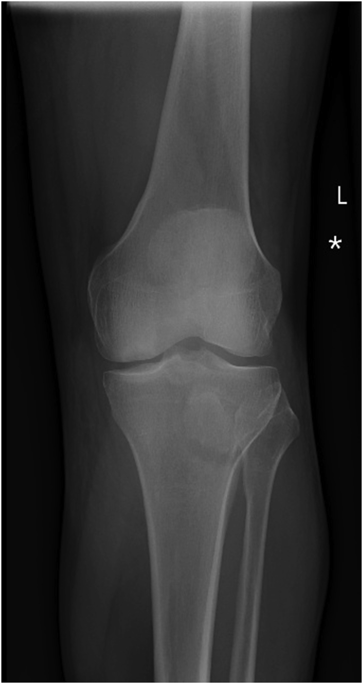
Anteroposterior (AP) plain radiograph on presentation to the emergency department.
Fig. 2.

Lateral plain radiograph on presentation to the emergency department.
The following day a plan was made for operative fixation of the tibial tuberosity; with the intention of treating the patella fracture non-operatively unless there was displacement on fluoroscopic screening. Had the patella fracture displaced more than 2 mm in either articular step or gapping the plan was to treat with tension band wire fixation.
The patient was taken to theatre 24 h post injury. Two partially threaded 6.5 mm cannulated screws were chosen to provide robust fixation of the anterior tuberosity fragment. The fracture was reduced percutaneously using a combination of large clamps (Fig. 3) and a Kirschner wires, with check x-rays to confirm adequate reduction (Fig. 4 and Fig. 5). The screw hold was good and washers were judged not to be required.
Fig. 3.

Photograph from theatre showing fracture reduction with a large clamp.
Fig. 4.

AP intraoperative image intensifier (II) image taken at time of surgery.
Fig. 5.

Lateral intraoperative image intensifier (II) image taken at time of surgery.
The patella fracture was then screened, the fracture did not displace on knee flexion. On deep palpation the patella retinaculum was judged to be intact. A decision was hence made to treat the patella fracture with a cylinder cast for 2 weeks and subsequent hinged knee bracing with an increase in flexion of 30° at two weekly intervals for a total of eight weeks.
The patient was allowed to touch weight-bear with crutches in for the first 4 weeks and then allowed to progressively increase the weight placed on the left leg.
At review in the clinic 8 weeks after injury, patient was able to walk comfortably without aids or crutches and had a full range of movement as compared with the other side. At 1 year the patient was back to all daily activities including walking distances of several miles and was very satisfied with the result with no pain in the knee at all. He could fully extend the leg and straight leg raise as normal without any problems. The wound healed without complication.
3. Discussion
Our patient’s fall onto a flexed knee and poor bone quality are likely to be the combined causes of this unusual injury pattern. Classical teaching is that a tendon will never avulse at both its origin and insertion points.7 In this case the fact that both the patella fractured (origin of the patella tendon) and the tibial tuberosity avulsed (insertion of the patella tendon) is due to the relatively higher tensile strength of the patella tendon compared with that of the bone.
The site of maximal stress in the extensor mechanism is dependent on the degree of knee flexion at time of injury with the greatest forces on the patella tendon being when the knee is flexed greater than 60°. Kinematic studies have shown the patella tendon does not experience large changes in length between 30 and 110° of flexion.8 This will therefore increase the strain at the tendon-bone interface which has been shown to be 3–4 times the strain experienced in the tendon’s midsubstance. In patients with poor bone quality this strain may result in fracture. Our patient had a significant past history of smoking as well as previous treatment with antipsychotics, both which have been shown to reduce bone density.9,10 This patient is currently under investigation by the rheumatologist for osteoporosis; a DEXA scan was organised but unfortunately 1 year down the line the patient has not managed to attend yet.
Chautems et al.6 in their report of a similar injury concluded that a main contributing factor was underlying bone disease; osteoporosis in a 90-year-old lady. They also established that treatment should be fixation of both fractures.
We have however managed to get an excellent result by fixation of only the tibial tuberosity; particularly as the patella fracture was undisplaced and the surrounding soft tissues and retinaculum was intact.
In conclusion, bifocal detachment of the patella tendon is an extremely rare injury. When it does occur osteoporosis should be considered as a contributing factor. A successful outcome can be achieved by fixation of the tibial tuberosity alone, as long as the patella fracture is undisplaced and the retinaculum intact.
Funding
No sources of financial support were received for the production of this article.
Ethical adherence
Ethical review was not sought for the production of this article.
Declaration of competing interest
No conflict of Interest is declared.
References
- 1.Ogden J.A., Tross R.B., Murphy M.J. Fractures of the tibial tuberosity in adolescents. J Bone Joint Surg Am. 1980;62(2):205–215. [PubMed] [Google Scholar]
- 2.Hamilton S.W., Gibson P.H. Simultaneous bilateral avulsion fractures of the tibial tuberosity in adolescence: a case report and review of over 50 years of literature. Knee. 2006;13(5):404–407. doi: 10.1016/j.knee.2006.04.008. [DOI] [PubMed] [Google Scholar]
- 3.Aterkar M.V., Mahajan U.D., Somani A.M. Tibial tuberosity avulsion fracture in an adult- a rare case report. Int J Med Res Health Sci. 2014;3:1016–1018. doi: 10.5958/2319-5886.2014.00042.3. [DOI] [Google Scholar]
- 4.Mounasamy V., Brown T.E. Avulsion fracture of the tibial tuberosity with articular extension in an adult: a novel method of fixation. Eur J Orthop Surg Traumatol. 2008;18:157–159. [Google Scholar]
- 5.Vella D., Peretti G., Fra F. One case of fracture of the tibial tuberosity in the adult. Chir Organi Mov. 1992;77:299–301. [PubMed] [Google Scholar]
- 6.Chautems R., Michel J. Bifocal avulsion of the patellar tendon in an adult. Rev Chir Orthop Reparatrice Appar Mot. 2001;87(4):388–391. [PubMed] [Google Scholar]
- 7.William L.H., Charles R.H., William D.A. Avulsion fractures of the tibial tubercle. J Bone Joint Surg Am The Journal Of Bone & Joint Surgery. 1971;53(8):1579–1583. [PubMed] [Google Scholar]
- 8.Defrate L.E., Nha K.W., Papannagari R., Moses J.M., Gill T.J., Li G. The biomechanical function of the patellar tendon during in-vivo weight-bearing flexion. J Biomech. 2007;40(8):1716–1722. doi: 10.1016/j.jbiomech.2006.08.009. [DOI] [PMC free article] [PubMed] [Google Scholar]
- 9.Law M.R., Hackshaw A.K. A meta-analysis of cigarette smoking, bone mineral density and risk of hip fracture: recognition of a major effect. BMJ. 1997;315:841. doi: 10.1136/bmj.315.7112.841. [DOI] [PMC free article] [PubMed] [Google Scholar]
- 10.Wang M., Hou R., Jian J. Effects of antipsychotics on bone mineral density and prolactin levels in patients with schizophrenia: a 12-month prospective study. Hum Psychopharmacol. Mar. 2014;29(2):183–189. doi: 10.1002/hup.2387. [DOI] [PubMed] [Google Scholar]