Abstract
Purpose
Molecular-based targeted therapy has improved life expectancy for advanced non-small cell lung cancer (NSCLC). However, it does not have to be inevitable that patients receiving third-generation EGFR-TKIs become drug resistant. EGFR C797S and MET amplification are common mechanisms of osimertinib. However, a large part of these potential drug mechanisms remains unknown, and further research is needed.
Methods
Tumour and blood samples from forty advanced NSCLC patients were identified as acquired drug resistant to osimertinib. The NGS panel was applied to detect EGFR C797S and MET amplification in tumour tissues or plasma samples. Whole-exome sequencing was conducted in five pairs of tumour tissues obtained before osimertinib administration and after osimertinib resistance in patients without C797S/cMET amplification.
Results
The overall C797S mutation rate was 20%, and MET amplification was detected in six out of sixteen C797S-negative samples. PDGFRA in the PI3K-AKT-mTOR signalling pathway, RASAL2, RIN3 and SOS2 in the RAS-Raf-ERK signalling pathway, PTK2 in the ERBB signalling pathway and ABCC1 and ABCB5 in the ABC membrane pump system were identified as candidate crucial genes of drug resistance to osimertinib.
Conclusion
EGFR C797S mutation and MET amplification are leading mechanisms for osimertinib resistance in lung cancer. The crucial potential mutated genes defined in our present study may need further validation in a considerable number of lung cancer patients.
Keywords: osimertinib, drug resistance, whole-exome sequencing, lung cancer, targeted therapy
Introduction
Lung cancer is the leading cause of cancer-related mortality in the world, and non-small-cell lung cancer (NSCLC) accounts for 85% of lung cancer cases.1 Targeted therapy based on molecular alterations has increased the survival time and quality of life of patients with advanced NSCLC in recent decades.2,3
Osimertinib, a third-generation EGFR tyrosine kinase inhibitor, is a representative targeted therapy in NSCLC, which prolongs progression-free survival to 19 months and overall survival to 39 months as the first line of therapy.4 As the precise choice for patients with the T790 M mutation who acquire drug resistance to the first generation of EGFR-TKIs, patients taking osimertinib can reach a PFS of 10.1 months and OS of 26.8 months when this drug is used as a second line therapy.5 However, similar to other targeted therapies, osimertinib undoubtedly results in drug resistance.6 The next line of treatment must be based on overcoming the potential drug resistance mechanism. Studies have found that multiple mechanisms account for its resistance, and EGFR C797S and MET amplification are the main causes, accounting for approximately 20% and 15%, respectively.7–9 As a second line of therapy in T790M patients with lung cancer, almost 40% of patients acquiring drug resistance to osimertinib remain unidentified.8,10 Therefore, further explorations are urgently needed to elaborate the potential novel mechanisms for acquiring drug resistance to osimertinib.
Methods
Patients
Tumour tissue and plasma samples were provided by patients in the single centre of our respiratory department in PLA General Hospital from 2017 to 2019. Enrolled patients were required to meet the following requirements: 1. All patients had acquired drug resistance after receiving the first generation of EGFR-TKIs and proved to have the T790M mutation. 2. All patients took osimertinib as second-line therapy. 3. All of these patients agreed to provide tumour tissue or plasma before osimertinib initiation (BOI) and after osimertinib resistance (AOR). Our research was approved by the ethics committee of PLA General Hospital, and signed informed consent was provided by the patients. And the conduction of whole research was carried out in accordance with the Declaration of Helsinki.
Sample Collection
Enrolled patients provided tumour tissue or plasma before receiving osimertinib, and plasma samples were also given every 6 weeks during the course of osimertinib treatment. Finally, tumour tissue or plasma samples were supplied after osimertinib resistance. Tumour tissue obtained from bronchoscope biopsy and CT-guided needle lung biopsy was stored in liquid nitrogen. Approximately 6 mL of plasma was stored at −80°C, and cfDNA was extracted for next-generation sequencing.
EGFR C797S Detection and MET Analysis with NGS Panel
Tumour DNA and cfDNA were quantified by a Nanodrop 2000 (Thermo Fisher Scientific, Waltham, MA) and Qubit 3.0 using a dsDNA HS Assay kit (Life Technologies, Waltham, MA) according to the manufacturer’s protocols. All of these procedures were performed at a CAP/CLIA certified clinical diagnostic laboratory. The following analysis was performed according to our previous research using the same NGS platform and NGS panel covering 140 genes, including EGFR, MET, and ALK.11
Whole-Exome Sequencing and Bioinformatic Analysis of C797S/MET‑Negative Tumour Tissue and Blood Cell Samples in Paired
Genomic DNA from tumour tissue and paired blood cells of C797S/MET‑negative patients was cut into fragment sizes of 150 ~ 200 bp with Bioruptor_NGS (Diagenode SA, Denville, USA). Samples of 200 ng of genomic DNA were performed with a process of end repair and ligation to hybridise to the captured library. Each of the captured libraries was sequenced on an Illumina HiSeq X Ten (Illumina, San Diego, CA, USA) to average target exome coverage of 100× in reference blood cell DNA and 200× in tumour tissue.
High-quality reads were mapped to the NCBI reference human genome (hg19). Then, duplicates were merged and removed with SAMtools. Local realignment and base quality score recalibration were processed with the Genome Analysis Toolkit (GATK). Taking the corresponding blood cells as a control, SNVs and INDELs were predicted with GATK and annotated with the ANNOVAR software tool. Moreover, those mutations only in tumour tissue AOR but not in BOI of the same patients were selected and further analysed with the R project for statistical computing (version 3.1.3) and pathway analysis.
Results
Study Design and Clinical Characteristics of Patients
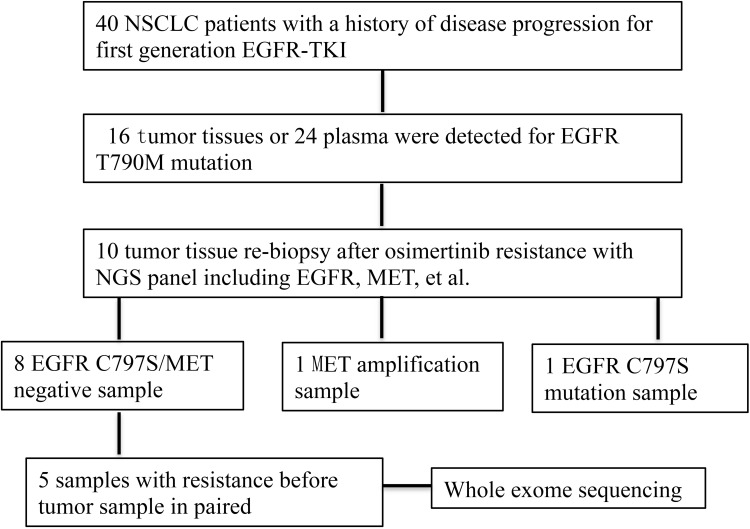
The flowchart of our research is presented in Figure 1. In total, 40 patients were proven to have EGFR T790 M by tumour biopsy in 16 patients and liquid biopsy in 24 patients after acquiring drug resistance to the first generation of EGFR-TKIs. All of these patients received osimertinib as second-line therapy and reached a PFS of 3–39 months for osimertinib. Ten patients underwent rebiopsy, including lung and lymph node biopsy, to analyse resistance-related mutations, including C797S and MET, by NGS panel. Eight patients were C797S/MET-negative, but only 5 patients had paired tumour tissue before taking osimertinib. The clinical characteristics of these 5 patients are listed in Table 1.
Figure 1.
Flowchart of research design.
Table 1.
Clinical Characteristics of Patients Receiving Whole-Exome Sequencing
| ID | Age | Sex | Histology | Smoking History | Clinical Stage | Biopsy Site | PFS |
|---|---|---|---|---|---|---|---|
| 6 | 62 | Female | Adenocarcinoma | No | IVB | Lung | 9 |
| 12 | 64 | Male | Adenocarcinoma | Yes | IVA | Lymph | 11 |
| 15 | 67 | Female | Adenocarcinoma | No | IVB | Lung | 7 |
| 29 | 61 | Female | Adenocarcinoma | No | IVA | Lung | 9 |
| 35 | 65 | Female | Adenocarcinoma | No | IVB | Lymph | 13 |
Detection of EGFR C797S Mutation and MET Amplification by NGS Panel
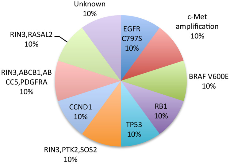
The C797S mutation rate was 15% (6/40) in patients who acquired drug resistance to osimertinib. Among the 10 patients who received rebiopsy, only 1 patient was C797S-positive and the rest of the patients were proven to be C797S-negative by both tumour tissue and plasma. One patient was identified with MET amplification among the 9 C797S-negative patients (Figure 2).
Figure 2.
Pie chart of 10 patients with drug resistance of osimertinib. Only 5 of which received whole-exome sequencing.
Potential Novel Resistance Mechanism of Osimertinib Identified by Exome Sequencing
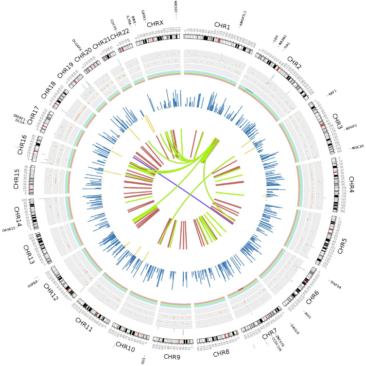
Five patients proved C797S/MET-negative and provided tumour tissue from BOI and AOR in paired finishing of the whole-exome sequencing. All sequencing data were analysed according to the standard protocol.12 In the whole sequencing, the captured targeted region was almost 60.46 MB, the clean read rate was more than 92%, and more than 99% of reads mapped successfully. The mean depth of targeted region was 137.36-fold. Somatic SNVs were defined with sequencing data analysed by MuTect2 (Cibulskis et al, 2013) and ANNOVAR (Wang, K. et al, 2010), taking blood cells as the normal control and mutations in BOI tumour tissue as the time control. A total of 2160 SNVs were identified from these 5 patients and after filtering with the database, including 1000 Genomes Project database and the Exome Aggregation Consortium database, analysing with SIFT and Polyphen2 for pathogenicity, final novo key mutated genes were minimized to 227. For inDel evaluation, the same procedures were processed as for SNV analysis. The final number of InDels was 1387 in these 5 patients, and this number decreased to 370 significant genetic mutations after filtering with a common database. Variations obtained from whole-exome sequencing are presented in Figure 3.
Figure 3.
Variations detected by whole-exome sequencing in all samples.
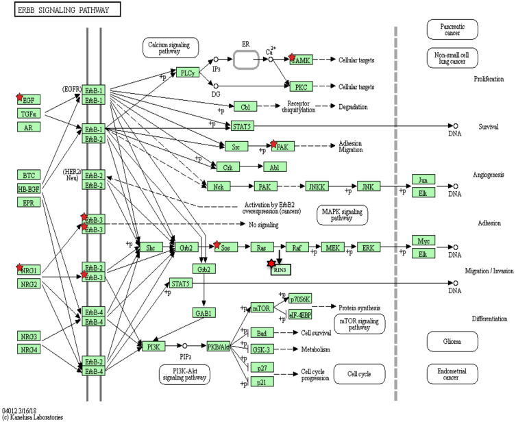
The total SNVs and InDels screened via public tumour-related databases were analysed with the KEGG pathway and R project to find final key candidate mutations accumulated in key signalling pathways for acquiring drug resistance in osimertinib (Figure 4). Four cancer-related signalling pathways were identified to account for drug resistance: 1. PDGFRA in the PI3K-AKT-mTOR signal pathway; 2. RASAL2, RIN3 and SOS2 in the RAS-Raf-ERK signal pathway; 3. PTK2 in the ERBB signal pathway; 4. ABCC1, ABCB5 in the ABC membrane pump system. To our surprise, there was 1 specific gene, RIN3, the mutations of which appeared in 3 of 5 patients, including the same 2 deletions and 1 SNP.
Figure 4.
Potential candidate genes for osimertinib resistance presented within cancer-related signal pathway.
Discussion
The purpose of our research was to reveal the genomic variations resulting in third-generation EGFR-TKI-osimertinib resistance in addition to common EGFR C797S and c-Met amplification. Whole-exome sequencing was applied to determine the potential gene alterations leading to resistance in paired tumour tissues before osimertinib administration and after osimertinib resistance in 5 patients without C797S or c-Met amplification.
Many studies have focused on the variations in resistance to osimertinib, including EGFR C797S, c-Met, BRAF, CCND1, and PI3KCA.13–15 However, most of them were performed with the NGS panel method, which was made up of multiple genes proven to be associated with lung cancer or cancer therapy. To the best of our knowledge, this is the first study to explore candidate genomic variations for osimertinib resistance with whole-exome sequencing in paired tumour tissues. A number of potential key functional genetic variations were identified in our research.
PDGFRA, as a key factor in the PI3K-AKT-mTOR signalling pathway, plays a role in promoting cell proliferation, migration, and differentiation.16 The CNVs of PDGFRA were associated with the survival time of ALK-TKIs in NSCLC,17 and were also proven to cause ALK-TKI resistance in advanced NSCLC.18 Although PDGFRA variations have been defined to be activated in many solid tumours and drug therapies, this is the first study to illuminate the influence of PDGFRA alteration on osimertinib resistance. In our present research, a PDGFRA gene exon-17 missense of L796 W was detected in osimertinib-resistant, C797S-negative/MET-unamplified tumour tissue. The PDGFRA alteration defined in our research was an SNV, a common type of mutation in oncogene activation, which we took as a potential secondary mutation induced by osimertinib to promote drug resistance. Currently, several PDGFRA inhibitors, eg, imatinib and sunitinib, have been developed as anticancer agents.19 A deep understanding of PDGFRA alterations would benefit subsequent therapy after drug resistance to osimertinib as these PDGFRA inhibitors are widely applied to treat multiple cancers and might also offer potential treatment options for patients with acquired drug resistance driven by PDGFRA activation.
SOS2, RIN3 and RASAL2 were significant members downstream of the RAS-Raf-ERK signalling pathway. It was also shown that SOS2 deletion could activate RAS, which can initiate downstream signalling for cell proliferation and survival via the Raf/MEK/ERK pathway.20,21 However, little is known about the relationship between SOS2 and drug resistance in cancer therapy. RIN3 (Ras and Rab interactor 3), as a member of the RIN family, shares functional domains with other RIN members, including RIN1 and RIN2-a stabilizer and stimulator of GTP-Rab5.22 Previous studies have demonstrated that RIN1 expression is associated with NSCLC prognosis and survival,23 and a decrease in cell proliferation caused by Rin1 depletion is correlated with a decrease in epidermal growth factor receptor (EGFR) signalling in A549 lung cancer cells.24 However, the association of RIN3 with lung cancer, especially drug resistance, remains unknown. In particular, RIN3 alterations were found in 3 out of 5 patients in our research. Further exploration is warranted to illuminate the association of RIN3 and resistance to osimertinib. RASAL2 was also identified in our research as a potential mechanism of osimertinib resistance. It has been proven that RASL2 plays inconsistent functions in different cancers. Li et al found that inactivation of RASAL2 enhanced cell migration via the promotion of the epithelial mesenchymal transition (EMT) in lung cancer, and Yi et al proved that higher expression of RASAL2 was significantly associated with disease metastasis in patients with CRC.25–27 Further research is needed to analyse the role of RASAL2 in drug resistance to osimertinib.
We also found a PTK2 (FAK) mutation in a patient with drug resistance to osimertinib. It has been reported that PTK2 hyperphosphorylation occurs in EGFR-TKI-resistant NSCLC cell lines and that the combination of a PTK2 inhibitor (defactinib) and EGFR-TKIs (erlotinib or osimertinib) could recover EGFR-TKI (erlotinib or osimertinib) sensitivity in EGFR-TKI-resistant NSCLC with PTK2 hyperphosphorylation in vivo and vitro.28 However, the role of PTK2 mutation in drug resistance to osimertinib needs further exploration. ABCC1 and ABCB5 are members of the membrane translation pump system and have been defined to be associated with drug resistance to chemotherapy in multiple cancers.29,30 However, the association of these 2 gene variations and resistance to osimertinib also requires deep and further research.
There were also some limitations in our research. First, the number of samples needed to be large to validate the result, and the second one is that further study concerning the deep further mechanism validation by vitro and vivo experiments for these potential candidate key genes of drug resistance is warranted. And we have planned to finish the validation of our preliminary findings on mechanisms of drug resistance for Osimertinib.
Putative mechanisms for osimertinib were found in 5 patients via whole-exome sequencing. Panel sequencing based on known genes will reveal some mechanisms, and broader genetic detection is warranted to develop new mechanisms and therapies for osimertinib drug resistance.
Acknowledgment
We thank all patients and their families’ supports to our work.
Disclosure
The authors report no conflicts of interest in this work.
References
- 1.Howlader NNA, Krapcho M, Miller D, et al. eds. SEER Cancer Statistics Review, 1975–2014, National Cancer Institute. Bethesda, MD; 2017. [Google Scholar]
- 2.Maemondo M, Inoue A, Kobayashi K, et al. Gefitinib or chemotherapy for non-small-cell lung cancer with mutated EGFR. N Engl J Med. 2010;362(25):2380–2388. doi: 10.1056/NEJMoa0909530 [DOI] [PubMed] [Google Scholar]
- 3.Solomon BJ, Mok T, Kim DW, et al. First-line crizotinib versus chemotherapy in ALK-positive lung cancer. N Engl J Med. 2015;373:1582. [DOI] [PubMed] [Google Scholar]
- 4.Ramalingam SS, Vansteenkiste J, Planchard D, et al. Overall survival with osimertinib in untreated, EGFR -mutated advanced NSCLC. N Engl J Med. 2020;382(1):41–50. doi: 10.1056/NEJMoa1913662 [DOI] [PubMed] [Google Scholar]
- 5.Papadimitrakopoulou VA, Mok TS, Han JY, et al. Osimertinib versus platinum-pemetrexed for patients with EGFR T790M advanced NSCLC and progression on a prior EGFR-tyrosine kinase inhibitor: AURA3 overall survival analysis. Ann Oncol. 2020;31(11):1536–1544. doi: 10.1016/j.annonc.2020.08.2100 [DOI] [PubMed] [Google Scholar]
- 6.Oxnard GR, Hu Y, Mileham KF, et al. Assessment of resistance mechanisms and clinical implications in patients with EGFR T790M–positive lung cancer and acquired resistance to osimertinib. JAMA Oncol. 2018;4(11):1527–1534. doi: 10.1001/jamaoncol.2018.2969 [DOI] [PMC free article] [PubMed] [Google Scholar]
- 7.Tang ZH, Lu JJ. Osimertinib resistance in non-small cell lung cancer: mechanisms and therapeutic strategies. Cancer Lett. 2018;420:242–246. doi: 10.1016/j.canlet.2018.02.004 [DOI] [PubMed] [Google Scholar]
- 8.Le X, Puri S, Negrao MV, et al. Landscape of EGFR-dependent and -independent resistance mechanisms to osimertinib and continuation therapy beyond progression in EGFR -mutant NSCLC. Clin Cancer Res. 2018;24(24):6195–6203. doi: 10.1158/1078-0432.CCR-18-1542 [DOI] [PMC free article] [PubMed] [Google Scholar]
- 9.Mehlman C, Cadranel J, Rousseau-Bussac G, et al. Resistance mechanisms to osimertinib in EGFR-mutated advanced non-small-cell lung cancer: a multicentric retrospective French study. Lung Cancer. 2019;137:149–156. doi: 10.1016/j.lungcan.2019.09.019 [DOI] [PubMed] [Google Scholar]
- 10.Del Re M, Crucitta S, Gianfilippo G, et al. Understanding the mechanisms of resistance in EGFR-positive NSCLC: from tissue to liquid biopsy to guide treatment strategy. Int J Mol Sci. 2019;20(16):20. doi: 10.3390/ijms20163951 [DOI] [PMC free article] [PubMed] [Google Scholar]
- 11.Wu Z, Yang Z, Li CS, et al. Differences in the genomic profiles of cell-free DNA between plasma, sputum, urine, and tumor tissue in advanced NSCLC. Cancer Med. 2019;8(3):910–919. doi: 10.1002/cam4.1935 [DOI] [PMC free article] [PubMed] [Google Scholar]
- 12.Cao W, Wu W, Yan M, et al. Multiple region whole-exome sequencing reveals dramatically evolving intratumor genomic heterogeneity in esophageal squamous cell carcinoma. Oncogenesis. 2015;4(11):e175. doi: 10.1038/oncsis.2015.34 [DOI] [PMC free article] [PubMed] [Google Scholar]
- 13.Ho CC, Liao WY, Lin CA, et al. Acquired BRAF V600E mutation as resistant mechanism after treatment with osimertinib. J Thorac Oncol. 2017;12(3):567–572. doi: 10.1016/j.jtho.2016.11.2231 [DOI] [PubMed] [Google Scholar]
- 14.Chabon JJ, Simmons AD, Lovejoy AF, et al. Circulating tumour DNA profiling reveals heterogeneity of EGFR inhibitor resistance mechanisms in lung cancer patients. Nat Commun. 2016;7(1):11815. doi: 10.1038/ncomms11815 [DOI] [PMC free article] [PubMed] [Google Scholar]
- 15.Lee J, Shim JH, Park WY, et al. Rare mechanism of acquired resistance to osimertinib in korean patients with EGFR-mutated non-small cell lung cancer. Cancer Res Treat. 2019;51(1):408–412. doi: 10.4143/crt.2018.138 [DOI] [PMC free article] [PubMed] [Google Scholar]
- 16.Andrae J, Gallini R, Betsholtz C. Role of platelet-derived growth factors in physiology and medicine. Genes Dev. 2008;22(10):1276–1312. doi: 10.1101/gad.1653708 [DOI] [PMC free article] [PubMed] [Google Scholar]
- 17.Yang H, Wang F, Deng Q, et al. Predictive and prognostic value of phosphorylated c-KIT and PDGFRA in advanced non-small cell lung cancer harboring ALK fusion. Oncol Lett. 2019;17(3):3071–3076. doi: 10.3892/ol.2019.9972 [DOI] [PMC free article] [PubMed] [Google Scholar]
- 18.McCoach CE, Le AT, Gowan K, et al. Resistance mechanisms to targeted therapies in ROS1 + and ALK + non–small cell lung cancer. Clin Cancer Res. 2018;24(14):3334–3347. doi: 10.1158/1078-0432.CCR-17-2452 [DOI] [PMC free article] [PubMed] [Google Scholar]
- 19.Antonescu CR, DeMatteo RP. CCR 20th anniversary commentary: a genetic mechanism of imatinib resistance in gastrointestinal stromal tumor—where are we a decade later? Clin Cancer Res. 2015;21(15):3363–3365. doi: 10.1158/1078-0432.CCR-14-3120 [DOI] [PMC free article] [PubMed] [Google Scholar]
- 20.McCubrey JA, Steelman LS, Chappell WH, et al. Roles of the Raf/MEK/ERK pathway in cell growth, malignant transformation and drug resistance. Biochim Biophys Acta. 2007;1773:1263–1284. [DOI] [PMC free article] [PubMed] [Google Scholar]
- 21.Steelman LS, Franklin RA, Abrams SL, et al. Roles of the Ras/Raf/MEK/ERK pathway in leukemia therapy. Leukemia. 2011;25(7):1080–1094. doi: 10.1038/leu.2011.66 [DOI] [PubMed] [Google Scholar]
- 22.Yoshikawa M, Kajiho H, Sakurai K, et al. Tyr-phosphorylation signals translocate RIN3, the small GTPase Rab5-GEF, to early endocytic vesicles. Biochem Biophys Res Commun. 2008;372(1):168–172. doi: 10.1016/j.bbrc.2008.05.027 [DOI] [PubMed] [Google Scholar]
- 23.Wang Q, Gao Y, Tang Y, et al. Prognostic significance of RIN1 gene expression in human non-small cell lung cancer. Acta Histochem. 2012;114(5):463–468. doi: 10.1016/j.acthis.2011.08.008 [DOI] [PubMed] [Google Scholar]
- 24.Tomshine JC, Severson SR, Wigle DA, et al. Cell proliferation and epidermal growth factor signaling in non-small cell lung adenocarcinoma cell lines are dependent on Rin1. J Biol Chem. 2009;284(39):26331–26339. doi: 10.1074/jbc.M109.033514 [DOI] [PMC free article] [PubMed] [Google Scholar]
- 25.Zhou B, Zhu W, Jiang X, et al. RASAL2 plays inconsistent roles in different cancers. Front Oncol. 2019;9:1235. doi: 10.3389/fonc.2019.01235 [DOI] [PMC free article] [PubMed] [Google Scholar]
- 26.Pan Y, Tong JHM, Lung RWM, et al. RASAL2 promotes tumor progression through LATS2/YAP1 axis of hippo signaling pathway in colorectal cancer. Mol Cancer. 2018;17(1):102. doi: 10.1186/s12943-018-0853-6 [DOI] [PMC free article] [PubMed] [Google Scholar]
- 27.Li N, Li S. RASAL2 promotes lung cancer metastasis through epithelial-mesenchymal transition. Biochem Biophys Res Commun. 2014;455(3–4):358–362. doi: 10.1016/j.bbrc.2014.11.020 [DOI] [PubMed] [Google Scholar]
- 28.Tong X, Tanino R, Sun R, et al. Protein tyrosine kinase 2: a novel therapeutic target to overcome acquired EGFR-TKI resistance in non-small cell lung cancer. Respir Res. 2019;20(1):270. doi: 10.1186/s12931-019-1244-2 [DOI] [PMC free article] [PubMed] [Google Scholar]
- 29.Si X, Gao Z, Xu F, et al. SOX2 upregulates side population cells and enhances their chemoresistant ability by transactivating ABCC1 expression contributing to intrinsic resistance to paclitaxel in melanoma. Mol Carcinog. 2020;59(3):257–264. doi: 10.1002/mc.23148 [DOI] [PubMed] [Google Scholar]
- 30.Li Y, Liu Y, Ren J, et al. miR-1268a regulates ABCC1 expression to mediate temozolomide resistance in glioblastoma. J Neurooncol. 2018;138(3):499–508. doi: 10.1007/s11060-018-2835-3 [DOI] [PubMed] [Google Scholar]


