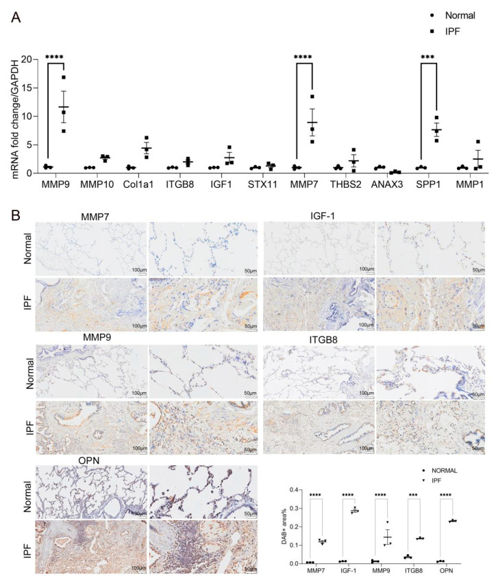
Figure 4.
(A) Quantitative PCR was used to evaluate the relative expression of candidate genes (ANXA3, STX11, THBS2, MMP1, MMP9, MMP7, MMP10, SPP1, COL1A1, ITGB8, IGF1) in human normal lung tissues (n = 3) and IPF lung tissues (n = 3), and the relative quantification of the expression of the candidate genes was measured using glyseraldehyde-3-phosphate dehydrogenase (GAPDH) mRNA as an internal control. Data are presented as mean ± SEM (n = 3; *** p < 0.001, **** p < 0.0001; by two-way ANOVA with Duncan’s post hoc test). (B) The expression of MMP7, IGF1, SPP1, MMP9, and ITGB8 in human normal lung tissues (n = 3) and IPF lung tissues (n = 3) was determined by immunohistochemistry (scale bars: left panels = 100 μm, right panels = 50 μm). The DAB Substrate System (DAKO) was used to reveal the immunohistochemical staining; the positive areas in each image were analyzed by Image J software; the percentages of MMP7, IGF1, SPP1, MMP9, and ITGB8 areas were quantified (*** p < 0.001, **** p < 0.0001).

