. 2021 Jan 29;10:e65443. doi: 10.7554/eLife.65443

Figure 1. Patient-specific Walker-Warburg syndrome (WWS) induced pluripotent stem (iPS) cell-derived myotubes display reduced functional glycosylation of α-dystroglycan (α-DG).

(A) Representative immunostaining of wild type (WT) and FP4 iPS cell-derived myotubes for myosin heavy chain (MHC) and IIH6 (in red). DAPI stains nuclei (in blue). Scale bar, 200 μm. (B) Representative western blot for α-DG core and α-DG functional glycosylation (IIH6) in WT and FP4 myotubes. β-DG was used as loading control. Lower panel shows wheat germ agglutinin (WGA) pull-down for these samples and representative laminin overlay assay (LOA) of WGA elutes shows laminin detection only in WT samples. (C) Graph bars show respective quantification of IIH6 (B) normalized to β-DG and shown as the fold difference of WT. Error bars represent standard errors of five independent experiments. Significance was evaluated by the unpaired Student’s t test. ****p<0.0001.

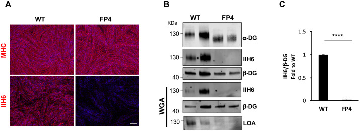
Figure 1.

Figure 1—figure supplement 1. Pluripotency characterization of reprogrammed FP4 induced pluripotent stem (iPS) cell line.

Figure 1—figure supplement 1.

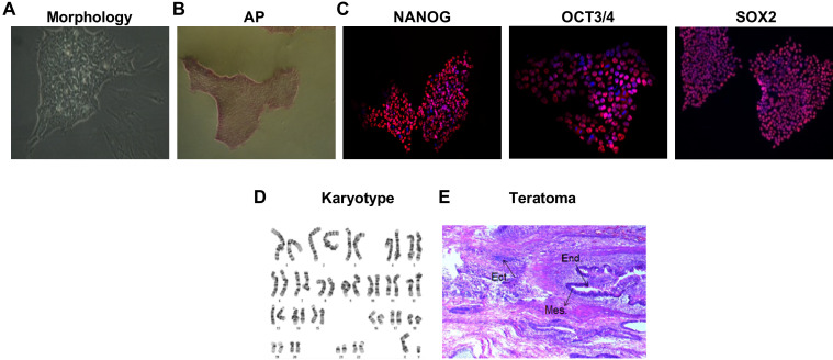
(A–C) Representative images show typical (A) pluripotent colony morphology, (B) alkaline phosphatase activity, and (C) immunostaining for OCT3/4, SOX2, and NANOG (red). DAPI stains nuclei (in blue). Scale bar is 200 μm. (D) Cytogenetic analyses show normal karyotype. (E) Subcutaneous injection of FP4 iPS cells into NOD scid gamma (NSG) mice results in teratoma formation. Representative image shows hematoxylin-eosin staining of a teratoma denoting the presence of tissues derived from all three germ layers.
Figure 1—figure supplement 2. Fukutin-related protein (FKRP) overexpression rescues functional glycosylation of α-dystroglycan (α-DG) in Walker-Warburg syndrome (WWS) FP4 induced pluripotent stem (iPS) cell-derived myotubes.

Figure 1—figure supplement 2.

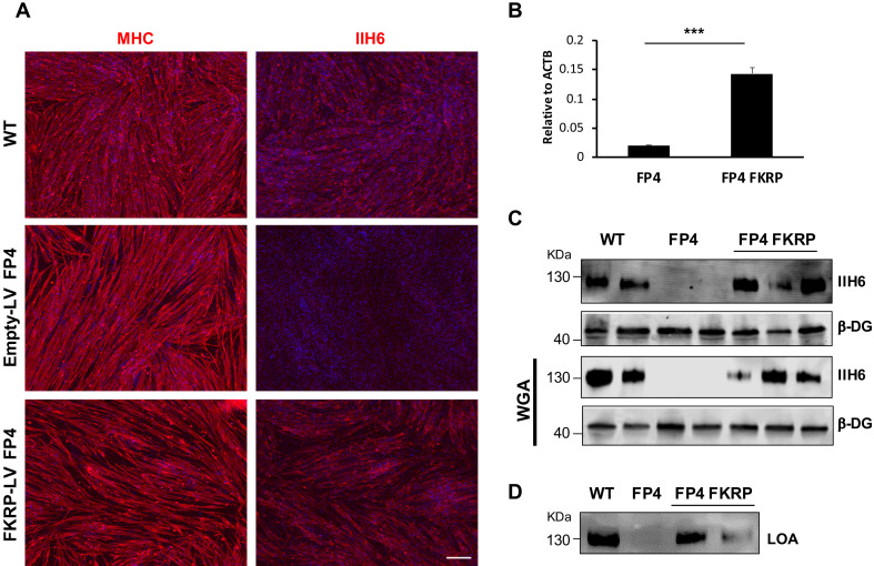
(A) Representative immunofluorescence staining of control (empty vector LV) and FKRP-LV FP4 iPS cell-derived myotubes for myosin heavy chain (MHC) and IIH6 (in red). Wild type (WT) myotubes were used as reference. DAPI stains nuclei (in blue). Scale bar, 200 μm. (B) RT-qPCR analysis shows increased expression levels of FKRP in FKRP-LV FP4 iPS cell-derived myotubes compared to control. Error bars represent standard errors of three independent experiments. (C–D) Introduction of FKRP rescues impaired functional glycosylation of α-DG in FP4 FKRP myotubes, as shown by western blot for IIH6 (C) and laminin overlay assay (LOA) denoting laminin detection (D). β-DG was used as loading control. Lower panel shows representative wheat germ agglutinin (WGA) pull-down. Significance was evaluated by the unpaired Student’s t test. ***p<0.001.