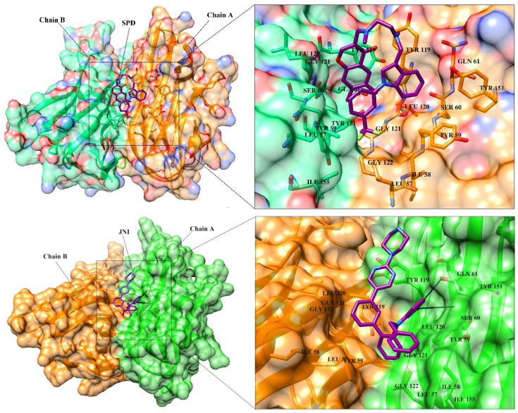
Figure 3.
The 3D-structure of TNF-α is presented in dimeric form in complex with SPD (A) and JNJ525 (B). The ligand binding residues at dimer interface are highlighted. Chain A and B are presented in orange and green color, respectively. The binding site residues are depicted in stick model in their respective chain colors. The co-crystallized ligands (SPD and JNJ525) are shown in magenta color (stick model). SPD is predominantly bound with hydrophobic interactions, however JNJ525 is bound with hydrophobic interactions as well as H-bonding with Ser60 and Tyr151. Hydrogen bonds are depicted in black lines.

