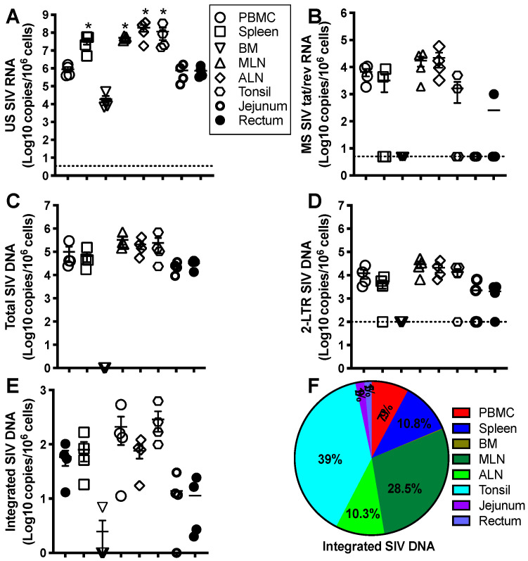
Figure 2.
Representative levels and anatomical tissue distribution of cell-associated SIV RNA/DNA in SIV-infected macaques. Adult Indian-origin rhesus macaques (Macaca mulatta) were intravenously inoculated with 100 TCID50 SIVmac251. After 8 weeks, these animals received three anti-HIV drugs (TFV 20 mg/kg/day; FTC 30 mg/kg/day and DTG 2.5 mg/kg/day) for 20 months. The levels of cell-associated unspliced (US) SIV RNA (A), multiply spliced (MS) SIV tat/rev RNA (B), total SIV DNA (C), circular SIV 2-long terminal repeat (LTR) (D), and integrated proviral DNA (E), in blood, spleen, mesenteric lymph node, axillary lymph node, jejunum, and rectum from SIV-infected animals 3 months after ATI, reaching the levels prior to treatment. (F) Distribution of proviral reservoir in tissues examined. Note that integrated proviral DNA was predominantly distributed in peripheral blood and lymphoid compartments, and rapidly increased to pre-treatment levels after ATI. Cell-associated SIV RNA/DNA are expressed as copies per one-million cells. * p < 0.01, compared with PBMCs.

