Congenital cytomegalovirus (HCMV) infection may cause significant fetal malformation, lifelong disease, and, in severe cases, fetal or neonatal death. Placental infection with HCMV is the major mechanism of mother-to-child transmission (MTCT) and fetal injury. Thus, any pharmaceutical antiviral interference to reduce viral load may reduce placental damage, MTCT, and fetal disease. However, there is currently no licensed HCMV antiviral for use during pregnancy. In this study, aciclovir and the HCMV-specific antivirals letermovir, maribavir, and cidofovir were compared with ganciclovir for antiviral effects in model systems of pregnancy, including first-trimester TEV-1 trophoblast cell cultures and third-trimester ex vivo placental explant histocultures.
KEYWORDS: cytomegalovirus, congenital infection, pregnancy, antiviral drugs, placenta, ex vivo models, letermovir, maribavir, cidofovir, valaciclovir
ABSTRACT
Congenital cytomegalovirus (HCMV) infection may cause significant fetal malformation, lifelong disease, and, in severe cases, fetal or neonatal death. Placental infection with HCMV is the major mechanism of mother-to-child transmission (MTCT) and fetal injury. Thus, any pharmaceutical antiviral interference to reduce viral load may reduce placental damage, MTCT, and fetal disease. However, there is currently no licensed HCMV antiviral for use during pregnancy. In this study, aciclovir and the HCMV-specific antivirals letermovir, maribavir, and cidofovir were compared with ganciclovir for antiviral effects in model systems of pregnancy, including first-trimester TEV-1 trophoblast cell cultures and third-trimester ex vivo placental explant histocultures. HCMV-infected trophoblasts at 7 days postinfection (dpi) showed an EC50 of 21 μM for aciclovir, 0.0007 μM for letermovir, 0.11 μM for maribavir, and 0.29 μM for cidofovir, relative to 0.42 μM for ganciclovir. Antivirals added at 10 μM showed no cytotoxic effects and did not affect trophoblast cell proliferation (P > 0.9999). Multiple-round HCMV replication measured at 7 dpi showed letermovir, maribavir, and cidofovir treatment inhibited immediate early, early, and true late viral protein expression as assayed on Western blots. Antiviral treatment of HCMV-infected placental explants showed significant inhibition (P < 0.05) of viral replication with letermovir (83.3%), maribavir (83.6%), cidofovir (89.3%), and ganciclovir (82.4%), but not aciclovir (P > 0.9999). In ex vivo model systems, recently trialed HCMV antivirals letermovir and maribavir were effective at inhibiting HCMV replication. They partly fulfil requirements for use as safe and effective therapeutics during pregnancy to control congenital HCMV. Clinical trials of these newer agents would assist assessment of their utility in pregnancy.
INTRODUCTION
Congenital cytomegalovirus (HCMV) infection is the most common nongenetic cause of fetal malformation in developed countries, with a mean global incidence of 0.64% (1). Congenital HCMV (cCMV) may case neonatal death, prematurity, and chronic illness from lung, liver, and neurological disease. Approximately 10% of HCMV-infected babies will be symptomatic at birth, presenting with chronic conditions such as sensorineural hearing loss, vision loss, prematurity, intrauterine growth restriction, microcephaly, and motor defects, with ∼10% among these dying of multiorgan dysfunction. A significant proportion (∼15%) of initially asymptomatic HCMV-infected babies additionally develop disease between birth and 5 years of age.
Maternal primary infection, reinfection, or reactivation of latent virus during pregnancy can lead to HCMV infecting and replicating within the placenta, crossing the materno-fetal interface, and subsequently infecting the fetus—known as mother-to-child transmission (MTCT). Fetal injury results from direct viral cytopathic damage, although placental infection may also cause fetal injury via HCMV-induced immunomodulation, dysregulation of placental development, and placental dysfunction, as we and others have shown (2–7). The placenta is the life support system for the developing fetus, providing for exchange of oxygen, nutrients, antibodies, hormonal compounds, and excretory products between mother and fetus. An underdeveloped or abnormal placenta can lead to placental insufficiency and subsequent fetal injury. The placenta therefore represents the main organ of cCMV pathogenesis and an important target for therapy to prevent fetal damage.
Despite the clinical and social importance of cCMV, there are no licensed therapeutics available for use during pregnancy to treat HCMV placental infection and prevent subsequent transplacental transmission and congenital disease (8, 9). This is partly due to systemic drug toxicity, potential teratogenicity, or limited evidence for efficacy of the established and licensed HCMV antiviral drugs. The licensed HCMV-specific direct-acting antivirals (DAAs) of the type nucleoside/nucleotide/pyrophosphate analogues, such as ganciclovir (GCV), cidofovir (CDV), and foscarnet (FOS), have significant toxicity issues, which make them unlikely to be used during pregnancy.
There are four antivirals of potential interest in the prevention and treatment of cCMV during pregnancy: valaciclovir (VACV), letermovir (LMV), maribavir (MBV), and brincidofovir (BCV). VACV, the prodrug of the nucleoside analogue aciclovir (ACV), is the only HCMV antiviral to be investigated in pregnant women to date (10–12), due to its established high safety profile. ACV is first converted by viral thymidine kinases to ACV monophosphate; however, HCMV lacks this kinase and so the exact mechanism of action against HCMV has not been deciphered in detail, but appears to be dependent on the nucleoside-phosphorylating activity of the HCMV protein kinase pUL97 (13, 14). LMV is a 3,4-dihydro-quinazoline and targets the HCMV terminase complex, interfering with HCMV DNA concatemer cleavage and packaging of HCMV DNA into capsids. LMV was recently approved by the FDA for prophylaxis to prevent HCMV infection and disease in hematopoietic stem cell transplant (HSCT) recipients. MBV is a riboside benzimidazole that binds to the HCMV-encoded kinase pUL97, inhibiting viral nuclear egress and virus production efficiency. MBV has recently been granted Orphan Drug Designation by the European Commission and Breakthrough Therapy Designation by the FDA as a treatment for HCMV in transplant patients. BCV is an alkoxyalkyl ester analogue of the nucleoside analogue CDV, designed to release CDV intracellularly, allowing for higher intracellular concentrations and lower toxicities. A recent phase III trial failed to meet the primary endpoint of reduction in clinically significant HCMV infection in HCT recipients (15).
The efficacy and toxicity of these HCMV antivirals has yet to be investigated in detail and in parallel in model systems of pregnancy. Given some very promising findings in other settings of antivirals such as LMV (16), MBV (17), CDV/BCV (18), and VACV (11), we investigated the efficacy and toxicity of these antivirals in first-trimester extravillous trophoblast cells and third-trimester ex vivo placental explant histocultures.
RESULTS
HCMV replication in first-trimester placental trophoblast cells is inhibited by antiviral treatment.
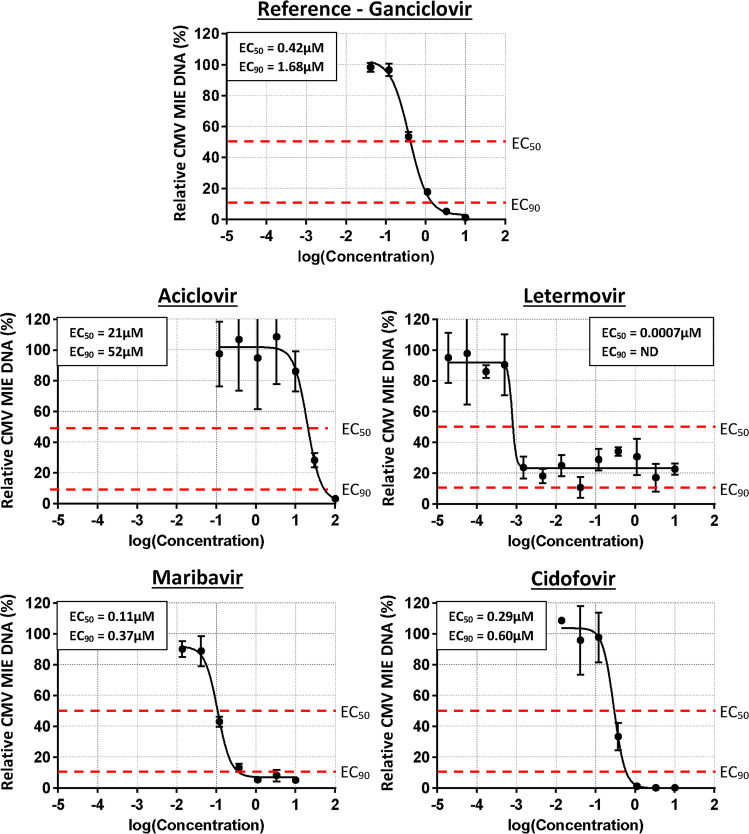
The effects of antiviral treatment on HCMV replication in placental trophoblast cells was investigated in Merlin-infected first-trimester extravillous trophoblast (TEV-1) cell culture supernatant at 7 dpi using quantitative real-time PCR (RT-qPCR) (Fig. 1). Treatment with the reference antiviral GCV showed a 50% effective concentration (EC50) value of 0.43 μM and an EC90 value of 1.68 μM with a 98.80% reduction in HCMV DNA at 10 μM. Treatment with ACV as a surrogate for VACV showed an EC50 value of 21 μM and an EC90 value of 52 μM with a 13.8% reduction in HCMV DNA at 10 μM and a 96.84% reduction at 100 μM. Treatment with LMV showed an EC50 value of 0.0007 μM but an EC90 value could not be determined, with a 77.31% reduction in HCMV DNA at 10 μM. Treatment with MBV showed an EC50 value of 0.11 μM and an EC90 value of 0.37 μM, with a 98.35% reduction in HCMV DNA at 10 μM. Treatment with CDV as a surrogate for BCV showed an EC50 value of 0.29 μM and an EC90 value of 0.60 μM, with a 99.83% reduction in HCMV DNA at 10 μM.
FIG 1.
Efficacy of experimental HCMV antivirals in first-trimester placental trophoblast cells compared with reference antiviral ganciclovir. TEV-1 first-trimester placental trophoblasts were infected with the genetically intact HCMV Merlin strain (2 PFU/cell) and treated with serial dilutions of antiviral compounds at 4 h postinfection. HCMV viral titers in culture supernatants were measured at 7 days postinfection by RT-qPCR and are presented relative to untreated cells. Representative data are presented as mean ± standard deviation (SD) with the corresponding EC50 and EC90 values given (n = 3; experimental triplicates); ND, not determined.
Presence of extracellular HCMV concatemeric DNA in settings of LMV-treated cells may influence the results using RT-qPCR.
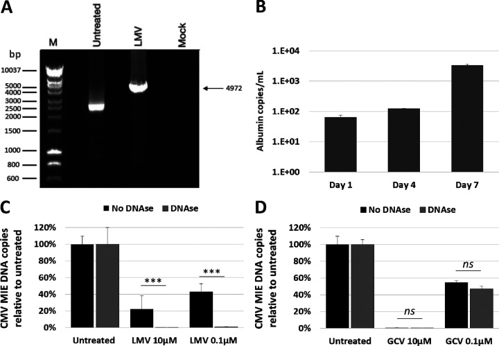
Despite LMV being a highly potent antiviral with a reported EC50 of 4.5 nM and EC90 of 6.1 nM using fluorescence reduction assays (AiCuris), we were unable to determine an EC90 value using RT-qPCR. Cells treated with LMV have previously been shown to result in the accumulation of uncleaved HCMV genome concatemers using Southern blotting (19). We hypothesized that the inability to determine an EC90 value using RT-qPCR may be due to unencapsulated and unenveloped HCMV concatemers in LMV-treated trophoblast cell culture supernatant, thereby confounding the results. HCMV concatemers were detected in cell lysates using a novel PCR assay spanning the HCMV head to tail genomes (Fig. 2A). Sanger sequencing showed the product was specific to the proposed amplified region. We also observed the increasing release of cellular DNA using albumin as a marker in trophoblast cell culture supernatants over the seven day time course (∼1.5 log increase from day 1 to day 7 postinfection) (Fig. 2B). DNase is unable to penetrate the HCMV capsid but will digest any unencapsulated DNA, including unpackaged HCMV concatemers, prior to nucleic acid extraction. Merlin-infected TEV-1 cells treated with 10 μM or 0.1 μM concentrations of LMV showed a 77.6% and 57.0% reduction in viral load relative to untreated cells, respectively (Fig. 2C). When the supernatants were treated with DNase prior to nucleic acid extraction, Merlin-infected TEV-1 cells treated with 10 μM or 0.1 μM concentrations of LMV showed a 99.5% and 98.9% reduction in viral load relative to DNase-treated, LMV-untreated cells, respectively. There was a significant difference in HCMV inhibition observed between the DNase-treated and DNase-untreated samples (P < 0.0001) (Fig. 2C). As a negative control, Merlin-infected TEV-1 cells treated with 10 μM or 0.1 μM concentrations of GCV showed a 99.6% and 45.4% reduction in viral load relative to untreated cells, respectively (Fig. 2D). When the supernatants were treated with DNase prior to nucleic acid extraction, Merlin-infected TEV-1 cells treated with 10 μM or 0.1 μM concentrations of GCV showed a 99.7% and 53.0% reduction in viral load relative to DNase-treated, GCV-untreated cells, respectively. No differences in HCMV inhibition were observed between the DNase-treated and DNase-untreated samples (P > 0.05) (Fig. 2D). These findings support our hypothesis that unencapsulated and unenveloped HCMV concatemers in LMV-treated trophoblast cell culture supernatant artificially inflate viral load measurements by RT-qPCR.
FIG 2.
The detection and elimination of concatemeric HCMV DNA. (A) HCMV DNA concatemers detected in Merlin-infected, letermovir (LMV)-treated (10 μM) cells (4,972-bp product). (B) Release of cellular DNA (albumin) into cell culture supernatant over a 7 day time course. (C and D) Cell culture supernatants treated with DNase prior to qPCR analysis shows elimination of unencapsulated concatemer HCMV DNA in LMV-treated cell cultures (C) but not in ganciclovir (GCV)-treated cell cultures (D). ***, P < 0.001; ns, not significant. Representative data are presented as mean ± SD (n = 3; experimental triplicates).
Digesting HCMV concatemers in LMV-treated cells prior to nucleic acid extraction provides a more accurate EC90 value using RT-qPCR.
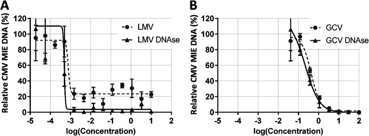
Supernatants from LMV-treated, GCV-treated, and untreated Merlin-infected TEV-1 cultures were treated with DNase prior to nucleic acid extraction or left untreated (Fig. 3). LMV-treated cells with a pre-DNase digestion step showed an EC50 of 0.5 nM and an EC90 of 0.7 nM compared to an EC50 of 0.8 nM and an undetermined EC90 value without DNase treatment. GCV-treated cells with a pre-DNase digestion step showed an EC50 of 0.30 μM and an EC90 of 1.28 μM compared to an EC50 of 0.42 μM and an EC90 of 1.44 μM. There was a statistically significant difference between the LMV DNase-treated and DNase-untreated samples (P = 0.0398), which was not observed between the GCV DNase-treated and DNase-untreated samples (P = 0.1953).
FIG 3.
Efficacy of experimental HCMV antiviral letermovir in first-trimester placental trophoblast cells compared with reference antiviral ganciclovir with DNase treatment. (A) Degrading unencapsulated, unenveloped HCMV DNA with DNase in letermovir-treated cell culture supernatant removed confounding concatemer detection and resulted in the ability to determine EC90 values. (B) DNase treatment of ganciclovir-treated cells had no significant effect on the EC50 and EC90 values (P = 0.1953). Representative data are presented as mean ± SD (n = 3; experimental triplicates).
Antiviral treatment was nontoxic and did not affect placental trophoblast cell proliferation.
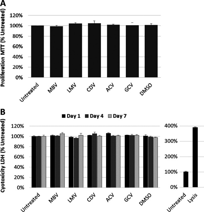
Placental trophoblast cells were treated with the highest antiviral concentration range of 10 μM and 3-(4,5-dimethyl-2-thiazolyl)-2,5-diphenyl-2H-tetrazolium bromide (MTT) proliferation assays and lactate dehydrogenase (LDH) cytotoxicity assays were performed (Fig. 4). Cells treated with antivirals or dimethyl sulfoxide (DMSO) at 50% confluence showed no significant differences in trophoblast cell proliferation at 24 h post treatment relative to untreated cells (P > 0.9999) (Fig. 4A). There were no significant differences in LDH release in antiviral-treated cells or DMSO-treated cells compared with untreated cells at days 1, 4, and 7 after antiviral treatment (P > 0.9999) (Fig. 4B). To monitor the reliability of our assay conditions, positive controls of cytotoxicity (lysing of cells) were routinely used (Fig. 4B).
FIG 4.
Antiviral treatment of placental trophoblast cells does not show any cytotoxicity at high inhibitory concentrations. TEV-1 first-trimester placental trophoblast cells were treated with 10 μM antivirals. (A) MTT cell proliferation assay on antiviral-treated cells at 24 h posttreatment relative to untreated cells. (B) LDH release measured in cell culture supernatant at 1, 4, and 7 days after antiviral treatment relative to untreated cells, with cell lysis acting as a positive control. Representative data are presented as mean ± SD (n = 3; experimental triplicates; P > 0.05).
Antiviral treatment inhibits viral protein expression in placental trophoblast cells after multiple rounds of HCMV replication.
The effects of the leading antiviral treatments (MBV, LMV, and CDV) on HCMV protein expression in placental trophoblast cells were investigated in Merlin-infected first-trimester extravillous trophoblast (TEV-1) cell culture lysates after multiple rounds of replication at 7 dpi using Western blotting (Fig. 5). At 25-μM concentrations, MBV strongly inhibited expression of immediate early (IE1p72), early (pUL44 and pp65), and true-late (pp28) viral proteins relative to untreated cells (98.5%, 97.9%, 89.6%, and 98.5% reductions, respectively). LMV treatment resulted in a reduction of 67.2% for IE1p72, 36.0% for pUL44, 64.4% for pp65, and 95.5% for pp28 expression relative to untreated cells. CDV treatment resulted in a reduction of 76.4% for IE1p72, 94.5% for pUL44, 68.3% for pp65, and 95.8% for pp28 expression relative to untreated cells. It should be emphasized that these antivirals, although acting through different modes of antiviral activity, as explained above, show similarly high efficacy in this read-out system. Thus, the results indicate a strong inhibitory effect of the three analyzed drugs on viral protein production in this extravillous trophoblast in vitro model, as translating into an overall IE-E-L suppressive effect in a clinically relevant, multiple round infection.
FIG 5.
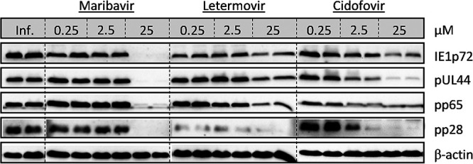
Antiviral treatment inhibits viral protein expression after multiple rounds of replication. Merlin-infected TEV-1 placental trophoblast cells at 7 dpi treated with maribavir, letermovir, or cidofovir show inhibition of HCMV immediate early (IE1p72), early (pUL44 and pp65), and true-late (pp28) protein expression relative to untreated cells at the highest antiviral concentrations (25 μM).
Antiviral treatment inhibits viral replication in HCMV-infected ex vivo placental explant histocultures.
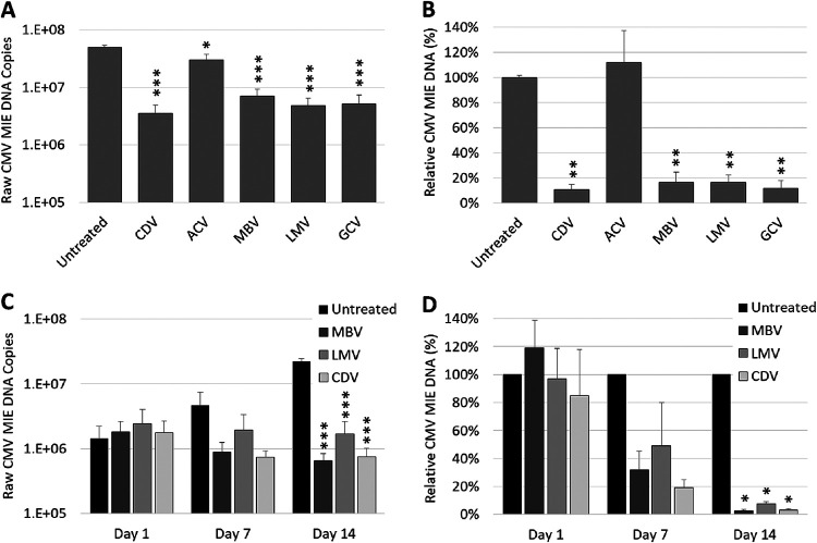
Human placental explant histocultures were infected with the HCMV strain Merlin and treated with 25-μM antivirals at 5 dpi (Fig. 6A). A concentration of 25 μM was chosen based on the first observational study of VACV treatment for symptomatic intrauterine HCMV infection during pregnancy, which showed a mean maternal blood concentration of 24.63 μM after maternal oral administration of 8 g/day VACV over 4 to 6 weeks (10, 20). The concentration also corresponded to a 10-fold higher concentration of the published average EC50 values for GCV (2.5 μM) in in vitro cell culture models.
FIG 6.
Antiviral inhibition of HCMV replication in third-trimester ex vivo placental explant histocultures. Merlin-infected placental explants were treated at 5 dpi with 25 μM concentration of antivirals or left untreated. (A) Raw HCMV viral load was measured 14 days post treatment. (B) Viral load was normalized against cellular albumin copy numbers and presented relative to untreated cells. Antiviral efficacy kinetics of lead antiviral candidates maribavir, letermovir, and cidofovir were assessed at days 1, 7, and 14 days after treatment. (C and D) Raw HCMV viral load was measured (C) or viral load was normalized against cellular albumin copy numbers (D) and presented relative to untreated cells. *, P < 0.01; **, P < 0.001; ***, P < 0.0001. Representative data are presented as mean ± SD (n = 3; experimental triplicates).
At 19 dpi (14 days posttreatment) relative to untreated placental explant histocultures, there was significant inhibition of viral replication in explants treated with ACV (38.5% reduction; P = 0.0049), LMV (90.4% reduction; P < 0.0001), MBV (85.7% reduction; P < 0.0001), CDV (92.9% reduction; P < 0.0001), and GCV (89.6% reduction; P < 0.0001). When HCMV DNA copies were normalized against cellular albumin copies to control for differences in placental explant cell numbers (Fig. 6B), there was no inhibition of viral replication in explants treated with ACV (P > 0.9999), but significant inhibition was found in HCMV-infected explants treated with LMV (83.3% reduction; P = 0.0003), MBV (83.6% reduction; P = 0.0003), CDV (89.3% reduction; P = 0.0002), and GCV (82.4% reduction; P = 0.0002) relative to untreated explants.
To characterize the antiviral effects in more detail for the three leading candidate antivirals MBV, LMV, and CDV, HCMV replication kinetics were quantitated at days 1, 7, and 14 after antiviral treatment (25 μM) (Fig. 6C). HCMV DNA copies normalized against cellular albumin copies showed there was no significant difference found between untreated and antiviral-treated cells at day 1 posttreatment (P > 0.9999). At day 7 posttreatment, there was a nonsignificant reduction in viral loads for treatment with LMV (50.9% reduction; P = 0.3272), MBV (68.0% reduction; P = 0.1273), and CDV (80.6% reduction; P = 0.0636) relative to untreated cells. At day 14 posttreatment, there was significant inhibition of viral load for treatment with LMV (95.8% reduction; P = 0.0022), MBV (98.6% reduction; P = 0.0018), and CDV (97.0% reduction; P = 0.0020) relative to untreated cells (Fig. 6D). Compared to LMV-treated cells, MBV and CDV treatment resulted in a 66.5% and 29.2% reduction in viral replication at 14 days posttreatment, respectively (P > 0.05). Combined, these data demonstrate the efficacy of the recently developed HCMV antivirals LMV and MBV and the potential of BCV in inhibiting HCMV replication in robust multicellular ex vivo placental models of pregnancy.
DISCUSSION
The antiviral efficacy and toxicity of established and more recent antivirals was studied in human placental cell models and ex vivo placental tissue relevant to assessing their future use during pregnancies with congenital HCMV infection. Recently developed HCMV antivirals (MBV and LMV), compared with the reference antiviral drug GCV, displayed high efficacy and low placental cell toxicity profiles in pregnancy model systems.
The antiviral EC50 values obtained from placental trophoblasts infected with the genetically intact Merlin strain of HCMV showed LMV displayed the most potent antiviral efficacy, followed by MBV, CDV, GCV, and lastly ACV. The relative EC50 values obtained are consistent with data from human fibroblast cell cultures (19, 21–27). The higher EC50 and EC90 values observed for ACV treatment is unsurprising given that HCMV lacks the thymidine kinase enzyme to convert ACV to ACV monophosphate and as such is not a specific antiviral for HCMV. Paradoxically, a nonrandomized, single group assignment phase IV clinical trial (12) and a recent prospective, randomized, double-blind, placebo-controlled phase II/III trial (11) showed that VACV treatment resulted in a reduction in the number of symptomatic children at birth as well as the number of terminations of pregnancy for fetal anomalies, and a reduction in the rate of fetal infection after maternal primary infection acquired early in pregnancy, respectively. There is some evidence that the HCMV pUL97 kinase phosphorylates ACV in the absence of the thymidine kinase, albeit with lesser efficiency (13, 14). This may be a possible explanation for the efficacy observed in our study at high antiviral concentrations and the efficacy observed in vivo in clinical trials.
The inability of LMV to inhibit viral replication by more than 80% relative to untreated cells at the high concentration of 10 μM (77.3% reduction) and the results obtained being similar to treatment at 0.0015 μM (76.3% reduction) was surprising given the known high potency of LMV in fibroblast cell culture models using plaque reduction and fluorescence reduction assays (19, 23, 24). As these assays measure encapsulated, enveloped, replication-competent viral particles, whereas RT-qPCR measures viral DNA, the accumulation of replication-incompetent HCMV concatemers from LMV treatment could be a possible explanation for this discrepancy. As DNase digests any unencapsulated, unenveloped DNA, but is unable to penetrate the viral capsid, we treated supernatants with DNase prior to nucleic acid extraction to eliminate any unencapsulated, unenveloped HCMV concatemers from the RT-qPCR measurement of viral load. Consistent with our hypothesis, DNase treatment of LMV-treated cell culture supernatants resulted in significantly lower estimates of viral loads than observed in DNase treatment of GCV-treated cell culture supernatants. This novel method can be of use clinically, where viral loads may be artificially inflated by RT-qPCR detection of free floating concatemers in patients undergoing LMV treatment. Using this method, the viral loads will more accurately reflect both the true result and clinical evaluation of LMV efficacy.
Antiviral treatment at 10 μM, which is much higher than the EC50 values (all <0.42 μM) for all the HCMV-specific antivirals, did not show any cytotoxic effects. This is consistent with limited animal model studies that showed both LMV and MBV have low toxicity and no teratogenic effect or maternal toxicity (28–30). Several studies in animal guinea pig models also showed CDV and BCV have potential benefits in preventing HCMV transmission during pregnancy without toxicity (31–33). Clearly, some of these antivirals can have significant toxicity issues in vivo (e.g., GCV and CDV) which do not manifest in our cell proliferation or LDH assays. While these assays do not fully demonstrate potential safety of antiviral treatment during pregnancy, they do suggest these antivirals are nontoxic to the placental organ. Further animal studies and phase II safety studies on antiviral toxicity and potential teratogenicity would provide additional safety data for the potential use of these therapeutics during pregnancy.
The viral kinetics in HCMV-infected placental explants in response to treatment with LMV, MBV, and CDV showed viral replication was inhibited over the 14-day time course, whereas there was a consistent increase in viral DNA in untreated placental explants. A recent study investigating the placental transfer of LMV and MBV using a placental perfusion model showed both antivirals crossed the placenta at a low to moderate rate, with the mean concentration in the fetal compartment being superior to the EC50 for both molecules (30). Furthermore, they showed some accumulation of the antivirals in the placental tissue where HCMV replicates, causes placental damage, and where MTCT occurs. These treatments could therefore limit HCMV replication within the placenta, which can be an indirect mechanism of HCMV causing adverse pregnancy outcomes through placental dysfunction, and potentially prevent HCMV from entering the fetal circulation and causing direct cytopathic damage to the developing fetal organs.
These data from ex vivo experiments show recently licensed and studied antivirals, particularly LMV and MBV, display high efficacy and low toxicity profiles in first-trimester placental trophoblast cell cultures and third-trimester placental explant histocultures. Further investigations are warranted to characterize these antivirals more profoundly in terms of applicability for use in extended regimens of antiviral treatment. It will be particularly exciting to learn more about their potential for use specifically during pregnancy, or in other applications, to support the control of congenital HCMV disease.
MATERIALS AND METHODS
Antiviral compounds.
Analytical grade aciclovir was obtained from Sigma-Aldrich. Letermovir and maribavir were obtained from MedChemExpress. Analytical grade cidofovir and ganciclovir was obtained from Sigma-Aldrich. Stock aliquots were prepared in dimethyl sulfoxide (DMSO) and stored at –80°C.
Cultured cells and viruses.
Human first-trimester TEV-1 extravillous trophoblast cells (34, 35) were maintained in Ham’s F10 nutrient mix (Life Technologies) supplemented with 10% fetal bovine serum (FBS) and 100 U/ml penicillin G, 100 μg/ml streptomycin, and 29.2 μg/ml l-glutamine (1XPSG), (Life Technologies) at 37°C with 5% CO2. Genetically intact HCMV strain Merlin (UL128+, RL132−) was derived from a Merlin-BAC recombinant, pAL1120, kindly provided by Richard Stanton (University of Cardiff, United Kingdom) (36) and propagated in RPE-1 cells kindly provided by Barry Slobedman. Titer of virus stocks was determined using standard plaque assays.
TEV-1 culture assays.
TEV-1 cells were seeded in 24-well plates and inoculated with virus in triplicate at a multiplicity of infection (MOI) of 2 PFU/cell. Mock-infected cultures were established concurrently. Plates were centrifuged at 770 × g for 30 min followed by 2 h of incubation at 37°C with 5% CO2. Supernatant was removed and replaced with fresh medium with or without antivirals and incubated at 37°C with 5% CO2.
Clinical placentae and placental villous explant histocultures.
Term placentae were collected with consent from women undergoing elective Caesarean section delivery who had had a healthy pregnancy and were not in labor, under ethics approval SESIAHS HREC 09/126. Placental villous explant histocultures were established as previously described (37). Briefly, placental explants were inoculated with 1 × 10⁷ PFU of HCMV strain Merlin and incubated for 5 days at 37°C supplemented with 5% CO2. At day five postinfection, explants were washed in 1× PBS and transferred to fresh plates with fresh medium. The explants were then treated with 25 μM antivirals and incubated for a further 7 days. At day 12 postinfection (7 days postinhibitor treatment), medium was again replaced with fresh medium and fresh compounds and histocultures were incubated until explant harvest at 19 days postinfection (14 days postinhibitor treatment).
Nucleic acid extraction and quantitative real-time PCR.
Total nucleic acid from the TEV-1 trophoblast cell culture supernatants and the placental explant tissues were extracted using MagnaPure LC Total Nucleic Acid kit according to the manufacturer’s protocol (Roche) as previously described (37). Quantitative real-time PCR was performed using a Roche 480 LightCycler with Kapa Sybr Fast qPCR master mix (Merck). The number of cell-associated HCMV major immediate early (MIE) DNA copies was normalized against cellular albumin copies as previously described (3, 21), using previously published oligonucleotide primers (3). Reactions were carried out under the following conditions: denaturation at 95°C for 5 min, followed by 45 cycles of denaturation at 94°C for 15 s, annealing at 60°C for 20 s, and elongation at 72°C for 15 s. Product specificity was determined using melt peak analysis.
HCMV genome detection.
The TEV-1 trophoblasts were infected with HCMV Merlin strain (2 PFU/cell) or mock infected and either treated with 10 μM letermovir or left untreated. Total nucleic acid from TEV-1 trophoblast cell lysates was extracted using DNeasy blood and tissue kit according to the manufacturer’s protocol (Qiagen). Concatemer HCMV DNA was amplified between nucleotide 232,815 (forward primer: 5′-GCACGTCCCAAACTGGCTTGAGGAG-3′) within the TRS region of one genome and nucleotide 2,136 (reverse primer: 5′-GGAAAGAGCGTGTGTGATCTGGCCGAG-3′) within the RL1 region of the next genome, yielding a 4,967-bp product spanning the HCMV genomes (Fig. 7). PCR amplification of the 4,967-bp region would not be able to occur on cleaved genomes, as cleavage occurs in the middle of the amplified region between the “a” sequences of two concatemer genomes, as shown in Fig. 7. PCR was performed using the Expand High Fidelity PCR system as previously described (38). PCR products were detected by electrophoresis on 1% agarose gel in 0.5× Tris-borate-EDTA (TBE) and visualized with Sybr Safe. Specificity of product was determined by Sanger sequencing.
FIG 7.
Schematic diagram of HCMV genome concatemer detection by PCR.
DNase treatment.
Merlin-infected TEV-1 trophoblast cell culture supernatants treated with letermovir, ganciclovir, or left untreated were incubated with amplification-grade DNase I (Merck) or H2O at room temperature for 15 min. Stop solution was added and samples heated at 70°C for 10 min to denature the DNase enzyme, and then placed on ice for qRT-PCR analysis.
Western blot analysis.
Western blot analysis was performed by standard procedures as described previously (39). Immunostaining was performed with the antibodies mouse MAb-β-actin (Ac-15, Sigma), mouse MAb-IE1p72/pUL44 (IE/E; clones DDG9 and CCH2, Dako), mouse MAb-pp65 (Abcam), mouse MAb-pp28 (Abcam), and horseradish peroxidase (HRP)-conjugated anti-mouse secondary antibody (Pierce). Densitometry of immunostaining was performed using ImageJ software. The mean densitometry values for control-infected cells normalized against β-actin were assumed to be 100% and this value was used to calculate relative protein expression.
MTT assay.
TEV-1 cells were treated with 10 μM antivirals at 40 to 50% confluence and incubated for 24 h at 37°C supplemented with 5% CO2. Medium was removed and replaced with medium containing 1 mg/ml MTT (3-[4,5-dimethyl-2-thiazolyl]-2,5-diphenyl-2H-tetrazolium bromide) and cells were incubated for 2 h. Cells were washed with 100% isopropanol for 10 min to dissolve the formazan and plates were read at an optical density of 570 nm (OD570).
Cytotoxicity assay.
Cell damage was measured using LDH release assays performed using the CytoTox 96 nonradioactive cytotoxicity assay (Promega), with the supernatant of placental cells cultured for 1, 4, and 7 days in the presence of inhibitors at 10 μM according to the manufacturer’s protocols.
Statistical analysis.
The EC50 and EC90 values for antivirals were determined using nonlinear regression and dose-response inhibition. For comparison between two groups, a Student’s t test was performed. For comparisons between three groups or more, the nonparametric Kruskal-Wallis test was initially performed to identify the presence of a difference between treated and nontreated groups. Where a significant difference was detected (P < 0.05), a one-way ANOVA was performed with post hoc Bonferroni correction applied for multiple comparisons. Statistical analysis was performed using GraphPad Prism v7.0.
ACKNOWLEDGMENTS
The study was funded by an Early Career Award from the Thrasher Research Fund (grant RG181876-Hamilton) and was supported by grants from the Australian National Health and Medical Research Council (grant APP1127717-Hamilton), the Australia-Germany Joint Research Cooperation Scheme (grants 2017-18/RG162050, 2020-21/RG192195), the DAAD-Go8 (grants 2017-18/MM-WDR-STH-JM, 2020-21/MM-WDR), the Wilhelm Sander-Stiftung (grant 2018.121.1/MM-SBT), and the Interdisciplinary Center for Clinical Research (IZKF) of the Faculty of Medicine of FAU (grant A88/MM-HS). The funders had no role in study design, data collection and interpretation, or the decision to submit the work for publication.
The authors are grateful to Richard Stanton (University of Cardiff, Wales, UK) and Barry Slobedman (University of Sydney, Australia) for the supply of valuable materials. We thank Joanna Youngson, Maria Jimenez, and Rebecca Anderson (Sydney Cord Blood Bank, Sydney Children’s Hospital, Sydney, Australia) for arranging consent for woman to donate placental tissue. We thank Megan Lenardon and Lynn Tran for designing HCMV concatemer primers.
W.D.R. and M.M supervised the project; S.T.H., M.M., and W.D.R. designed the research; S.T.H. performed experiments; S.T.H. collected and analyzed data; S.T.H., M.M., and W.D.R. wrote the manuscript.
REFERENCES
- 1.Kenneson A, Cannon MJ. 2007. Review and meta-analysis of the epidemiology of congenital cytomegalovirus (CMV) infection. Rev Med Virol 17:253–276. doi: 10.1002/rmv.535. [DOI] [PubMed] [Google Scholar]
- 2.Hamilton ST, Scott G, Naing Z, Iwasenko J, Hall B, Graf N, Arbuckle S, Craig ME, Rawlinson WD. 2012. Human cytomegalovirus-induces cytokine changes in the placenta with implications for adverse pregnancy outcomes. PLoS One 7:e52899. doi: 10.1371/journal.pone.0052899. [DOI] [PMC free article] [PubMed] [Google Scholar]
- 3.Hamilton ST, Hutterer C, Egilmezer E, Steingruber M, Milbradt J, Marschall M, Rawlinson WD. 2018. Human cytomegalovirus utilises cellular dual-specificity tyrosine phosphorylation-regulated kinases during placental replication. Placenta 72–73:10–19. doi: 10.1016/j.placenta.2018.10.002. [DOI] [PubMed] [Google Scholar]
- 4.Scott GM, Chow SS, Craig ME, Pang CN, Hall B, Wilkins MR, Jones CA, Lloyd AR, Rawlinson WD. 2012. Cytomegalovirus infection during pregnancy with maternofetal transmission induces a proinflammatory cytokine bias in placenta and amniotic fluid. J Infect Dis 205:1305–1310. doi: 10.1093/infdis/jis186. [DOI] [PubMed] [Google Scholar]
- 5.van Zuylen WJ, Ford CE, Wong DD, Rawlinson WD. 2016. Human cytomegalovirus modulates expression of noncanonical Wnt receptor ROR2 to alter trophoblast migration. J Virol 90:1108–1115. doi: 10.1128/JVI.02588-15. [DOI] [PMC free article] [PubMed] [Google Scholar]
- 6.Maidji E, Nigro G, Tabata T, McDonagh S, Nozawa N, Shiboski S, Muci S, Anceschi MM, Aziz N, Adler SP, Pereira L. 2010. Antibody treatment promotes compensation for human cytomegalovirus-induced pathogenesis and a hypoxia-like condition in placentas with congenital infection. Am J Pathol 177:1298–1310. doi: 10.2353/ajpath.2010.091210. [DOI] [PMC free article] [PubMed] [Google Scholar]
- 7.Hamilton ST, Scott GM, Naing Z, Rawlinson WD. 2013. Human cytomegalovirus directly modulates expression of chemokine CCL2 (MCP-1) during viral replication. J Gen Virol 94:2495–2503. doi: 10.1099/vir.0.052878-0. [DOI] [PubMed] [Google Scholar]
- 8.Hamilton ST, van Zuylen W, Shand A, Scott GM, Naing Z, Hall B, Craig ME, Rawlinson WD. 2014. Prevention of congenital cytomegalovirus complications by maternal and neonatal treatments: a systematic review. Rev Med Virol 24:420–433. doi: 10.1002/rmv.1814. [DOI] [PubMed] [Google Scholar]
- 9.Rawlinson WD, Boppana SB, Fowler KB, Kimberlin DW, Lazzarotto T, Alain S, Daly K, Doutre S, Gibson L, Giles ML, Greenlee J, Hamilton ST, Harrison GJ, Hui L, Jones CA, Palasanthiran P, Schleiss MR, Shand AW, van Zuylen WJ. 2017. Congenital cytomegalovirus infection in pregnancy and the neonate: consensus recommendations for prevention, diagnosis, and therapy. Lancet Infect Dis 17:e177–e188. doi: 10.1016/S1473-3099(17)30143-3. [DOI] [PubMed] [Google Scholar]
- 10.Jacquemard F, Yamamoto M, Costa JM, Romand S, Jaqz-Aigrain E, Dejean A, Daffos F, Ville Y. 2007. Maternal administration of valaciclovir in symptomatic intrauterine cytomegalovirus infection. BJOG 114:1113–1121. doi: 10.1111/j.1471-0528.2007.01308.x. [DOI] [PubMed] [Google Scholar]
- 11.Shahar-Nissan K, Pardo J, Peled O, Krause I, Bilavsky E, Wiznitzer A, Hadar E, Amir J. 2020. Valaciclovir to prevent vertical transmission of cytomegalovirus after maternal primary infection during pregnancy: a randomised, double-blind, placebo-controlled trial. Lancet 396:779–785. doi: 10.1016/S0140-6736(20)31868-7. [DOI] [PubMed] [Google Scholar]
- 12.Leruez-Ville M, Ghout I, Bussieres L, Stirnemann J, Magny JF, Couderc S, Salomon LJ, Guilleminot T, Aegerter P, Benoist G, Winer N, Picone O, Jacquemard F, Ville Y. 2016. In utero treatment of congenital cytomegalovirus infection with valacyclovir in a multicenter, open-label, phase II study. Am J Obstet Gynecol 215:462.e1–462.e10. doi: 10.1016/j.ajog.2016.04.003.]. [DOI] [PubMed] [Google Scholar]
- 13.Zimmermann A, Michel D, Pavic I, Hampl W, Luske A, Neyts J, De Clercq E, Mertens T. 1997. Phosphorylation of aciclovir, ganciclovir, penciclovir and S2242 by the cytomegalovirus UL97 protein: a quantitative analysis using recombinant vaccinia viruses. Antiviral Res 36:35–42. doi: 10.1016/s0166-3542(97)00034-x. [DOI] [PubMed] [Google Scholar]
- 14.Talarico CL, Burnette TC, Miller WH, Smith SL, Davis MG, Stanat SC, Ng TI, He Z, Coen DM, Roizman B, Biron KK. 1999. Acyclovir is phosphorylated by the human cytomegalovirus UL97 protein. Antimicrob Agents Chemother 43:1941–1946. doi: 10.1128/AAC.43.8.1941. [DOI] [PMC free article] [PubMed] [Google Scholar]
- 15.Marty FM, Winston DJ, Chemaly RF, Mullane KM, Shore TB, Papanicolaou GA, Chittick G, Brundage TM, Wilson C, Morrison ME, Foster SA, Nichols WG, Boeckh MJ, Group STCS. 2019. A Randomized, double-blind, placebo-controlled phase 3 trial of oral brincidofovir for cytomegalovirus prophylaxis in allogeneic hematopoietic cell transplantation. Biol Blood Marrow Transplant 25:369–381. doi: 10.1016/j.bbmt.2018.09.038. [DOI] [PMC free article] [PubMed] [Google Scholar]
- 16.Marty FM, Ljungman P, Chemaly RF, Maertens J, Dadwal SS, Duarte RF, Haider S, Ullmann AJ, Katayama Y, Brown J, Mullane KM, Boeckh M, Blumberg EA, Einsele H, Snydman DR, Kanda Y, DiNubile MJ, Teal VL, Wan H, Murata Y, Kartsonis NA, Leavitt RY, Badshah C. 2017. Letermovir prophylaxis for cytomegalovirus in hematopoietic-cell transplantation. N Engl J Med 377:2433–2444. doi: 10.1056/NEJMoa1706640. [DOI] [PubMed] [Google Scholar]
- 17.Papanicolaou GA, Silveira FP, Langston AA, Pereira MR, Avery RK, Uknis M, Wijatyk A, Wu J, Boeckh M, Marty FM, Villano S. 2019. Maribavir for refractory or resistant cytomegalovirus infections in hematopoietic-cell or solid-organ transplant recipients: a randomized, dose-ranging, double-blind, phase 2 study. Clin Infect Dis 68:1255–1264. doi: 10.1093/cid/ciy706. [DOI] [PMC free article] [PubMed] [Google Scholar]
- 18.Marty FM, Winston DJ, Rowley SD, Vance E, Papanicolaou GA, Mullane KM, Brundage TM, Robertson AT, Godkin S, Mommeja-Marin H, Boeckh M, CMX001-201 Clinical Study Group. 2013. CMX001 to prevent cytomegalovirus disease in hematopoietic-cell transplantation. N Engl J Med 369:1227–1236. doi: 10.1056/NEJMoa1303688. [DOI] [PubMed] [Google Scholar]
- 19.Goldner T, Hewlett G, Ettischer N, Ruebsamen-Schaeff H, Zimmermann H, Lischka P. 2011. The novel anticytomegalovirus compound AIC246 (Letermovir) inhibits human cytomegalovirus replication through a specific antiviral mechanism that involves the viral terminase. J Virol 85:10884–10893. doi: 10.1128/JVI.05265-11. [DOI] [PMC free article] [PubMed] [Google Scholar]
- 20.Leruez-Ville M, Ville Y. 2016. Optimum treatment of congenital cytomegalovirus infection. Expert Rev Anti Infect Ther 14:479–488. doi: 10.1586/14787210.2016.1173540. [DOI] [PubMed] [Google Scholar]
- 21.Morere L, Andouard D, Labrousse F, Saade F, Calliste CA, Cotin S, Aubard Y, Rawlinson WD, Esclaire F, Hantz S, Ploy MC, Alain S. 2015. Ex vivo model of congenital cytomegalovirus infection and new combination therapies. Placenta 36:41–47. doi: 10.1016/j.placenta.2014.11.003. [DOI] [PubMed] [Google Scholar]
- 22.Chou S, Wechel LC, Marousek GI. 2007. Cytomegalovirus UL97 kinase mutations that confer maribavir resistance. J Infect Dis 196:91–94. doi: 10.1086/518514. [DOI] [PubMed] [Google Scholar]
- 23.Marschall M, Stamminger T, Urban A, Wildum S, Ruebsamen-Schaeff H, Zimmermann H, Lischka P. 2012. In vitro evaluation of the activities of the novel anticytomegalovirus compound AIC246 (letermovir) against herpesviruses and other human pathogenic viruses. Antimicrob Agents Chemother 56:1135–1137. doi: 10.1128/AAC.05908-11. [DOI] [PMC free article] [PubMed] [Google Scholar]
- 24.Lischka P, Hewlett G, Wunberg T, Baumeister J, Paulsen D, Goldner T, Ruebsamen-Schaeff H, Zimmermann H. 2010. In vitro and in vivo activities of the novel anticytomegalovirus compound AIC246. Antimicrob Agents Chemother 54:1290–1297. doi: 10.1128/AAC.01596-09. [DOI] [PMC free article] [PubMed] [Google Scholar]
- 25.Drew WL, Miner RC, Marousek GI, Chou S. 2006. Maribavir sensitivity of cytomegalovirus isolates resistant to ganciclovir, cidofovir or foscarnet. J Clin Virol 37:124–127. doi: 10.1016/j.jcv.2006.07.010. [DOI] [PubMed] [Google Scholar]
- 26.Scott GM, Weinberg A, Rawlinson WD, Chou S. 2007. Multidrug resistance conferred by novel DNA polymerase mutations in human cytomegalovirus isolates. Antimicrob Agents Chemother 51:89–94. doi: 10.1128/AAC.00633-06. [DOI] [PMC free article] [PubMed] [Google Scholar]
- 27.Snoeck R, Andrei G, Schols D, Balzarini J, De Clercq E. 1992. Activity of different antiviral drug combinations against human cytomegalovirus replication in vitro. Eur J Clin Microbiol Infect Dis 11:1144–1155. doi: 10.1007/BF01961133. [DOI] [PubMed] [Google Scholar]
- 28.Biron KK, Harvey RJ, Chamberlain SC, Good SS, Smith AA, 3rd, Davis MG, Talarico CL, Miller WH, Ferris R, Dornsife RE, Stanat SC, Drach JC, Townsend LB, Koszalka GW. 2002. Potent and selective inhibition of human cytomegalovirus replication by 1263W94, a benzimidazole L-riboside with a unique mode of action. Antimicrob Agents Chemother 46:2365–2372. doi: 10.1128/aac.46.8.2365-2372.2002. [DOI] [PMC free article] [PubMed] [Google Scholar]
- 29.Bigley TM, McGivern JV, Ebert AD, Terhune SS. 2016. Impact of a cytomegalovirus kinase inhibitor on infection and neuronal progenitor cell differentiation. Antiviral Res 129:67–73. doi: 10.1016/j.antiviral.2016.02.007. [DOI] [PMC free article] [PubMed] [Google Scholar]
- 30.Faure Bardon V, Peytavin G, Le MP, Guilleminot T, Elefant E, Stirnemann J, Leruez-Ville M, Ville Y. 2020. Placental transfer of Letermovir & Maribavir in the ex vivo human cotyledon perfusion model. New perspectives for in utero treatment of congenital cytomegalovirus infection. PLoS One 15:e0232140. doi: 10.1371/journal.pone.0232140. [DOI] [PMC free article] [PubMed] [Google Scholar]
- 31.Bravo FJ, Cardin RD, Bernstein DI. 2006. Effect of maternal treatment with cyclic HPMPC in the guinea pig model of congenital cytomegalovirus infection. J Infect Dis 193:591–597. doi: 10.1086/499603. [DOI] [PubMed] [Google Scholar]
- 32.Schleiss MR, Anderson JL, McGregor A. 2006. Cyclic cidofovir (cHPMPC) prevents congenital cytomegalovirus infection in a guinea pig model. Virol J 3:9. doi: 10.1186/1743-422X-3-9. [DOI] [PMC free article] [PubMed] [Google Scholar]
- 33.Bravo FJ, Bernstein DI, Beadle JR, Hostetler KY, Cardin RD. 2011. Oral hexadecyloxypropyl-cidofovir therapy in pregnant guinea pigs improves outcome in the congenital model of cytomegalovirus infection. Antimicrob Agents Chemother 55:35–41. doi: 10.1128/AAC.00971-10. [DOI] [PMC free article] [PubMed] [Google Scholar]
- 34.Feng HC, Choy MY, Deng W, Wong HL, Lau WM, Cheung AN, Ngan HY, Tsao SW. 2005. Establishment and characterization of a human first-trimester extravillous trophoblast cell line (TEV-1). J Soc Gynecol Invest 12:e21-32–e32. doi: 10.1016/j.jsgi.2005.02.008. [DOI] [PubMed] [Google Scholar]
- 35.Wu D, Hong H, Huang X, Huang L, He Z, Fang Q, Luo Y. 2016. CXCR2 is decreased in preeclamptic placentas and promotes human trophoblast invasion through the Akt signaling pathway. Placenta 43:17–25. doi: 10.1016/j.placenta.2016.04.016. [DOI] [PubMed] [Google Scholar]
- 36.Stanton RJ, Baluchova K, Dargan DJ, Cunningham C, Sheehy O, Seirafian S, McSharry BP, Neale ML, Davies JA, Tomasec P, Davison AJ, Wilkinson GW. 2010. Reconstruction of the complete human cytomegalovirus genome in a BAC reveals RL13 to be a potent inhibitor of replication. J Clin Invest 120:3191–3208. doi: 10.1172/JCI42955. [DOI] [PMC free article] [PubMed] [Google Scholar]
- 37.Hutterer C, Hamilton S, Steingruber M, Zeittrager I, Bahsi H, Thuma N, Naing Z, Orfi Z, Orfi L, Socher E, Sticht H, Rawlinson W, Chou S, Haupt VJ, Marschall M. 2016. The chemical class of quinazoline compounds provides a core structure for the design of anticytomegaloviral kinase inhibitors. Antiviral Res 134:130–143. doi: 10.1016/j.antiviral.2016.08.005. [DOI] [PubMed] [Google Scholar]
- 38.Champier G, Hantz S, Couvreux A, Stuppfler S, Mazeron MC, Bouaziz S, Denis F, Alain S. 2007. New functional domains of human cytomegalovirus pUL89 predicted by sequence analysis and three-dimensional modelling of the catalytic site DEXDc. Antivir Ther 12:217–232. [PubMed] [Google Scholar]
- 39.Hamilton ST, Milbradt J, Marschall M, Rawlinson WD. 2014. Human cytomegalovirus replication is strictly inhibited by siRNAs targeting UL54, UL97 or UL122/123 gene transcripts. PLoS One 9:e97231. doi: 10.1371/journal.pone.0097231. [DOI] [PMC free article] [PubMed] [Google Scholar]


