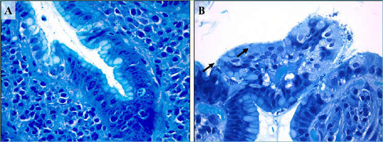
Figure 1.

(A-B). Helicobacter pylori active gastritis (Giemsa staining). Lymphocytic inflammation and neutrophilic epithelium infiltration with conventional spiral-shaped H. pylori (A; 630x magnification). Dormant or stressed coccoid microorganisms form (arrows, B; 630 x magnification).
