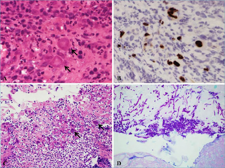
Figure 1.

(A) Cytomegalovirus esophagitis (magnification 40x). Nucleomegaly with intranuclear inclusions (black arrow). (B) Cytomegalovirus esophagitis (magnification 40x). Immunohistochemistry with anti-CMV antibody, showing sparse positive nuclei. (C) Herpes Simplex Virus esophagitis (magnification 10x). Multinucleated giant cells and ground glass intranuclear inclusions (black arrow) with necrotic debris and inflammation. (D) Candida Albicans esophagitis (magnification 20x). Alcian Blu PAS staining showing hyphae and spores on the surface of squamous epithelium.
