

Abstract
Airway management in children with craniofacial anomalies can be complicated and may require multiple attempts with conventional direct laryngoscopy (DL). Videolaryngoscopes (VLs) have a well-established role in difficult airway management in adults; however, their role remains to be fully elucidated in paediatric age group. There is a relative paucity in the literature regarding the role of VLs in cases of syndromic children, and it is not clear whether they should be used as an initial option or as a rescue device. Herein, we report a series of cases of children with Pierre Robin sequence, Beckwith–Wiedemann syndrome, and Hurler’s syndrome wherein VLs proved beneficial after multiple failed DL attempts. Following initial failed attempts to intubate using DL, these children were subsequently intubated using VLs. Therefore, VLs should be used for initial intubation attempts in syndromic children with potential difficult airways.
Keywords: Airway management, child, congenital abnormality, intubation, videolaryngoscopy
Introduction
Airway management is a crucial skill for an anaesthesiologist and a fundamental part of general anaesthesia (GA). Paediatric tracheal intubations may be difficult and require multiple attempts, especially in those with craniofacial anomalies (1, 2); however, each additional attempt may increase the airway morbidity (3). There is a growing interest for videolaryngoscopes (VLs) among paediatric anaesthesiologists since it has the potential to facilitate endotracheal intubation and decrease adverse consequences (4, 5). Although the use of VLs has become more prevalent in paediatric anaesthesia practice, documentation of its use, especially for rescue in syndromic children with craniofacial anomalies, remains scarce. This case series describes children with craniofacial anomalies posted for emergency surgery where the use of VLs helped us successfully manage ‘difficult-to-intubate’ scenarios.
Case Presentations
Written informed consent was obtained from the parents of all the three following cases.
Case 1
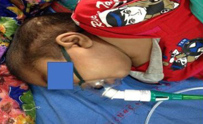
A 2-day-old, 1.8-kg full-term Pierre Robin sequence (PRS) neonate with micrognathia, retrognathia, glossoptosis, high-arched palate and right-hand deformity presented for tracheoesophageal fistula (TEF) repair (Figure 1). The child was anaesthetised using incremental induction of sevoflurane. Initial two attempts with direct laryngoscopy (DL) using Miller and Macintosh blades, respectively, revealed a Cormack and Lehane (C&L) grade-IV view. Since bag and mask ventilation (BMV) was adequate, 3 mg succinylcholine was administered. The third attempt by a senior anaesthesiologist using a paraglossal approach with a Macintosh blade and optimum external laryngeal manipulation (OELM) could only improve the view to CLgrade IIIb. The arterial oxygen saturation momentarily reduced to low 80 s but improved immediately on BMV. The CMAC Miller size ‘0’ blade was then introduced, which resulted in a CL grade IIb view that improved to grade IIa with OELM, and intubation with styletted 2.5-mm internal diameter (ID) endotracheal tube (ETT) was successful.
Figure 1.
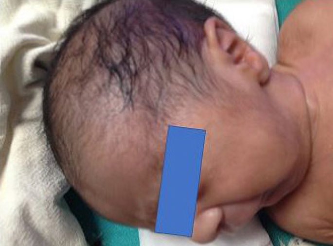
Pierre Robin sequence neonate
Case 2
A 2-day-old male child with Beckwith–Wiedemann syndrome (BWS) was referred to our hospital after normal vaginal delivery for omphalocele repair under GA (Figures 2 and 3). There was a history of hypoglycaemia following birth, and the child was receiving 10% dextrose. Examination revealed peculiar facies, prominent eyes, deformed ears and macroglossia. Midline DL with a size ‘0’ Miller blade revealed a CLgrade IIIb view. Another attempt by a senior anaesthetist using a Macintosh blade with shoulder roll yielded a CLgrade IIIa view with OELM. The use of Truview-PCD VL (Truphatek International Ltd., Netanya, Israel), with 5 L min oxygen flow, yielded a grade IIb view that improved to CLgradeI with OELM, and the child was intubated with a styletted 3-mm ID ETT.
Figure 2.
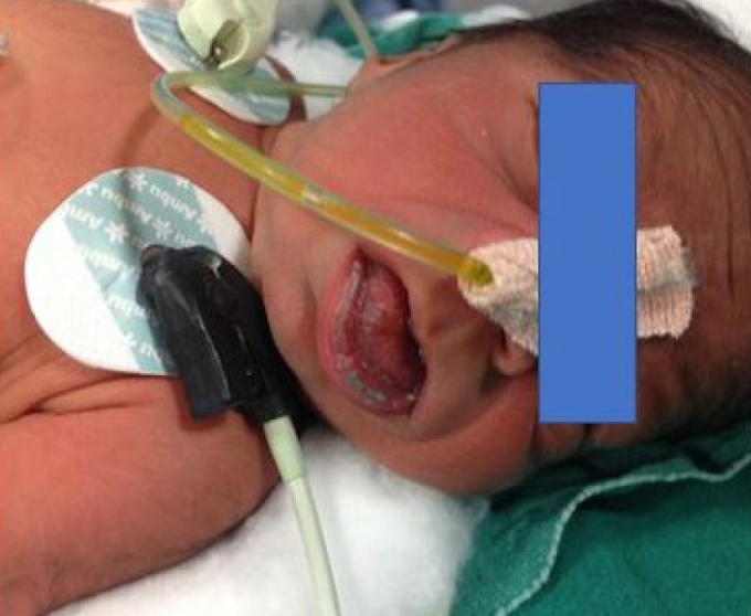
Beckwith–Wiedemann syndrome neonate
Figure 3.
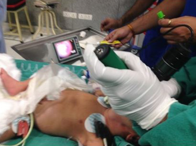
Beckwith–Wiedemann syndrome neonate being intubated with Truview-PCD VL, and glottic view on the monitor
Case 3
A 10-year-old, male Hurler’s syndrome child with bilateral pneumonitis was posted for an emergency incarcerated umbilical hernia repair. He had a large head, coarse features, cloudy cornea, delayed milestones, short stature (106 cm), speech deficit and mental retardation (Figures 4 and 5). His parents provided a history of snoring and frequent awakening during sleep. Characteristic foot and rib deformities were present. Airway examination showed short neck, macroglossia, high-arched palate, Mallampati class IV, enlarged tonsils and poor submandibular compliance. Sevoflurane-based inhalational induction was planned while preserving spontaneous breaths. The first attempt using a Macintosh blade resulted in a CLgrade IIIb view with OELM. Second attempt with a McCoy blade revealed grade IIb view; however, since the child started desaturating, the attempt was terminated. The BMV was successful only after applying a two-handed technique with the use of oral and nasal airways. Intraoral tissues and pharyngeal space were revealed to be non-compliant and narrow, respectively. Repeat attempt with Truview-PCD VL (with oxygen insufflation of 5 L min−1) resulted in a CLgrade IIb view with OELM, but intubation (with Optistylet use) failed. A 10Fr bougie could be inserted into the trachea, and a 5.5-mm ETT could be railroaded over it.
Figure 4.
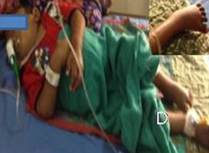
Hurlers syndrome child with foot deformity (inset)
Figure 5.
Hurler’s child (side profile)
Discussion
This series describes the successful management of difficult airway (DA) in syndromic children using VLs. The anatomical and physiological differences in paediatric patients make their airway management challenging. The presence of craniofacial anomalies further aggravated the difficulty in management of airways in the child with PRS (small anterior mandibular space, tongue pushed back to the hypopharynx, and high-arched palate increasing the difficulty in intubation), BWS (large protuberant tongue) and Hurler’s (coarse facies, enlarged tongue, small mandible, large tonsils, large tongue, micrognathia, Mallampati class IV, reduced submandibular compliance, short neck, and limited neck and jaw movement). The problem is further aggravated in those with craniofacial anomalies, especially in emergency scenarios, such as our cases, wherein time for evaluation is limited and waking-up and deferring the case for a later date is not a feasible option.
Airway management in such patients requires careful planning and may need advanced techniques for securing the airway (6). Awake fiberoptic intubation is the safest technique for securing the airway in DA cases but has its limitations in this age group, e.g., the lack of cooperation, the unavailability of expertise and/or age-related equipment, and the need for sedation/GA. Therefore, it was not considered in our cases (7). Macroglossia hindered intubation by making it difficult to displace the tongue during laryngoscopy (8). A DA cart consisting of appropriately sized ETTs, curved and straight DLs, oral airways, bougie, supraglottic devices (SGDs), face masks, VL and fiberoptic bronchoscope (FOB) was arranged.
Sevoflurane inhalational induction with preserved spontaneous respiration is the most favoured technique among paediatric anaesthesiologists for managing DA cases, because the child’s airway can be established by a graded onset of anaesthesia during induction. We managed all the three cases with sevoflurane-based inhalational technique.
Muscle relaxants might improve intubation conditions and decrease the incidence of laryngospasm. Hence, after initial failed attempts in the PRC child, we administered succinylcholine after ensuring adequate BMV (9). In patients with a history of snoring during sleep, BMV may be difficult; therefore, we avoided muscle relaxants in the Hurler’s child.
Neonates with PRS have micrognathia (small jaw) and glossoptosis (the tongue falls to the back of the throat) that leads to airway obstruction and respiratory distress. Various intubation techniques including McGrath VL, Airtraq™ VL, FOB and intubation via SGDs have been described; however, CMAC VL has not been reported (10–12).
Airway management of a BWS child may be difficult due to macroglossia hampering with BMV and laryngoscopy (13). Glidescope VL has been used to facilitate intubation in a 4-year-old BWS child with a history of multiple failed attempts at intubation; however, the use of Truview-PCD VL has not been previously reported. The Truview-PCD blade has a 48º anterior deflection, which helped us visualise the anteriorly placed larynx (14).
In Hurler’s syndrome, mucopolysaccharides accumulate throughout the body. The infiltration of these deposits in the airway leads to macroglossia, thickened and non-compliant soft tissues of the oropharynx and blockage of nasal passages. This may lead to progressive airway obstruction, difficult BMV and difficult intubation (DI). A review of airway management in Hurler’s children reported DI in five out of ten children while intubation failed in three. These three cases were rescued by the use of SGD, FOB and Airtraq VL (15).
A multi-centric registry of children with DA demonstrated that more than two DL attempts were associated with high failure rates and increased complications (transient hypoxemia was the most common) (2). In addition, this study identified that weight <10 kg, micrognathia and three DL attempts before indirect technique increased the risk of complications. Our case with Hurler’s syndrome also desaturated despite apnoeic oxygenation, probably because of the pre-existing pneumonitis and prolonged intubation attempts. Therefore, paediatric anaesthesiologists should minimise the number of DL attempts, use an indirect technique (VL/FOB) early, and consider oxygenation during intubation attempts.
Although a range of VLs has been introduced, they are yet to be approved as a first-line device for routine intubation in children. Some studies have shown that VL is better than conventional DL for routine or DI in children (4, 5). However, none of them have been carried out in syndromic children. In our cases, early use of a VL could have reduced intubation attempts and multiple failures could have been averted. The CMAC Miller blade comes intuitively to those familiar with conventional DL. Furthermore, it does not require special preparation and can be used in case of emergency.
It may be difficult to intubate despite a good glottic view with VL. Hence, a styletted ETT is recommended for intubation. Truview VL is provided with its dedicated Optistylet that has a fixed curvature, and it may be difficult to direct the ETT. We also experienced similar difficulties in one of our patients and had to use a bougie.
In syndromic children, back-up plans such as tracheostomy and SGD can be technically difficult. This reiterates the fact that advanced airway management techniques such as VL should be used during initial intubation attempts.
Videolaryngoscopy is an evolutionary step that involves the use of video and optical technology to provide a non-line-of-sight view of the larynx. It yields a superior image of the larynx with a higher success rate, especially in difficult situations. Children with craniofacial anomalies may have additional airway lesions, which can hamper intubation attempts. Hence, initial attempts with VLs will provide quick and real-time assessment of airway anomalies and help us modify our plan accordingly.
Conclusion
VLs have revolutionised the management of paediatric DA. At present, the choice of a VL depends on the availability, the clinical scenario, individual preference and expertise. Anaesthesiologists should have a multi-layered contingency plan to handle the airway of a syndromic child. Indirect visualisation devices should be used for initial intubation attempts as DL is likely to fail in these patients.
Main Points:
Airway management using conventional DL may be challenging in cases of syndromic children.
A series of cases of children with Pierre Robin sequence, Beckwith–Wiedemann syndrome, and Hurler’s syndrome are reported.
Initial attempts at intubation using DL failed; intubation using VLs succeeded.
VLs should be used for initial intubation attempts in syndromic children with potential difficult airways.
Footnotes
Informed Consent: Written informed consent was obtained from the parents of the patients who participated in this study.
Peer-review: Externally peer-reviewed.
Author Contributions: Concept – A.G., N.G.; Design – A.G., N.G.; Supervision – G.K., K.K.G.; Resources – A.G., N.G.; Materials – A.G., N.G., G.K., K.K.G.; Data Collection and/or Processing – A.G.; Analysis and/or Interpretation – A.G., N.G.; Literature Search – A.G., N.G.; Writing Manuscript – A.G., N.G.; Critical Review – A.G., N.G., G.K., K.K.G.
Conflict of Interest: The authors have no conflicts of interest to declare.
Financial Disclosure: The authors declared that this study has received no financial support.
References
- 1.Holm-Knudsen RJ, Rasmussen LS. Pediatric airway management: basic aspects. Acta Anaesthesiol Scand. 2009;53:1–9. doi: 10.1111/j.1399-6576.2008.01794.x. [DOI] [PubMed] [Google Scholar]
- 2.Fiadjoe JE, Nishisaki A, Jagannathan N, Hunyady AI, Greenberg RS, Reynolds PI, et al. Airway management complications in children with difficult tracheal intubation from the Pediatric Difficult Intubation (PeDI) registry: a prospective cohort analysis. Lancet Respir Med. 2016;4:37–48. doi: 10.1016/S2213-2600(15)00508-1. [DOI] [PubMed] [Google Scholar]
- 3.Graciano AL, Tamburro R, Thompson AE, Fiadjoe J, Nadkarni VM, Nishisaki A. Incidence and associated factors of difficult tracheal intubations in pediatric ICUs: A report from National Emergency Airway Registry for Children. Intensive Care Med. 2014;40:1659–69. doi: 10.1007/s00134-014-3407-4. [DOI] [PubMed] [Google Scholar]
- 4.Sun Y, Lu Y, Huang Y, Jiang H. Pediatric video laryngoscope versus direct laryngoscope: A meta-analysis of randomized controlled trials. Paediatr Anaesth. 2014;24:1056–65. doi: 10.1111/pan.12458. [DOI] [PubMed] [Google Scholar]
- 5.Lingappan K, Arnold JL, Shaw TL, Fernandes CJ, Pammi M. Videolaryngoscopy versus direct laryngoscopy for tracheal intubation in neonates. Cochrane Database Syst Rev. 2015:CD009975. doi: 10.1002/14651858.CD009975.pub2. [DOI] [PubMed] [Google Scholar]
- 6.Shprintzen RJ. Pierre Robin, micrognathia and airway obstruction: the dependency of treatment on accurate diagnosis. Int Anesthesiol Clin. 1985;26:64–71. doi: 10.1097/00004311-198802610-00014. [DOI] [PubMed] [Google Scholar]
- 7.Asai T, Shingu K. Difficulty in advancing a tracheal tube over a fibreoptic bronchoscope: incidence, causes, and solutions. Br J Anaesth. 2004;92:870–81. doi: 10.1093/bja/aeh136. [DOI] [PubMed] [Google Scholar]
- 8.Shar AE. Mechanisms of airway obstruction in Robin se-quence: implications for treatment. Cleft Palate J. 1992;29:224–31. doi: 10.1597/1545-1569_1992_029_0224_moaoir_2.3.co_2. [DOI] [PubMed] [Google Scholar]
- 9.Priebe HJ. Should anesthesiologists have to confirm effective facemask ventilation before administering the muscle relaxant? J Anesth. 2016;30:132–7. doi: 10.1007/s00540-015-2072-2. [DOI] [PubMed] [Google Scholar]
- 10.Kim Y, Kim JE, Jeong DH, Lee J. Combined use of a Mc-Grath® MAC video laryngoscope and Frova Intubating Intro-ducer in a patient with Pierre Robin syndrome: a case report. Korean J Anesthesiol. 2014;66:310–3. doi: 10.4097/kjae.2014.66.4.310. [DOI] [PMC free article] [PubMed] [Google Scholar]
- 11.Vlatten A, Soder C. Airtraq optical laryngoscope intubation in a 5-month-old infant with a difficult airway because of Robin Sequence. Paediatr Anaesth. 2009;19:699–700. doi: 10.1111/j.1460-9592.2009.03038.x. [DOI] [PubMed] [Google Scholar]
- 12.Marston AP, Lander TA, Tibesar RJ, Sidman JD. Airway management for intubation in newborns with Pierre Robine sequence. Laryngoscope. 2012;122:1401–4. doi: 10.1002/lary.23260. [DOI] [PubMed] [Google Scholar]
- 13.Channabasapa SM, Pradeep SH, Dharmapa S, Sarji D. Anesthetic management of a neonate with Beckwith-Wiedemannsyndrome posted for repair of exomphalos. Saudi J Anaesth. 2016;10:249–50. doi: 10.4103/1658-354X.169490. [DOI] [PMC free article] [PubMed] [Google Scholar]
- 14.Eaton J, Atiles R, Tuchman JB. Glidescope for management ofthe difficult airway in a child with Beckwith-Wiedemann syndrome. Paediatr Anaesth. 2009;19:696–8. doi: 10.1111/j.1460-9592.2009.03031.x. [DOI] [PubMed] [Google Scholar]
- 15.Osthaus WA, Harendza T, Witt LH, Jüttner B, Dieck T, Grigull L, et al. Paediatric airway management in mucopolysaccharidosis 1: a retrospective case review. Eur J Anaesthesiol. 2012;29:204–7. doi: 10.1097/EJA.0b013e328350677b. [DOI] [PubMed] [Google Scholar]