Figure 4.
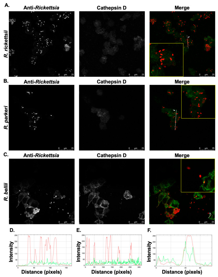
Differences in the co-localization of SFG rickettsial species with the activated, mature form of the lysosomal marker Cathepsin D. PMA-differentiated THP-1 cells were infected with (A) R. rickettsii Sheila Smith, (B) R. parkeri Portsmouth, and (C) R. bellii Yolo at MOIs of 10 for 24 h and then processed for immunofluorescence confocal microscopy analyses. Representative slices from z stacks of infected THP-1-derived macrophage are shown. (D–F) A generated RGB profile plot documents the relative fluorescence intensity along the indicated white line. Putative co-localization events for R. rickettsii (D), R. parkeri (E), and R. bellii (F) were deemed positive when fluorescence intensities from the green and red channels overlap at a given point in the image. Areas of interest that were used for determination of co-localization are enlarged to show detail (inset). Scale bar = 25 μm.

