PLOS ONE logoLink to PLOS ONE
. 2021 Mar 5;16(3):e0248206. doi: 10.1371/journal.pone.0248206

Small RNA sequencing to differentiate lung squamous cell carcinomas from metastatic lung tumors from head and neck cancers

Yoshihisa Shimada 1,*, Jun Matsubayashi 2, Akira Saito 3, Tatsuo Ohira 1, Masahiko Kuroda 3, Norihiko Ikeda 1
Editor: Sumitra Deb4
PMCID: PMC7935561  PMID: 33668046

Abstract

Distinguishing lung squamous cell carcinoma (LSQCC) from a solitary metastatic lung tumor (MSQCC) from head and neck squamous cell carcinoma (HNSQCC) presents a difficult diagnostic challenge even after detailed pathological assessment. Treatment options and estimated survival outcomes after pulmonary resection differ between patients with LSQCC and MSQCC. This study aimed to investigate whether microRNA (miRNA) profiling by RNA sequencing of HNSQCC, MSQCC, and LSQCC was useful for differential diagnosis of MSQCC and LSQCC. RNA sequencing was performed to identify bioinformatically significant miRNAs from a formalin-fixed paraffin-embedded (FFPE) block from a derivation set. MiRNA levels were confirmed by validation sets using FFPE samples and serum extracellular vesicles from patients. Step-wise discriminant analysis and canonical discriminant analysis identified 13 miRNAs by which the different expression patterns of LSQCC, MSQCC, and HNSQCC groups were demonstrated. Six miRNAs (miR-10a/28/141/320b/3120) were assessed in validation sets, and 4 miRNAs (miR-10a/28/141/3120) were significantly upregulated in LSQCCs compared with MSQCCs and HNSQCCs. Serum extracellular vesicles from LSQCC patients demonstrated significantly elevated miR-10a (p = .042), miR-28 (p = .041), and miR-3120 (p = .047) levels compared with those from MSQCC patients. RNA sequencing is useful for differential diagnosis of LSQCC and MSQCC, and the expression level of miR-10a, miR-28, and miR-3120 in serum extracellular vesicles are promising noninvasive tools for this purpose.

Introduction

Smoking acts as a significant risk factor for lung squamous cell carcinoma (LSQCC) and head and neck squamous cell carcinoma (HNSQCC). The patient’s clinical profiles and the histological appearance of both tumors are similar [1]. As it is often shown for both tumors to arise in the same patient, distinguishing an independent LSQCC from a solitary metastatic lung tumor (MSQCC) from HNSQCC presents a difficult diagnostic challenge even after detailed pathological assessment. In patients with MSQCC, a less invasive surgical procedure such as wedge lung resection is indicated as a palliative procedure, and the following systemic therapies are often available based on its pathological results and disease course. By contrast, curative-intent anatomical resection, including a segmentectomy, lobectomy, or pneumonectomy is the primary surgical procedure for LSQCC. However, those procedures have more considerable postoperative morbidities compared with wedge lung resection [2]. Treatment options and estimated survival outcomes differ between patients with LSQCC and MSQCC. Therefore, a reliable method for the differential diagnosis of these histological types before pulmonary resection is in higher demand.

MicroRNAs (miRNAs), which are endogenous and small non-coding RNAs that can function as potential oncogenes and tumor suppressors, have emerged as promising biomarkers for diagnostic purposes [3, 4]. The identification of discriminative and differentially expressed miRNA as a signature is a crucial task for cancer diagnosis and therapy [4, 5]. MiRNAs are readily detected in a formalin-fixed paraffin-embedded (FFPE) block and in various body fluids such as blood, urine, and saliva [6]. Recent reports have shown that miRNAs are transported in body fluids within extracellular vesicles [7]. Extracellular vesicles, including exosomes, are known as intercellular messengers and can shuttle cargos, such as mRNA, proteins, miRNA, and lipids between cells [810]. They have been studied in the cancer diagnostic setting because cancer-derived extracellular vesicles are found to be involved in metastatic cascades, such as invasion, migration, and the priming of metastatic niches [1118]. As LSQCC and MSQCC are biologically different entities of local and metastatic disease, patient’ serum-derived extracellular vesicles containing unique miRNAs can serve as noninvasive tools for their differential diagnosis.

In this study, we aimed to identify specific miRNAs for the differential diagnosis of MSQCC and LSQCC by small RNA sequencing and to quantify its miRNA levels in FFPE blocks and serum extracellular vesicles from patients with MSQCC and LSQCC for the validation analysis.

Materials and methods

Patients and clinical samples

LSQCC, MSQCC, and HNSQCC specimens were obtained from FFPE blocks of surgically resected tissue from patients at Tokyo Medical University Hospital. HNSQCC included patients with pharyngeal cancer, laryngeal cancer, and gingival cancer. There were a total of 28 patients who underwent surgical resection for solitary SQCC arising from the lung between January 2008 and December 2018. Of these, clinico-pathological diagnosis showed 16 patients with LSQCC and a history of HNSQCC and 12 patients with MSQCC. All 28 patients had previously undergone surgical resection for HNSQCC. For each case, pathologists reached a diagnosis of either LSQCC or MSQCC on the basis of clinical backgrounds and histologic features, including the degree of squamous differentiation and particular histological patterns (the presence of basaloid or clear cell morphology). The derivation set included 17 samples (8 LSQCCs, 6 MSQCCs, and 3 HNSQCCs) from 14 patients undergoing pulmonary resection between 2008 and 2014 that were used for small RNA sequencing (Table 1). The remaining 14 samples (8 LSQCCs and 6 MSQCCs) from 14 patients undergoing surgery between 2015 and 2018 were used for the validation study, and blood samples were also collected from the 14 patients before surgery. Each blood sample was centrifuged at 3,000 rpm for 5 minutes to separate the serum and stored at −80 <C until RNA extraction. The data of additional 41 patients (20 with stage I-II LSQCCs and 21 with stage I-III HNSQCCs) who underwent surgical resection between January 2017 and December 2018 were used for the other validation study. This study was approved by the Institutional Review Board of Tokyo Medical University (study approval no. 3868). Written informed consent for the use and analyses of clinical data was obtained preoperatively from each patient.

Table 1. Samples from 14 patients in the derivation for small RNA sequencing.

No. Type of samples Age, sex Primary organ Stage of LSQCC Type of HNSQCC Tumor differentiation Lung tumor location
1 LSQCC 70, male Lung IB Glottic cancer Poor Right upper
2 LSQCC 60, male Lung IIIB Pharyngeal cancer Moderate Right middle
3 LSQCC 65, male Lung IA Pharyngeal cancer Poor Right lower
4 LSQCC 72, male Lung IIIA Laryngeal cancer Moderate Right lower
5 LSQCC 71, male Lung IIIB Glottic cancer Poor Left upper
6 LSQCC 76, male Lung IIIB Tongue cancer Moderate Left upper
7 LSQCC 75, male Lung IA Laryngeal cancer Moderate Right upper
8 LSQCC 80, male Lung IIIA Gingival cancer Moderate Left upper
HNSQCC Same as No.8 patient Moderate -
9 MSQCC 66, male Pharynx - Pharyngeal cancer Moderate Left upper
10 MSQCC 69, female Pharynx - Pharyngeal cancer Moderate Right middle
HNSQCC Same as No.10 patient Moderate -
11 MSQCC 78, female Pharynx - Pharyngeal cancer Poor Right lower
12 MSQCC 74, male Larynx - Laryngeal cancer Moderate Left upper
13 MSQCC 75, male Tongue - Tongue cancer Moderate Left lower
HNSQCC Same as No.13 patient Moderate -
14 MSQCC 74, male Pharynx - Pharyngeal cancer Poor Right lower

LSQCC, lung squamous cell carcinoma; HNQCC, head and neck squamous cell carcinoma; MSQCC, metastatic tumors from head and neck squamous cell carcinoma.

Small RNA library preparation and sequencing

Total RNA of the 17 samples in the derivation set was isolated using NucleoSpin total RNA FFPE XS (Macherey-Nagel Inc. PA, USA) and was sent to Takara Bio Inc., where small RNA library preparation and sequencing were performed. Small RNA libraries were prepared using SMARTer smRNA-Seq Kit (Takara Bio Inc. Japan) for Illumina, and yields were evaluated using an Agilent 2100 BioAnalyzer. Small RNA sequencing was performed using the Illumina HiSeq 2500 system with HiSeq SBS Kit v4 reagents.

Isolation of extracellular vesicles

Extracellular vesicles were recovered by a sequential centrifugation procedure using Exosome Isolation Kit PS (MagCapture, Fujifilm Wako. Japan). Cells were grown in T75 culture flasks for 3 to 4 days, and the conditioned media was removed from the flasks. In the case of both cell culture media and patients serum, cells were pelleted from the media by centrifugation at 300 g for 5 minutes, followed by 1,200 g for 20 minutes. To eliminate other cellular debris, the supernatant was spun at 10,000 g for 30 minutes. The sample was concentrated by filtration (Vivaspin 20; Sartorius). After sample preparation, extracellular vesicles were purified by MagCapture according to the manufacturer’s instructions. Extracellular vesicles were verified by electron microscopy. Extracellular vesicle size and particle numbers were analyzed using the LM10 Nanoparticle Characterization System (NanoSight, Malvern Instruments, UK). The final extracellular vesicle pellet was eluted with elution buffer.

qRT-PCR

MiRNAs were isolated using miRNeasy FFPE Kit (QIAGEN, Netherland) for FFPE samples and Total Exosome RNA and Protein Isolation Kit (Thermo Fisher Scientific) for extracellular vesicles. cDNAs were generated using the TaqMan MicroRNA Reverse Transcription Kit (Thermo Fisher Scientific). Gene-specific TaqMan MicroRNA probes (Thermo Fisher Scientific) were utilized for quantitative analyses of miRNA transcript levels of miR-10a, miR-28, miR-99b, miR-141, miR-320b, and miR-3120. Cel-miR-39 was used as internal references. Polymerase chain reactions (PCR) were performed using StepOne (Thermo Fisher Scientific), and relative expression levels were calculated using the 2−ΔΔCT method.

Western blotting

Extracellular vesicles were lysed in RIPA lysis buffer (Thermo Fisher Scientific) containing a protease inhibitor cocktail (Roche, Switzerland). Equal amounts of total protein were loaded onto a 4% - 20% SDS-PAGE gel and then transferred onto PVDF membranes. Membranes were blocked with 5% milk and then incubated in Tris-buffered saline with Tween 20 with a CD9 primary antibody (EXOAB-CD9A-1; SBI) overnight, followed by incubation with horseradish peroxidase-conjugated secondary antibody (Sigma-Aldrich, MO, USA). Membranes were imaged on the ChemiDoc Touch Imaging System (BIO-RAD, CA, USA).

Statistical and bioinformatic analysis

Step-wise discriminant analysis was performed to determine potential groupings of miRNAs that maximally distinguish among LSQCC, MSQCC, and HNSQCC by using known miRNAs obtained from small RNA sequencing. Then, the canonical discriminant analysis was implemented to determine the discriminant coefficient sets 1 and 2. The absolute values of coefficients correspond to variables with more exceptional discriminant ability. The coefficient values meaning indicates how much a given miRNA contributed to a discriminant function. These analyses were carried out with the Statistical Analysis Software Package R (R Project for Statistical Computing; http://www.r-project.org). The Student t-test was performed to compare miRNA expression levels between 2 groups with the SPSS statistical software package (version 25.0; DDR3 RDIMM, SPSS). All tests were 2-sided, and p-values of less than 0.05 were considered to indicate a statistically significant difference between 2 groups.

Results

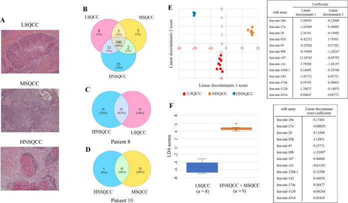
Representative pictures of LSQCC, MSQCC, and HNSQCC in the derivation set were shown in Fig 1A. A total of 160 known miRNAs and 2,079 novel non-coding RNAs were detected by small RNA sequencing. Expression changes of known miRNA among the 3 histological groups were identified by overlapping the differential gene sets, as shown in the Venn diagram in Fig 1B. We reveal 2 representative Venn diagrams of a patient with LSQCC with a past treatment history of HNSQCC (patient 8, Fig 1C), and the other patient with MSQCC with a history of HNSQCC (patient 10, Fig 1D). Step-wise linear discriminant analysis identified 13 bioinformatically significant miRNAs, and discriminant functions were given by canonical discriminant analysis. The following formula estimated linear discriminant scores 1 and 2 obtained by the miRNA expression level of interest (fragments per kilobase of exon per million reads mapped; FPKM);

LDscore=A1*X1+A2*X2++A13*X13

Fig 1. MiRNA profiling by small RNA sequencing of LSQCC, MSQCC, and HNSQCC samples.

Fig 1

(A) Representative pathological pictures of LSQCC, MSQCC, and HNSQCC in the derivation set. (B) Venn diagram showing miRNAs that were differentially and commonly expressed in the 3 groups. (C) A diagram of a patient with LSQCC and past treatment history of HNSQCC. (D) A diagram of another patient with MSQCC and a history of HNSQCC. (E) The result of the classification with linear discriminant analysis with the list of coefficients of each miRNA’s linear discriminants. Step-wise discriminant analysis of the 2 linear discriminants demonstrating that expression profiles of the 13 bioinformatically significant miRNAs can distinguish LSQCC from MSQCC and HNSQCC. (F) Linear discriminant analysis with linear discriminant score coefficients demonstrates that the expression profiles of 13 miRNAs can distinguish LSQCC from the combined group of MSQCC and HNSQCC. LSQCC, lung squamous cell carcinoma; HNSQCC, head and neck squamous cell carcinoma; MSQCC, metastatic tumors from HNSQCC; LD, linear discriminant; miRNA, microRNA; FPKM, fragments per kilobase of exon per million reads mapped.

A: coefficients of linear discriminants, X: standardized miRNA FPKM value

Linear discriminants 1/ linear discriminants 2 values were on Fig 1D table. The result of the classification with linear discriminant analysis with the list of coefficients of each miRNA’s linear discriminants was shown in Fig 1E. To evaluate the performance of the model with the miRNAs to distinguish LSQCC from MSQCC and HNSQCC, 1 case (LSQCC-test, MSQCC-test, and HNSQCC-test) was randomly extracted from each group as the validation case, and the rest 14 cases were used to generate the new 13-miRNA-based models. Three-fold cross-validation identified that the miRNAs were capable of differentiating LSQCC and MSQCC. Type I and II errors and an error rate obtained by the random forest algorithm in the validation sets were shown in supplementary figure legends (S1A–S1C Fig). The other linear discriminant and canonical discriminant analyses with its linear discriminant score coefficients were performed, and that made a clear distinction between LSQCC and the combined group of MSQCC and HNSQCC (Fig 1F).

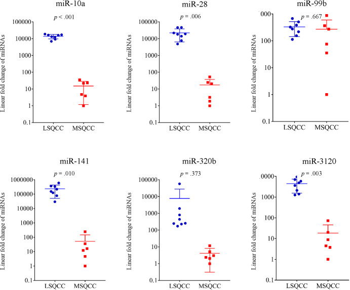
Of the 13 miRNAs, 6 genes (miR-10a, miR-28, miR-99b, miR-107, miR-320b, and miR-3120) were selected for further validation analyses because the 6 miRNAs obtained from a set of linear discriminant coefficients indicated an area in proximity of the LSQCC lineage. We analyzed the quantification of the expression levels of the 6 miRNAs in 14 samples (8 LSQCC and 6 MSQCC) as an initial validation set (Fig 2). We compared the level of the 6 miRNAs in FFPE samples in 14 patients with MSQCC or LSQCC. LSQCC patients had significantly higher miR-10a (p < .001), miR-28 (p = .006), miR-141 (p = .010), and miR-3120 (p = .003) levels than MSQCC patients (Fig 2). In the other validation study, the expression levels of miR-10a, miR-28, miR-141, and miR-3120 were validated between LSQCCs and HNSQCCs (Fig 3). MiR-10a (p < .001), miR-28 (p < .001), miR-141 (p = .002), and miR-3120 (p = .017) in LSQCCs are significantly higher than those in HNSQCCs (Fig 3).

Fig 2. The quantification of 6 miRNAs from FFPE samples in 14 patients for a validation study.

Fig 2

LSQCCs have significantly higher miR-10a (p < .001), miR-28 (p = .006), miR-141 (p = .009), and miR-3120 (p = .003) levels than HNSQCCs. miRNA, microRNA; FFPE, formalin-fixed paraffin-embedded; LSQCC, lung squamous cell carcinoma; MSQCC, metastatic tumors from head and neck squamous cell carcinoma.

Fig 3. Comparison of the expression of 4 miRNAs (miR-10a/28/141/3120) from FFPE block from 20 LSQCC and 21 HNSQCC patients in a validation dataset.

Fig 3

LSQCC samples had significantly higher miR-10a (p < .001), miR-28 (p < .001), miR-141 (p = .002), and miR-3120 (p = .017) levels than HNSQCC samples. miRNA, microRNA; FFPE, formalin-fixed paraffin-embedded; LSQCC, lung squamous cell carcinoma; HNSQCC, head and neck squamous cell carcinoma.

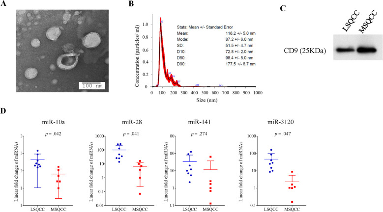
Fig 4A shows extracellular vesicles that were isolated from the serum of an LSQCC patient. Extracellular vesicles were identified on transmission electron microscopy as small spherical vesicles and were characterized by the nanoparticle characterization system (Fig 4B). We checked the identification of extracellular vesicles to ensure consistency of the results in a total of 4 patients. The protein contents of extracellular vesicles were assayed by western blotting and were confirmed to express the common exosome marker CD9 (Fig 4C). MiR-10a (p = .042), miR-28 (p = .041), and miR-3120 (p = .047) levels in extracellular vesicles in the initial validation set were significantly elevated in LSQCC patients compared with MSQCC patients (Fig 4D). S2 Fig demonstrated comparative results of the 4 miRNA levels (miR-10a/28/141/3120) in matched FFPE and serum extracellular vesicle samples.

Fig 4. Characteristics of serum extracellular vesicles from 14 patients and the quantification of selected miRNA levels in extracellular vesicles.

Fig 4

(A) A representative electron microscopic image of extracellular vesicles from the serum of an LSQCC patient. (B) Results of nanoparticle characterization analysis demonstrating the number of extracellular vesicles and their size distribution. (C) The protein contents of extracellular vesicles from an LSQCC and MSQCC patients were assayed by western blotting and were confirmed to express the common exosome marker CD9. (D) MiR-10a (p = .042), miR-28 (p = .041), and miR-3120 (p = .047) levels in extracellular vesicles were significantly elevated in LSQCC patients compared with MSQCC patients. miRNA, microRNA; LSQCC, lung squamous cell carcinoma; MSQCC, metastatic tumors from head and neck squamous cell carcinoma.

We checked the levels of the four miRNAs using extracellular vesicles derived from 4 LSQCC cell lines (EBA-1, H520, and LK-2 purchased from ATCC) and 1 HNSQCC cell line (BHY purchased from DSMZ) to examine whether the gene expressions in patients’ samples agree with those in cell lines. There were no significant differences in the expressions between the two subtypes except for miR-3120 (data not shown).

Discussion

When a pure-solid pulmonary nodule detected in patients with a history of HNSQCC, a differential diagnosis must be made between metastasis and primary lung cancer. Primary lung cancer, including LSQCC, justifies anatomical pulmonary resection with curative intent, whereas the preferred therapeutic approach for a solitary MSQCC might be limited resection followed by systemic therapies. There have been several studies performing molecular genetic approaches to address this diagnostic issue, such as loss of heterozygosity analysis, microsatellite analysis, p53 expression analysis, and miRNA profiling with array data using paired tumor samples from LSQCC and HNSQCC [1922]. Geurts et al. reported that loss of heterozygosity analysis might be useful if performed together with conventional clinico-histological assessments for the differential diagnosis of the 2 types of tumors [21]. Munoz et al. demonstrated that miR-34a levels and ratios of miR-10a/miR-10b were useful in differentiating between LSQCC and MSQCC [22]. Although these may be useful additional analyses when performed together with the gold standard histological assessments, the results have been obtained only from the evaluation of surgical specimens. That indicates that those results do not affect the therapeutic decision-making process, including the selection of surgical procedures before pulmonary resection. We thus assessed whether liquid biopsy with extracellular vesicles from patient’s serum was an easily-available noninvasive alternative to surgical specimens for supporting the differential diagnosis.

The validation dataset with FFPE from MSQCC and LSQCC demonstrated that miR-10a, miR-28, miR-141, and miR-3120 were significantly higher in LSQCCs than MSQCCs, while the other dataset with LSQCC and HNSQCC samples revealed that these 4 miRNAs were more abundant in LSQCCs than HNSQCCs. The expression levels of miR-10a, miR-28, and miR-3120 in serum extracellular vesicles from LSQCC patients were significantly higher than those from MSQCC patients.

MiR-10a has been reported to act as a tumor suppressor in various types of cancer, whereas a limited number of studies have reported it as an oncogene [2326]. Munoz et al. reported that miR-10a was expressed at higher levels in LSQCC than in HNSQCC [22]. MiR-28 is known to be a tumor-suppressive miRNA in several tumor tissues and shown to be involved in the inhibition of the migration, invasion, and metastasis of a variety of cancer [27]. MiR-141 is one of the common cancer-associated miRNAs, and ovarian cancer-secreted miR-141 was reported to act as a significant mediator of intercellular communication, promoting endothelial cell angiogenesis [28]. It has been reported that miR-141 may act as a tumor suppressor in some malignancies such as gastric cancer, pancreatic cancer, and colorectal cancer [2931]. On the other hand, miR-3120 was reported to promote stemness and invasiveness of colon cancer cells, although there have been few reports concerning the epigenetic significance of miR-3120 in cancer pathogenesis [32]. Thus, the role of miR-3120 in LSQCC and HNSQCC remains unclear. Three miRNAs, miR-10a, miR-28, and miR-141, identified by RNA sequencing in the present study, are considered tumor suppressor genes in a wide range of cancers. It can make good sense for LSQCCs as locally invasive tumors showing significantly higher expression levels of the tumor-suppressive genes compared with MSQCCs as metastatic tumors.

Extracellular vesicles, which contain a large amount of miRNA, are presently thought to be the main contributor to tumor progression and metastasis [1113, 18, 33]. As extracellular vesicles from LSQCCs in the present study contain a significantly larger amount of miR-10a, miR-28, and miR-3120 that were confirmed in the FFPE samples compared with those from MSQCCs, these nano-size particles released from cancer cells are likely to be ideal tools for liquid biopsy to differentiate LSQCC from MSQCC or HNSQCC. However, the methods of isolation and characterization of circulating extracellular vesicles are still a matter of debate. Further studies are hence required before this liquid biopsy approach can be applied in routine clinical practice.

Although LSQCC and MSQCC samples in the derivation set from patients diagnosed based on pathological review had the potential to yield biased results, miRNA profiles of 13 miRNAs could classify all the samples into 3 groups, LSQCC, MSQCC, HNSQCC, according to the epigenetic expression. Previous studies’ results were interpreted based on the premise that MSQCC and primary HNSQCC had genetically similar traits [19, 21, 22]. However, metastasis is considered to be a clonally selective process, and HNSQCC can contain multiple clonal subpopulations capable of forming MSQCC. Therefore, our study’s results support the notion that individual metastases express both different and common phenotypes, and genetic instability leads to clonal metastases becoming heterogeneous.

In conclusion, our study demonstrated that miRNA profiling by small RNA sequencing followed by validation analyses using qRT-PCR of the selected miRNAs in both FFPE samples and serum extracellular vesicles is useful for the differential diagnosis of LSQCC and MSQCC. These results led to the hypothesis that the quantification of miR-10a, miR-28, miR-141, and miR-3120 levels in not just FFPE samples but also serum extracellular vesicles may be a simple method to differentiate between HNSQCC and LSQCC lineages for the appropriate management of patients with a solitary SQCC arising in the lung before pulmonary resection even if they have a history of HNSQCC.

Supporting information

S1 Dataset. Minimal data set.

(DOCX)

S1 Fig. Three-fold cross-validation of our model with the 13 miRNAs.

Each different classification with linear discriminant analysis with the coefficients of each miRNA’s linear discriminants achieved the good separation of each histologic type (A, B, and C). Type I and II errors in the validation set were 11% and 1%, respectively. An error rate obtained by the random forest algorithm was 5.88%.

(TIF)

S2 Fig. Comparative results of the 4 miRNA levels (miR-10a/28/141/3120) in matched FFPE and serum extracellular vesicle samples.

Formalin-fixed paraffin-embedded samples from LSQCC patients had significantly higher miR-10a (p < .001), miR-28 (p = .006), miR-141 (p = .009), and miR-3120 (p = .004) levels than serum extracellular vesicle samples from the matched patients.

(TIF)

S1 Raw image

(TIF)

Acknowledgments

The authors are indebted to the medical editors from the Department of International Medical Communications of Tokyo Medical University, for editing the English manuscript. We thank Kumiko Nagase for her technical support.

Glossary of abbreviations

FFPE

formalin-fixed paraffin-embedded

HNSQCC

Head and neck squamous cell carcinoma

LSQCC

Lung squamous cell carcinoma

miRNA

MicroRNA

MSQCC

Metastatic tumors from head and neck squamous cell carcinoma

qRT-PCR

quantitative real-time polymerase chain reaction

SWD

step-wise discriminant

Data Availability

All relevant data are within the manuscript and Supporting Information files.

Funding Statement

This study was supported in part by a Grant-in-Aid for Cancer Research from the Ministry of Education, Culture, Sports, and Technology, Japan (#18K08799; Y. Shimada), and internal research funding from department of thoracic surgery, Tokyo Medical University. There was no additional external funding received for this study.

References

  • 1.Ezzati M, Henley SJ, Lopez AD, and Thun MJ. Role of smoking in global and regional cancer epidemiology: current patterns and data needs. Int J Cancer. 2005;116(6):963–71. 10.1002/ijc.21100 [DOI] [PubMed] [Google Scholar]
  • 2.Rotman JA, Plodkowski AJ, Hayes SA, de Groot PM, Shepard JA, Munden RF, et al. Postoperative complications after thoracic surgery for lung cancer. Clin Imaging. 2015;39(5):735–49. 10.1016/j.clinimag.2015.05.013 [DOI] [PubMed] [Google Scholar]
  • 3.Calin GA, Sevignani C, Dumitru CD, Hyslop T, Noch E, Yendamuri S, et al. Human microRNA genes are frequently located at fragile sites and genomic regions involved in cancers. Proc Natl Acad Sci U S A. 2004;101(9):2999–3004. 10.1073/pnas.0307323101 [DOI] [PMC free article] [PubMed] [Google Scholar]
  • 4.Peng Y, and Croce CM. The role of MicroRNAs in human cancer. Signal Transduct Target Ther. 2016;1:15004. 10.1038/sigtrans.2015.4 [DOI] [PMC free article] [PubMed] [Google Scholar]
  • 5.Datta A, Das P, Dey S, Ghuwalewala S, Ghatak D, Alam SK, et al. Genome-Wide Small RNA Sequencing Identifies MicroRNAs Deregulated in Non-Small Cell Lung Carcinoma Harboring Gain-of-Function Mutant p53. Genes (Basel). 2019;10(11). 10.3390/genes10110852 [DOI] [PMC free article] [PubMed] [Google Scholar]
  • 6.Halvaei S, Daryani S, Eslami SZ, Samadi T, Jafarbeik-Iravani N, Bakhshayesh TO, et al. Exosomes in Cancer Liquid Biopsy: A Focus on Breast Cancer. Mol Ther Nucleic Acids. 2018;10:131–41. 10.1016/j.omtn.2017.11.014 [DOI] [PMC free article] [PubMed] [Google Scholar]
  • 7.Zhang J, Li S, Li L, Li M, Guo C, Yao J, et al. Exosome and exosomal microRNA: trafficking, sorting, and function. Genomics Proteomics Bioinformatics. 2015;13(1):17–24. 10.1016/j.gpb.2015.02.001 [DOI] [PMC free article] [PubMed] [Google Scholar]
  • 8.Kahlert C, and Kalluri R. Exosomes in tumor microenvironment influence cancer progression and metastasis. J Mol Med (Berl). 2013;91(4):431–7. 10.1007/s00109-013-1020-6 [DOI] [PMC free article] [PubMed] [Google Scholar]
  • 9.Raposo G, and Stoorvogel W. Extracellular vesicles: exosomes, microvesicles, and friends. J Cell Biol. 2013;200(4):373–83. 10.1083/jcb.201211138 [DOI] [PMC free article] [PubMed] [Google Scholar]
  • 10.Thery C, Zitvogel L, and Amigorena S. Exosomes: composition, biogenesis and function. Nat Rev Immunol. 2002;2(8):569–79. 10.1038/nri855 [DOI] [PubMed] [Google Scholar]
  • 11.Al-Nedawi K, Meehan B, Kerbel RS, Allison AC, and Rak J. Endothelial expression of autocrine VEGF upon the uptake of tumor-derived microvesicles containing oncogenic EGFR. Proc Natl Acad Sci U S A. 2009;106(10):3794–9. 10.1073/pnas.0804543106 [DOI] [PMC free article] [PubMed] [Google Scholar]
  • 12.Whiteside TL. Exosomes carrying immunoinhibitory proteins and their role in cancer. Clin Exp Immunol. 2017;189(3):259–67. 10.1111/cei.12974 [DOI] [PMC free article] [PubMed] [Google Scholar]
  • 13.Paggetti J, Haderk F, Seiffert M, Janji B, Distler U, Ammerlaan W, et al. Exosomes released by chronic lymphocytic leukemia cells induce the transition of stromal cells into cancer-associated fibroblasts. Blood. 2015;126(9):1106–17. 10.1182/blood-2014-12-618025 [DOI] [PMC free article] [PubMed] [Google Scholar]
  • 14.Tickner JA, Urquhart AJ, Stephenson SA, Richard DJ, and O’Byrne KJ. Functions and therapeutic roles of exosomes in cancer. Front Oncol. 2014;4:127. 10.3389/fonc.2014.00127 [DOI] [PMC free article] [PubMed] [Google Scholar]
  • 15.Rahman MA, Barger JF, Lovat F, Gao M, Otterson GA, and Nana-Sinkam P. Lung cancer exosomes as drivers of epithelial mesenchymal transition. Oncotarget. 2016;7(34):54852–66. 10.18632/oncotarget.10243 [DOI] [PMC free article] [PubMed] [Google Scholar]
  • 16.Peinado H, Aleckovic M, Lavotshkin S, Matei I, Costa-Silva B, Moreno-Bueno G, et al. Melanoma exosomes educate bone marrow progenitor cells toward a pro-metastatic phenotype through MET. Nat Med. 2012;18(6):883–91. 10.1038/nm.2753 [DOI] [PMC free article] [PubMed] [Google Scholar]
  • 17.Thery C, Ostrowski M, and Segura E. Membrane vesicles as conveyors of immune responses. Nat Rev Immunol. 2009;9(8):581–93. 10.1038/nri2567 [DOI] [PubMed] [Google Scholar]
  • 18.Costa-Silva B, Aiello NM, Ocean AJ, Singh S, Zhang H, Thakur BK, et al. Pancreatic cancer exosomes initiate pre-metastatic niche formation in the liver. Nat Cell Biol. 2015;17(6):816–26. 10.1038/ncb3169 [DOI] [PMC free article] [PubMed] [Google Scholar]
  • 19.Leong PP, Rezai B, Koch WM, Reed A, Eisele D, Lee DJ, et al. Distinguishing second primary tumors from lung metastases in patients with head and neck squamous cell carcinoma. J Natl Cancer Inst. 1998;90(13):972–7. 10.1093/jnci/90.13.972 [DOI] [PubMed] [Google Scholar]
  • 20.Kandioler D, Dekan G, End A, Pasching E, Buchmayer H, Gnant M, et al. Molecular genetic differentiation between primary lung cancers and lung metastases of other tumors. J Thorac Cardiovasc Surg. 1996;111(4):827–31; discussion 32. 10.1016/s0022-5223(96)70343-4 [DOI] [PubMed] [Google Scholar]
  • 21.Geurts TW, Nederlof PM, van den Brekel MW, van’t Veer LJ, de Jong D, Hart AA, et al. Pulmonary squamous cell carcinoma following head and neck squamous cell carcinoma: metastasis or second primary? Clin Cancer Res. 2005;11(18):6608–14. 10.1158/1078-0432.CCR-05-0257 [DOI] [PubMed] [Google Scholar]
  • 22.Munoz-Largacha JA, Gower AC, Sridhar P, Deshpande A, O’Hara CJ, Yamada E, et al. miRNA profiling of primary lung and head and neck squamous cell carcinomas: Addressing a diagnostic dilemma. J Thorac Cardiovasc Surg. 2017;154(2):714–27. 10.1016/j.jtcvs.2017.02.071 [DOI] [PubMed] [Google Scholar]
  • 23.Lu X, Xue B, Zhang T, Zhou X, and Zhang Y. Down-regulation of microRNA-10a mediates the anti-tumor effect of icaritin in A549 cells via the PTEN/AKT and ERK pathway. Gen Physiol Biophys. 2019;38(6):525–33. 10.4149/gpb_2019041 [DOI] [PubMed] [Google Scholar]
  • 24.Mu H, Xiang L, Li S, Rao D, Wang S, and Yu K. MiR-10a functions as a tumor suppressor in prostate cancer via targeting KDM4A. J Cell Biochem. 2019;120(4):4987–97. 10.1002/jcb.27774 [DOI] [PubMed] [Google Scholar]
  • 25.Bao M, Pan S, Yang W, Chen S, Shan Y, and Shi H. Serum miR-10a-5p and miR-196a-5p as non-invasive biomarkers in non-small cell lung cancer. Int J Clin Exp Pathol. 2018;11(2):773–80. [PMC free article] [PubMed] [Google Scholar]
  • 26.Liu Y, Zhang Y, Wu H, Li Y, Zhang Y, Liu M, et al. miR-10a suppresses colorectal cancer metastasis by modulating the epithelial-to-mesenchymal transition and anoikis. Cell Death Dis. 2017;8(4):e2739. 10.1038/cddis.2017.61 [DOI] [PMC free article] [PubMed] [Google Scholar]
  • 27.Fazio S, Berti G, Russo F, Evangelista M, D’Aurizio R, Mercatanti A, et al. The miR-28-5p Targetome Discovery Identified SREBF2 as One of the Mediators of the miR-28-5p Tumor Suppressor Activity in Prostate Cancer Cells. Cells. 2020;9(2). 10.3390/cells9020354 [DOI] [PMC free article] [PubMed] [Google Scholar]
  • 28.Masoumi-Dehghi S, Babashah S, and Sadeghizadeh M. microRNA-141-3p-containing small extracellular vesicles derived from epithelial ovarian cancer cells promote endothelial cell angiogenesis through activating the JAK/STAT3 and NF-kappaB signaling pathways. J Cell Commun Signal. 2020. 10.1007/s12079-020-00548-5 [DOI] [PMC free article] [PubMed] [Google Scholar]
  • 29.Ma L, Zhai B, Zhu H, Li W, Jiang W, Lei L, et al. The miR-141/neuropilin-1 axis is associated with the clinicopathology and contributes to the growth and metastasis of pancreatic cancer. Cancer Cell Int. 2019;19:248. 10.1186/s12935-019-0963-2 [DOI] [PMC free article] [PubMed] [Google Scholar]
  • 30.Du F, Yu C, Li R, Ding D, He L, and Wen G. Expression of miR-141 and YAP1 in gastric carcinoma and modulation of cancer cell proliferation and apoptosis. Int J Clin Exp Pathol. 2019;12(2):559–67. [PMC free article] [PubMed] [Google Scholar]
  • 31.Long ZH, Bai ZG, Song JN, Zheng Z, Li J, Zhang J, et al. miR-141 Inhibits Proliferation and Migration of Colorectal Cancer SW480 Cells. Anticancer Res. 2017;37(8):4345–52. 10.21873/anticanres.11828 [DOI] [PubMed] [Google Scholar]
  • 32.Hongdan L, and Feng L. miR-3120-5p promotes colon cancer stem cell stemness and invasiveness through targeting Axin2. Biochem Biophys Res Commun. 2018;496(2):302–8. 10.1016/j.bbrc.2018.01.021 [DOI] [PubMed] [Google Scholar]
  • 33.Vlassov AV, Magdaleno S, Setterquist R, and Conrad R. Exosomes: current knowledge of their composition, biological functions, and diagnostic and therapeutic potentials. Biochim Biophys Acta. 2012;1820(7):940–8. 10.1016/j.bbagen.2012.03.017 [DOI] [PubMed] [Google Scholar]

Decision Letter 0

Sumitra Deb

1 Oct 2020

PONE-D-20-26356

Small RNA sequencing to differentiate lung squamous cell carcinomas from metastatic lung tumors from head and neck cancers

PLOS ONE

Dear Dr. Shimada,

Thank you for submitting your manuscript to PLOS ONE. After careful consideration, we feel that it has merit but does not fully meet PLOS ONE’s publication criteria as it currently stands. Therefore, we invite you to submit a revised version of the manuscript that addresses the points raised during the review process.

Please submit your revised manuscript by Nov 15 2020 11:59PM. If you will need more time than this to complete your revisions, please reply to this message or contact the journal office at plosone@plos.org. When you're ready to submit your revision, log on to https://www.editorialmanager.com/pone/ and select the 'Submissions Needing Revision' folder to locate your manuscript file.

Please include the following items when submitting your revised manuscript:

  • A rebuttal letter that responds to each point raised by the academic editor and reviewer(s). You should upload this letter as a separate file labeled 'Response to Reviewers'.

  • A marked-up copy of your manuscript that highlights changes made to the original version. You should upload this as a separate file labeled 'Revised Manuscript with Track Changes'.

  • An unmarked version of your revised paper without tracked changes. You should upload this as a separate file labeled 'Manuscript'.

If you would like to make changes to your financial disclosure, please include your updated statement in your cover letter. Guidelines for resubmitting your figure files are available below the reviewer comments at the end of this letter.

If applicable, we recommend that you deposit your laboratory protocols in protocols.io to enhance the reproducibility of your results. Protocols.io assigns your protocol its own identifier (DOI) so that it can be cited independently in the future. For instructions see: http://journals.plos.org/plosone/s/submission-guidelines#loc-laboratory-protocols

We look forward to receiving your revised manuscript.

Kind regards,

Sumitra Deb, PhD

Academic Editor

PLOS ONE

Journal Requirements:

When submitting your revision, we need you to address these additional requirements.

1. Please ensure that your manuscript meets PLOS ONE's style requirements, including those for file naming. The PLOS ONE style templates can be found at

https://journals.plos.org/plosone/s/file?id=wjVg/PLOSOne_formatting_sample_main_body.pdf and

https://journals.plos.org/plosone/s/file?id=ba62/PLOSOne_formatting_sample_title_authors_affiliations.pdf

2. Please provide additional details regarding participant consent. In the ethics statement in the Methods and online submission information, please ensure that you have specified what type you obtained (for instance, written or verbal, and if verbal, how it was documented and witnessed). If your study included minors, state whether you obtained consent from parents or guardians. If the need for consent was waived by the ethics committee, please include this information.

3. Thank you for stating in your Funding Statement:

"This study was supported in part by a Grant-in-Aid for Cancer Research from the Ministry of Education, Culture, Sports, and Technology, Japan (#18K08799; Y. Shimada)."

Please provide an amended statement that declares *all* the funding or sources of support (whether external or internal to your organization) received during this study, as detailed online in our guide for authors at http://journals.plos.org/plosone/s/submit-now.  Please also include the statement “There was no additional external funding received for this study.” in your updated Funding Statement.

Please include your amended Funding Statement within your cover letter. We will change the online submission form on your behalf.

4. In your Data Availability statement, you have not specified where the minimal data set underlying the results described in your manuscript can be found. PLOS defines a study's minimal data set as the underlying data used to reach the conclusions drawn in the manuscript and any additional data required to replicate the reported study findings in their entirety. All PLOS journals require that the minimal data set be made fully available. For more information about our data policy, please see http://journals.plos.org/plosone/s/data-availability.

Upon re-submitting your revised manuscript, please upload your study’s minimal underlying data set as either Supporting Information files or to a stable, public repository and include the relevant URLs, DOIs, or accession numbers within your revised cover letter. For a list of acceptable repositories, please see http://journals.plos.org/plosone/s/data-availability#loc-recommended-repositories. Any potentially identifying patient information must be fully anonymized.

Important: If there are ethical or legal restrictions to sharing your data publicly, please explain these restrictions in detail. Please see our guidelines for more information on what we consider unacceptable restrictions to publicly sharing data: http://journals.plos.org/plosone/s/data-availability#loc-unacceptable-data-access-restrictions. Note that it is not acceptable for the authors to be the sole named individuals responsible for ensuring data access.

We will update your Data Availability statement to reflect the information you provide in your cover letter.

5. PLOS ONE now requires that authors provide the original uncropped and unadjusted images underlying all blot or gel results reported in a submission’s figures or Supporting Information files. This policy and the journal’s other requirements for blot/gel reporting and figure preparation are described in detail at https://journals.plos.org/plosone/s/figures#loc-blot-and-gel-reporting-requirements and https://journals.plos.org/plosone/s/figures#loc-preparing-figures-from-image-files. When you submit your revised manuscript, please ensure that your figures adhere fully to these guidelines and provide the original underlying images for all blot or gel data reported in your submission. See the following link for instructions on providing the original image data: https://journals.plos.org/plosone/s/figures#loc-original-images-for-blots-and-gels.

In your cover letter, please note whether your blot/gel image data are in Supporting Information or posted at a public data repository, provide the repository URL if relevant, and provide specific details as to which raw blot/gel images, if any, are not available. Email us at plosone@plos.org if you have any questions.

6. PLOS requires an ORCID iD for the corresponding author in Editorial Manager on papers submitted after December 6th, 2016. Please ensure that you have an ORCID iD and that it is validated in Editorial Manager. To do this, go to ‘Update my Information’ (in the upper left-hand corner of the main menu), and click on the Fetch/Validate link next to the ORCID field. This will take you to the ORCID site and allow you to create a new iD or authenticate a pre-existing iD in Editorial Manager. Please see the following video for instructions on linking an ORCID iD to your Editorial Manager account: https://www.youtube.com/watch?v=_xcclfuvtxQ

[Note: HTML markup is below. Please do not edit.]

Reviewers' comments:

Reviewer's Responses to Questions

Comments to the Author

1. Is the manuscript technically sound, and do the data support the conclusions?

The manuscript must describe a technically sound piece of scientific research with data that supports the conclusions. Experiments must have been conducted rigorously, with appropriate controls, replication, and sample sizes. The conclusions must be drawn appropriately based on the data presented.

Reviewer #1: Yes

Reviewer #2: Partly

**********

2. Has the statistical analysis been performed appropriately and rigorously?

Reviewer #1: Yes

Reviewer #2: No

**********

3. Have the authors made all data underlying the findings in their manuscript fully available?

The PLOS Data policy requires authors to make all data underlying the findings described in their manuscript fully available without restriction, with rare exception (please refer to the Data Availability Statement in the manuscript PDF file). The data should be provided as part of the manuscript or its supporting information, or deposited to a public repository. For example, in addition to summary statistics, the data points behind means, medians and variance measures should be available. If there are restrictions on publicly sharing data—e.g. participant privacy or use of data from a third party—those must be specified.

Reviewer #1: Yes

Reviewer #2: Yes

**********

4. Is the manuscript presented in an intelligible fashion and written in standard English?

PLOS ONE does not copyedit accepted manuscripts, so the language in submitted articles must be clear, correct, and unambiguous. Any typographical or grammatical errors should be corrected at revision, so please note any specific errors here.

Reviewer #1: No

Reviewer #2: Yes

**********

5. Review Comments to the Author

Please use the space provided to explain your answers to the questions above. You may also include additional comments for the author, including concerns about dual publication, research ethics, or publication ethics. (Please upload your review as an attachment if it exceeds 20,000 characters)

Reviewer #1: In this study the authors have investigated if differential levels of miRNAs in lung squamous cell carcinoma (LSQCC), solitary metastatic lung tumor (MSQCC) and head and neck squamous cell carcinoma (HNSQCC) will enable differential diagnosis of these cancer types. By using RNA sequencing, they found that LSQCC patients had significantly high levels of miR-10a, miR-28, and miR-3120 in in patient FFPE samples and were successfully able to recapitulate the same in extracellular vesicles from patient serum samples. Through this study the authors have proposed the possibility of detecting miRNA in patient extracellular vesicles (serum) as an alternative and minimally invasive approach to differentiate between HNSQCC and LSQCC patients.

Minor Revisions:

1) For consistency if data in Fig 2 and Fig 4 D is presented as a scatter dot plot as done in in Fig 3 will be more informative.

2) It is unclear if all the isolated vesicles were characterized by nanoparticle characterizing system or just some. Is Figure 4B. just a representative of sample from one patient?

3) Have the authors compared the miRNA levels in matched FFPE and serum samples? If so, it would be informative have it as supplementary data.

4) Some sections if re-written or re-phrased will improve the flow and readability of the manuscript.

a) Discussion section: Page 16, last paragraph, first two sentences read the same and have the same conclusion.

b) Discussion section: Page 18. 2nd paragraph- “miRNA profiles of 13 miRNAs could identify the 3 cancer groups as having epigenetically distinct manifestations.” It is not very clear what the author is trying say.

Reviewer #2: This study is an investigation into models for differentiating between lung squamous cell cancers and head and neck squamous cell cancers that metastasize to the lung using gene expression profiles of microRNAs and serum extracellular vesicles as a source of miRNAs.

The results are the identification of miRNAs that are of potential value for the classification based on differential expression. The use of serum extracellular vesicles is a very useful aspect.

It would help the average reader to have an explanation of how to interpret the coefficients from the discriminant analysis models.

This study shows the development and use of the models, but does not test the models for robustness or assess the potential of over-fitting. This would be done by cross-validation such as with Leave-One-Out based on the small sample size. The Type I and Type II errors and typical metrics for evaluating models, such as sensitivity and specificity, would then be presented. Results from model validation were not presented and models cannot be evaluated without this validation.

**********

6. PLOS authors have the option to publish the peer review history of their article (what does this mean?). If published, this will include your full peer review and any attached files.

If you choose “no”, your identity will remain anonymous but your review may still be made public.

Do you want your identity to be public for this peer review? For information about this choice, including consent withdrawal, please see our Privacy Policy.

Reviewer #1: No

Reviewer #2: No

[NOTE: If reviewer comments were submitted as an attachment file, they will be attached to this email and accessible via the submission site. Please log into your account, locate the manuscript record, and check for the action link "View Attachments". If this link does not appear, there are no attachment files.]

While revising your submission, please upload your figure files to the Preflight Analysis and Conversion Engine (PACE) digital diagnostic tool, https://pacev2.apexcovantage.com/. PACE helps ensure that figures meet PLOS requirements. To use PACE, you must first register as a user. Registration is free. Then, login and navigate to the UPLOAD tab, where you will find detailed instructions on how to use the tool. If you encounter any issues or have any questions when using PACE, please email PLOS at figures@plos.org. Please note that Supporting Information files do not need this step.

PLoS One. 2021 Mar 5;16(3):e0248206. doi: 10.1371/journal.pone.0248206.r002

Author response to Decision Letter 0


20 Nov 2020

October 26, 2020

Joerg Heber

Editor-in-Chief

PLOS ONE

Sumitra Deb

Academic editor

PLOS ONE

Manuscript ID PONE-D-20-26356 entitled "Small RNA sequencing to differentiate lung squamous cell carcinomas from metastatic lung tumors from head and neck cancers"

Dear Dr. Heber, Deb

Thank you so much for your letter of 2-October-2020, stating your comments to our manuscript. We have carefully revised our manuscript and responded all the comments from reviewers as follows.

Academic Editor

-COMMENT 1

Please ensure that your manuscript meets PLOS ONE's style requirements, including those for file naming. The PLOS ONE style templates can be found at https://journals.plos.org/plosone/s/file?id=wjVg/PLOSOne_formatting_sample_main_body.pdf and https://journals.plos.org/plosone/s/file?id=ba62/PLOSOne_formatting_sample_title_authors_affiliations.pdf

-ANSWER to comment 1: Thank you for this notification. We rechecked our manuscript following the style requirements.

-COMMENT 2

Please provide additional details regarding participant consent. In the ethics statement in the Methods and online submission information, please ensure that you have specified what type you obtained (for instance, written or verbal, and if verbal, how it was documented and witnessed). If your study included minors, state whether you obtained consent from parents or guardians. If the need for consent was waived by the ethics committee, please include this information.

-ANSWER to comment 2: Revised description is shown in Methods accordingly.

-CHANGES: Written informed consent for the use and analyses of clinical data was obtained preoperatively from each patient. (Page 9, Line 6)

-COMMENT 3

Thank you for stating in your Funding Statement:

"This study was supported in part by a Grant-in-Aid for Cancer Research from the Ministry of Education, Culture, Sports, and Technology, Japan (#18K08799; Y. Shimada)."

Please provide an amended statement that declares *all* the funding or sources of support (whether external or internal to your organization) received during this study, as detailed online in our guide for authors at http://journals.plos.org/plosone/s/submit-now. Please also include the statement “There was no additional external funding received for this study.” in your updated Funding Statement.

Please include your amended Funding Statement within your cover letter. We will change the online submission form on your behalf.

-ANSWER to comment 3: According to the comment, we revised the funding statement as follows.

-CHANGES: This study was supported in part by a Grant-in-Aid for Cancer Research from the Ministry of Education, Culture, Sports, and Technology, Japan (#18K08799; Y. Shimada), and internal research funding from department of thoracic surgery, Tokyo Medical University. There was no additional external funding received for this study. (Page 2, Line 13)

-COMMENT 4

In your Data Availability statement, you have not specified where the minimal data set underlying the results described in your manuscript can be found. PLOS defines a study's minimal data set as the underlying data used to reach the conclusions drawn in the manuscript and any additional data required to replicate the reported study findings in their entirety. All PLOS journals require that the minimal data set be made fully available. For more information about our data policy, please see http://journals.plos.org/plosone/s/data-availability.

-ANSWER to comment 4: According to this requirement, we create a minimal data set document.

-COMMENT 5

PLOS ONE now requires that authors provide the original uncropped and unadjusted images underlying all blot or gel results reported in a submission’s figures or Supporting Information files. This policy and the journal’s other requirements for blot/gel reporting and figure preparation are described in detail at https://journals.plos.org/plosone/s/figures#loc-blot-and-gel-reporting-requirements and https://journals.plos.org/plosone/s/figures#loc-preparing-figures-from-image-files. When you submit your revised manuscript, please ensure that your figures adhere fully to these guidelines and provide the original underlying images for all blot or gel data reported in your submission. See the following link for instructions on providing the original image data: https://journals.plos.org/plosone/s/figures#loc-original-images-for-blots-and-gels.

In your cover letter, please note whether your blot/gel image data are in Supporting Information or posted at a public data repository, provide the repository URL if relevant, and provide specific details as to which raw blot/gel images, if any, are not available. Email us at plosone@plos.org if you have any questions.

-ANSWER to comment 5: According to the comment, we created a figure with an uncropped image of western blotting as follows.

-CHANGES:

-COMMENT 6

PLOS requires an ORCID iD for the corresponding author in Editorial Manager on papers submitted after December 6th, 2016. Please ensure that you have an ORCID iD and that it is validated in Editorial Manager. To do this, go to ‘Update my Information’ (in the upper left-hand corner of the main menu), and click on the Fetch/Validate link next to the ORCID field. This will take you to the ORCID site and allow you to create a new iD or authenticate a pre-existing iD in Editorial Manager. Please see the following video for instructions on linking an ORCID iD to your Editorial Manager account: https://www.youtube.com/watch?v=_xcclfuvtxQ

-ANSWER to comment 6: According to this, my ORCID iD was registered.

Reviewer’s comments

Reviewer #1

-COMMENT 1

For consistency if data in Fig 2 and Fig 4 D is presented as a scatter dot plot as done in in Fig 3 will be more informative.

-ANSWER to comment 1: Thank you for this advice. According to the comment, we revised figures as follows.

-CHANGES:

Figure 4D

-COMMENT 2

It is unclear if all the isolated vesicles were characterized by nanoparticle characterizing system or just some. Is Figure 4B. just a representative of sample from one patient?

-ANSWER to comment 2: Thank you for this comment. That is a representative of samples from one patient. We have not performed nanoparticle trafficking analysis (NTA) for all patients. As we have shown, an exosome isolation kit with membrane protein magnetic beads was used in this project, and we confirmed the identification of many small vesicles from a total of 4 patients whose size is around 100 nm in diameter in samples by NTA and electron microscope. We added the phrase as follows.

-CHANGES:

We checked the identification of extracellular vesicles to ensure consistency of the results in a total of 4 patients. (Page 14, Line 4)

-COMMENT 3

Have the authors compared the miRNA levels in matched FFPE and serum samples? If so, it would be informative have it as supplementary data.

-ANSWER to comment 3: Thank you for this important advice. We can show results for comparison of gene expression from two different sources. Supplementary figure 1 is added as follows.

-CHANGES:

-COMMENT 4

Some sections if re-written or re-phrased will improve the flow and readability of the manuscript.

a) Discussion section: Page 16, last paragraph, first two sentences read the same and have the same conclusion.

b) Discussion section: Page 18. 2nd paragraph- “miRNA profiles of 13 miRNAs could identify the 3 cancer groups as having epigenetically distinct manifestations.” It is not very clear what the author is trying say.

-ANSWER to comment 4: Thank you for checking out these points. According to the first comment, we revised the sentence as follows. We also revised the second issue for more readable as follows.

-CHANGES:

a) The validation dataset with FFPE from MSQCC and LSQCC demonstrated that miR-10a, miR-28, miR-141, and miR-3120 were significantly higher in LSQCCs than MSQCCs, while the other dataset revealed that these 4 miRNAs were more abundant in LSQCCs than HNSQCCs. The expression levels of miR-10a, miR-28, and miR-3120 in serum extracellular vesicles from LSQCC patients were significantly higher than those from MSQCC patients. (Page 17, Line 3)

b) Although LSQCC and MSQCC samples in the derivation set from patients diagnosed based on pathological review had the potential to yield biased results, miRNA profiles of 13 miRNAs could classify all the samples into 3 groups, LSQCC, MSQCC, HNSQCC, according to epigenetic expression. (Page 18, Line 18)

Reviewer #2

-COMMENT 1

It would help the average reader to have an explanation of how to interpret the coefficients from the discriminant analysis models.

-ANSWER to comment 1: Thank you for this important advice. According to the comment, we added the following phrase.

-CHANGES:

The absolute values of coefficients correspond to variables with more exceptional discriminant ability. The coefficient values meaning indicates how much a given miRNA contributed to a discriminant function. (Page 12, Line 11)

COMMENT 2

This study shows the development and use of the models, but does not test the models for robustness or assess the potential of over-fitting. This would be done by cross-validation such as with Leave-One-Out based on the small sample size. The Type I and Type II errors and typical metrics for evaluating models, such as sensitivity and specificity, would then be presented. Results from model validation were not presented and models cannot be evaluated without this validation.

-ANSWER to comment 2: Thank you for this critical point. For determining potential grouping of miRNA distinguishing three types of SQCC in this study, we performed a stepwise discriminant analysis by using known miRNAs. It might have been more understandable if we could test our discriminant model to ensure robustness and to assess the potential of over-fitting with the other different SQCC datasets or collect more SQCC cases before the initial small-RNA sequencing. However, because MSQCC is a relatively rare form of lung tumors, we could collect only 13 MSQCC cases and had to separate those samples into a training cohort and a validation cohort. Linear discriminant analysis is a method to evaluate how well a group of variables supports a priori grouping objects. We finally found significant differences of epigenetic expressions among the 3 groups by using a total of 17 SQCC samples used for small RNA-sequencing followed by the discriminant analysis (Fig 1). The model was created for just selecting the variable miRNAs set for tumor type discrimination. The selected miRNAs were tested and verified by following qRT-PCR process. Thirteen miRNAs were identified as specific variables with high discriminatory power. Because this single discriminant analysis enabled us to find out significant miRNAs for practical and mathematically analytical viewpoints, we conclude that there is no need for cross-validation of the stepwise discriminant analysis in this setting. I appreciate that the reviewer pointed it out and provided us with better opportunities to assess our model again.

The comments offered by the reviewers and editors have been helpful in formulating what we believe is a stronger paper. We appreciate these thoughtful comments, and hope that our manuscript is now suitable for publication in PLOS ONE.

All related correspondence should be sent to Yoshihisa Shimada, M.D., Ph.D.

Department of Surgery, Tokyo Medical University Hospital

6-7-1 Nishishinjuku, Shinjyuku-ku, Tokyo, 160-0023, Japan

Phone: +81-(0)3-3342-6111, Fax: +81-(0)3-3342-6203

E-male: zenkyu@za3.so-net.ne.jp

Sincerely yours,

Yoshihisa Shimada, M.D., Ph.D.

Attachment

Submitted filename: Response to Reviewers.doc

Decision Letter 1

Sumitra Deb

24 Nov 2020

PONE-D-20-26356R1

Small RNA sequencing to differentiate lung squamous cell carcinomas from metastatic lung tumors from head and neck cancers

PLOS ONE

Dear Dr. Shimada,

Thank you for submitting your manuscript to PLOS ONE. After careful consideration, we feel that it has merit but does not fully meet PLOS ONE’s publication criteria as it currently stands. Therefore, we invite you to submit a revised version of the manuscript that addresses the points raised during the review process.

Please submit your revised manuscript by Jan 08 2021 11:59PM. If you will need more time than this to complete your revisions, please reply to this message or contact the journal office at plosone@plos.org. When you're ready to submit your revision, log on to https://www.editorialmanager.com/pone/ and select the 'Submissions Needing Revision' folder to locate your manuscript file.

Please include the following items when submitting your revised manuscript:

  • A rebuttal letter that responds to each point raised by the academic editor and reviewer(s). You should upload this letter as a separate file labeled 'Response to Reviewers'.

  • A marked-up copy of your manuscript that highlights changes made to the original version. You should upload this as a separate file labeled 'Revised Manuscript with Track Changes'.

  • An unmarked version of your revised paper without tracked changes. You should upload this as a separate file labeled 'Manuscript'.

If you would like to make changes to your financial disclosure, please include your updated statement in your cover letter. Guidelines for resubmitting your figure files are available below the reviewer comments at the end of this letter.

If applicable, we recommend that you deposit your laboratory protocols in protocols.io to enhance the reproducibility of your results. Protocols.io assigns your protocol its own identifier (DOI) so that it can be cited independently in the future. For instructions see: http://journals.plos.org/plosone/s/submission-guidelines#loc-laboratory-protocols

We look forward to receiving your revised manuscript.

Kind regards,

Sumitra Deb, PhD

Academic Editor

PLOS ONE

[Note: HTML markup is below. Please do not edit.]

Reviewers' comments:

Reviewer's Responses to Questions

Comments to the Author

1. If the authors have adequately addressed your comments raised in a previous round of review and you feel that this manuscript is now acceptable for publication, you may indicate that here to bypass the “Comments to the Author” section, enter your conflict of interest statement in the “Confidential to Editor” section, and submit your "Accept" recommendation.

Reviewer #1: All comments have been addressed

Reviewer #2: (No Response)

**********

2. Is the manuscript technically sound, and do the data support the conclusions?

The manuscript must describe a technically sound piece of scientific research with data that supports the conclusions. Experiments must have been conducted rigorously, with appropriate controls, replication, and sample sizes. The conclusions must be drawn appropriately based on the data presented.

Reviewer #1: Yes

Reviewer #2: No

**********

3. Has the statistical analysis been performed appropriately and rigorously?

Reviewer #1: Yes

Reviewer #2: No

**********

4. Have the authors made all data underlying the findings in their manuscript fully available?

The PLOS Data policy requires authors to make all data underlying the findings described in their manuscript fully available without restriction, with rare exception (please refer to the Data Availability Statement in the manuscript PDF file). The data should be provided as part of the manuscript or its supporting information, or deposited to a public repository. For example, in addition to summary statistics, the data points behind means, medians and variance measures should be available. If there are restrictions on publicly sharing data—e.g. participant privacy or use of data from a third party—those must be specified.

Reviewer #1: Yes

Reviewer #2: Yes

**********

5. Is the manuscript presented in an intelligible fashion and written in standard English?

PLOS ONE does not copyedit accepted manuscripts, so the language in submitted articles must be clear, correct, and unambiguous. Any typographical or grammatical errors should be corrected at revision, so please note any specific errors here.

Reviewer #1: Yes

Reviewer #2: Yes

**********

6. Review Comments to the Author

Please use the space provided to explain your answers to the questions above. You may also include additional comments for the author, including concerns about dual publication, research ethics, or publication ethics. (Please upload your review as an attachment if it exceeds 20,000 characters)

Reviewer #1: (No Response)

Reviewer #2: This paper indicates that it shows differentiation between LSQCC and MSQCC using miRNAs. The title says miRNAs are used to differentiate between LSQCC and MSQCC, the Abstract and manuscript text says that the aim is to determine if miRNAs expression can be used to differentially diagnose LSQCC and MSQCC, and it’s concluded that miRNA sequencing and quantitation is useful for differential diagnosis of LSQCC and MSQCC. However, no evidence was presented that shows how well any single miRNA or combination differentiated between LSQCC and MSQCC. This problem was stated in the first review and it continues to be true in this authors’ revision. As stated in the first review, some form of metrics is required to assess if the miRNAs are capable of differentiating between LSQCC and MSQCC and thus might be useful for diagnosis. Without any assessment of how well or how poorly miRNA expression can be used to distinguish between LSQCC and MSQCC, the above-mentioned statements by the authors are incorrect. What this paper does show is the differential expression of miRNAs between LSQCC and MSQCC. If the authors restrict the paper to this topic, the paper is fine. Speculation that the identified miRNAs might be useful for distinguishing between LSQCC and MSQCC is fine.

**********

7. PLOS authors have the option to publish the peer review history of their article (what does this mean?). If published, this will include your full peer review and any attached files.

If you choose “no”, your identity will remain anonymous but your review may still be made public.

Do you want your identity to be public for this peer review? For information about this choice, including consent withdrawal, please see our Privacy Policy.

Reviewer #1: No

Reviewer #2: No

[NOTE: If reviewer comments were submitted as an attachment file, they will be attached to this email and accessible via the submission site. Please log into your account, locate the manuscript record, and check for the action link "View Attachments". If this link does not appear, there are no attachment files.]

While revising your submission, please upload your figure files to the Preflight Analysis and Conversion Engine (PACE) digital diagnostic tool, https://pacev2.apexcovantage.com/. PACE helps ensure that figures meet PLOS requirements. To use PACE, you must first register as a user. Registration is free. Then, login and navigate to the UPLOAD tab, where you will find detailed instructions on how to use the tool. If you encounter any issues or have any questions when using PACE, please email PLOS at figures@plos.org. Please note that Supporting Information files do not need this step.

PLoS One. 2021 Mar 5;16(3):e0248206. doi: 10.1371/journal.pone.0248206.r004

Author response to Decision Letter 1


4 Dec 2020

December 5, 2020

Joerg Heber

Editor-in-Chief

PLOS ONE

Sumitra Deb

Academic editor

PLOS ONE

Manuscript ID PONE-D-20-26356R1 entitled "Small RNA sequencing to differentiate lung squamous cell carcinomas from metastatic lung tumors from head and neck cancers"

Dear Dr. Heber, Deb

Thank you so much for your letter of 24-November-2020, stating your comments to our manuscript. We have carefully revised our manuscript and responded all the comments from reviewers as follows.

Reviewer #2

-COMMENT 1

This paper indicates that it shows differentiation between LSQCC and MSQCC using miRNAs. The title says miRNAs are used to differentiate between LSQCC and MSQCC, the Abstract and manuscript text says that the aim is to determine if miRNAs expression can be used to differentially diagnose LSQCC and MSQCC, and it’s concluded that miRNA sequencing and quantitation is useful for differential diagnosis of LSQCC and MSQCC. However, no evidence was presented that shows how well any single miRNA or combination differentiated between LSQCC and MSQCC. This problem was stated in the first review and it continues to be true in this authors’ revision. As stated in the first review, some form of metrics is required to assess if the miRNAs are capable of differentiating between LSQCC and MSQCC and thus might be useful for diagnosis. Without any assessment of how well or how poorly miRNA expression can be used to distinguish between LSQCC and MSQCC, the above-mentioned statements by the authors are incorrect. What this paper does show is the differential expression of miRNAs between LSQCC and MSQCC. If the authors restrict the paper to this topic, the paper is fine. Speculation that the identified miRNAs might be useful for distinguishing between LSQCC and MSQCC is fine.

-ANSWER to comment 1: Thank you for this critical comment. What the reviewer pointed out is totally true. The validation is required to assess if the miRNAs can differentiate MSQCC from LSQCC with different datasets. It is supposed to be better that the versatility of our model is evaluated with the other set of samples. However, we cannot perform the same dimensional validation because the current version of miRNA kit probe is different from the previous version of it that was used as our model. Therefore, we randomly extracted 1 case (LSQCC-test, MSQCC-test, and HNSQCC-test) each from the 3 groups as the validation case, and the rest 14 cases were used to generate the new 13-miRNA-based models. We added the following phrase and supplementary figure as follows.

-CHANGES: To evaluate the performance of the model with the miRNAs to distinguish LSQCC from MSQCC and HNSQCC, 1 case (LSQCC-test, MSQCC-test, and HNSQCC-test) was randomly extracted from each group as the validation case, and the rest 14 cases were used to generate the new 13-miRNA-based models. Three-fold cross-validation identified that the miRNAs were capable of differentiating LSQCC and MSQCC (S1 Fig A, B, C). (Page 15, Line 18)

The comments offered by the reviewers and editors have been helpful in formulating what we believe is a stronger paper. We appreciate these thoughtful comments, and hope that our manuscript is now suitable for publication in PLOS ONE.

All related correspondence should be sent to Yoshihisa Shimada, M.D., Ph.D.

Department of Surgery, Tokyo Medical University Hospital

6-7-1 Nishishinjuku, Shinjyuku-ku, Tokyo, 160-0023, Japan

Phone: +81-(0)3-3342-6111, Fax: +81-(0)3-3342-6203

E-male: zenkyu@za3.so-net.ne.jp

Sincerely yours,

Yoshihisa Shimada, M.D., Ph.D.

Attachment

Submitted filename: Response to Reviewers.doc

Decision Letter 2

Sumitra Deb

8 Dec 2020

PONE-D-20-26356R2

Small RNA sequencing to differentiate lung squamous cell carcinomas from metastatic lung tumors from head and neck cancers

PLOS ONE

Dear Dr. Shimada,

Thank you for submitting your manuscript to PLOS ONE. After careful consideration, we feel that it has merit but does not fully meet PLOS ONE’s publication criteria as it currently stands. Therefore, we invite you to submit a revised version of the manuscript that addresses the points raised during the review process.

Please submit your revised manuscript by Jan 22 2021 11:59PM. If you will need more time than this to complete your revisions, please reply to this message or contact the journal office at plosone@plos.org. When you're ready to submit your revision, log on to https://www.editorialmanager.com/pone/ and select the 'Submissions Needing Revision' folder to locate your manuscript file.

Please include the following items when submitting your revised manuscript:

  • A rebuttal letter that responds to each point raised by the academic editor and reviewer(s). You should upload this letter as a separate file labeled 'Response to Reviewers'.

  • A marked-up copy of your manuscript that highlights changes made to the original version. You should upload this as a separate file labeled 'Revised Manuscript with Track Changes'.

  • An unmarked version of your revised paper without tracked changes. You should upload this as a separate file labeled 'Manuscript'.

If you would like to make changes to your financial disclosure, please include your updated statement in your cover letter. Guidelines for resubmitting your figure files are available below the reviewer comments at the end of this letter.

If applicable, we recommend that you deposit your laboratory protocols in protocols.io to enhance the reproducibility of your results. Protocols.io assigns your protocol its own identifier (DOI) so that it can be cited independently in the future. For instructions see: http://journals.plos.org/plosone/s/submission-guidelines#loc-laboratory-protocols

We look forward to receiving your revised manuscript.

Kind regards,

Sumitra Deb, PhD

Academic Editor

PLOS ONE

[Note: HTML markup is below. Please do not edit.]

Reviewers' comments:

Reviewer's Responses to Questions

Comments to the Author

1. If the authors have adequately addressed your comments raised in a previous round of review and you feel that this manuscript is now acceptable for publication, you may indicate that here to bypass the “Comments to the Author” section, enter your conflict of interest statement in the “Confidential to Editor” section, and submit your "Accept" recommendation.

Reviewer #2: (No Response)

**********

2. Is the manuscript technically sound, and do the data support the conclusions?

The manuscript must describe a technically sound piece of scientific research with data that supports the conclusions. Experiments must have been conducted rigorously, with appropriate controls, replication, and sample sizes. The conclusions must be drawn appropriately based on the data presented.

Reviewer #2: Yes

**********

3. Has the statistical analysis been performed appropriately and rigorously?

Reviewer #2: No

**********

4. Have the authors made all data underlying the findings in their manuscript fully available?

The PLOS Data policy requires authors to make all data underlying the findings described in their manuscript fully available without restriction, with rare exception (please refer to the Data Availability Statement in the manuscript PDF file). The data should be provided as part of the manuscript or its supporting information, or deposited to a public repository. For example, in addition to summary statistics, the data points behind means, medians and variance measures should be available. If there are restrictions on publicly sharing data—e.g. participant privacy or use of data from a third party—those must be specified.

Reviewer #2: Yes

**********

5. Is the manuscript presented in an intelligible fashion and written in standard English?

PLOS ONE does not copyedit accepted manuscripts, so the language in submitted articles must be clear, correct, and unambiguous. Any typographical or grammatical errors should be corrected at revision, so please note any specific errors here.

Reviewer #2: Yes

**********

6. Review Comments to the Author

Please use the space provided to explain your answers to the questions above. You may also include additional comments for the author, including concerns about dual publication, research ethics, or publication ethics. (Please upload your review as an attachment if it exceeds 20,000 characters)

Reviewer #2: The cross-validation is limited. All samples must be validated to avoid bias.

Readers should not have to "eyeball" the results of the validation to ascertain how well the model performed.

The performance of the model should be quantitated by Type I and Type II errors.

**********

7. PLOS authors have the option to publish the peer review history of their article (what does this mean?). If published, this will include your full peer review and any attached files.

If you choose “no”, your identity will remain anonymous but your review may still be made public.

Do you want your identity to be public for this peer review? For information about this choice, including consent withdrawal, please see our Privacy Policy.

Reviewer #2: No

[NOTE: If reviewer comments were submitted as an attachment file, they will be attached to this email and accessible via the submission site. Please log into your account, locate the manuscript record, and check for the action link "View Attachments". If this link does not appear, there are no attachment files.]

While revising your submission, please upload your figure files to the Preflight Analysis and Conversion Engine (PACE) digital diagnostic tool, https://pacev2.apexcovantage.com/. PACE helps ensure that figures meet PLOS requirements. To use PACE, you must first register as a user. Registration is free. Then, login and navigate to the UPLOAD tab, where you will find detailed instructions on how to use the tool. If you encounter any issues or have any questions when using PACE, please email PLOS at figures@plos.org. Please note that Supporting Information files do not need this step.

PLoS One. 2021 Mar 5;16(3):e0248206. doi: 10.1371/journal.pone.0248206.r006

Author response to Decision Letter 2


15 Dec 2020

December 15, 2020

Joerg Heber

Editor-in-Chief

PLOS ONE

Sumitra Deb

Academic editor

PLOS ONE

Manuscript ID PONE-D-20-26356R2 entitled "Small RNA sequencing to differentiate lung squamous cell carcinomas from metastatic lung tumors from head and neck cancers"

Dear Dr. Heber, Deb

Thank you so much for your letter of 8-December-2020, stating your comments to our manuscript. We have carefully revised our manuscript and responded all the comments from reviewers as follows.

Reviewer #2

-COMMENT 1

The cross-validation is limited. All samples must be validated to avoid bias.

Readers should not have to "eyeball" the results of the validation to ascertain how well the model performed. The performance of the model should be quantitated by Type I and Type II errors.

-ANSWER to comment 1: Thank you for this comment. Because the validation would deviate from the main line of the study, we described it in supplementary figure legends as follows.

-CHANGES: S1 Fig. Three-fold cross-validation of our model with the 13 miRNAs. Each different classification with linear discriminant analysis with the coefficients of each miRNA’s linear discriminants achieved the good separation of each histologic type (A, B, and C). Type I and II errors in the validation set were 11% and 1%, respectively. An error rate obtained by the random forest algorithm was 5.88%.

The comments offered by the reviewers and editors have been helpful in formulating what we believe is a stronger paper. We appreciate these thoughtful comments, and hope that our manuscript is now suitable for publication in PLOS ONE.

All related correspondence should be sent to Yoshihisa Shimada, M.D., Ph.D.

Department of Surgery, Tokyo Medical University Hospital

6-7-1 Nishishinjuku, Shinjyuku-ku, Tokyo, 160-0023, Japan

Phone: +81-(0)3-3342-6111, Fax: +81-(0)3-3342-6203

E-male: zenkyu@za3.so-net.ne.jp

Sincerely yours,

Yoshihisa Shimada, M.D., Ph.D.

Attachment

Submitted filename: Response to Reviewers.doc

Decision Letter 3

Sumitra Deb

30 Dec 2020

PONE-D-20-26356R3

Small RNA sequencing to differentiate lung squamous cell carcinomas from metastatic lung tumors from head and neck cancers

PLOS ONE

Dear Dr. Shimada,

Thank you for submitting your manuscript to PLOS ONE. After careful consideration, we feel that it has merit but does not fully meet PLOS ONE’s publication criteria as it currently stands. Therefore, we invite you to submit a revised version of the manuscript that addresses the points raised during the review process.

Please submit your revised manuscript by Feb 13 2021 11:59PM. If you will need more time than this to complete your revisions, please reply to this message or contact the journal office at plosone@plos.org. When you're ready to submit your revision, log on to https://www.editorialmanager.com/pone/ and select the 'Submissions Needing Revision' folder to locate your manuscript file.

Please include the following items when submitting your revised manuscript:

  • A rebuttal letter that responds to each point raised by the academic editor and reviewer(s). You should upload this letter as a separate file labeled 'Response to Reviewers'.

  • A marked-up copy of your manuscript that highlights changes made to the original version. You should upload this as a separate file labeled 'Revised Manuscript with Track Changes'.

  • An unmarked version of your revised paper without tracked changes. You should upload this as a separate file labeled 'Manuscript'.

If you would like to make changes to your financial disclosure, please include your updated statement in your cover letter. Guidelines for resubmitting your figure files are available below the reviewer comments at the end of this letter.

If applicable, we recommend that you deposit your laboratory protocols in protocols.io to enhance the reproducibility of your results. Protocols.io assigns your protocol its own identifier (DOI) so that it can be cited independently in the future. For instructions see: http://journals.plos.org/plosone/s/submission-guidelines#loc-laboratory-protocols

We look forward to receiving your revised manuscript.

Kind regards,

Sumitra Deb, PhD

Academic Editor

PLOS ONE

[Note: HTML markup is below. Please do not edit.]

Reviewers' comments:

Reviewer's Responses to Questions

Comments to the Author

1. If the authors have adequately addressed your comments raised in a previous round of review and you feel that this manuscript is now acceptable for publication, you may indicate that here to bypass the “Comments to the Author” section, enter your conflict of interest statement in the “Confidential to Editor” section, and submit your "Accept" recommendation.

Reviewer #3: (No Response)

**********

2. Is the manuscript technically sound, and do the data support the conclusions?

The manuscript must describe a technically sound piece of scientific research with data that supports the conclusions. Experiments must have been conducted rigorously, with appropriate controls, replication, and sample sizes. The conclusions must be drawn appropriately based on the data presented.

Reviewer #3: Partly

**********

3. Has the statistical analysis been performed appropriately and rigorously?

Reviewer #3: No

**********

4. Have the authors made all data underlying the findings in their manuscript fully available?

The PLOS Data policy requires authors to make all data underlying the findings described in their manuscript fully available without restriction, with rare exception (please refer to the Data Availability Statement in the manuscript PDF file). The data should be provided as part of the manuscript or its supporting information, or deposited to a public repository. For example, in addition to summary statistics, the data points behind means, medians and variance measures should be available. If there are restrictions on publicly sharing data—e.g. participant privacy or use of data from a third party—those must be specified.

Reviewer #3: Yes

**********

5. Is the manuscript presented in an intelligible fashion and written in standard English?

PLOS ONE does not copyedit accepted manuscripts, so the language in submitted articles must be clear, correct, and unambiguous. Any typographical or grammatical errors should be corrected at revision, so please note any specific errors here.

Reviewer #3: Yes

**********

6. Review Comments to the Author

Please use the space provided to explain your answers to the questions above. You may also include additional comments for the author, including concerns about dual publication, research ethics, or publication ethics. (Please upload your review as an attachment if it exceeds 20,000 characters)

Reviewer #3: The authors need to provide statistical analysis for Fig 5b. Also since the cell lines used in Fig 5 are already established providing an image for cells as shown in 5a seems redundant. Instead authors may analyze more cell lines to strengthen their conclusion.

**********

7. PLOS authors have the option to publish the peer review history of their article (what does this mean?). If published, this will include your full peer review and any attached files.

If you choose “no”, your identity will remain anonymous but your review may still be made public.

Do you want your identity to be public for this peer review? For information about this choice, including consent withdrawal, please see our Privacy Policy.

Reviewer #3: No

[NOTE: If reviewer comments were submitted as an attachment file, they will be attached to this email and accessible via the submission site. Please log into your account, locate the manuscript record, and check for the action link "View Attachments". If this link does not appear, there are no attachment files.]

While revising your submission, please upload your figure files to the Preflight Analysis and Conversion Engine (PACE) digital diagnostic tool, https://pacev2.apexcovantage.com/. PACE helps ensure that figures meet PLOS requirements. To use PACE, you must first register as a user. Registration is free. Then, login and navigate to the UPLOAD tab, where you will find detailed instructions on how to use the tool. If you encounter any issues or have any questions when using PACE, please email PLOS at figures@plos.org. Please note that Supporting Information files do not need this step.

PLoS One. 2021 Mar 5;16(3):e0248206. doi: 10.1371/journal.pone.0248206.r008

Author response to Decision Letter 3


27 Jan 2021

January 27, 2021

Joerg Heber

Editor-in-Chief

PLOS ONE

Sumitra Deb

Academic editor

PLOS ONE

Manuscript ID PONE-D-20-26356R3 entitled "Small RNA sequencing to differentiate lung squamous cell carcinomas from metastatic lung tumors from head and neck cancers"

Dear Dr. Heber, Deb

Thank you so much for your letter of 30-December-2020, stating your comments to our manuscript. We have carefully revised our manuscript and responded all the comments from reviewers as follows.

Reviewer #3

The authors need to provide statistical analysis for Fig 5b. Also since the cell lines used in Fig 5 are already established providing an image for cells as shown in 5a seems redundant. Instead authors may analyze more cell lines to strengthen their conclusion.

Answer:

Thank you for your advice. We set up this in vitro analysis with several cell lines (3 lung cancers and 1 head and neck cancer) again. The miR3120 level in LSQCCs was significantly higher than that in the HNSQCC line, whereas the remaining gene levels were not significantly different between the 2 histological types. There is no consistency in the results by patient’s sample experiments and in vivo ones. Therefore, we exclude Figure 5 form our article. The following phrase is added.

Change:

We checked the levels of the 4 miRNAs using extracellular vesicles derived from 4 LSQCC cell lines (EBA-1, H520, and LK-2 purchased from ATCC) and 1 HNSQCC cell line (BHY purchased from DSMZ) to examine whether the gene expressions in patients’ samples agree with those in cell lines. There were no significant differences in the expressions between two subtypes except for miR-3120 (data not shown).

The comments offered by the reviewers and editors have been helpful in formulating what we believe is a stronger paper. We appreciate these thoughtful comments, and hope that our manuscript is now suitable for publication in PLOS ONE.

All related correspondence should be sent to Yoshihisa Shimada, M.D., Ph.D.

Department of Surgery, Tokyo Medical University Hospital

6-7-1 Nishishinjuku, Shinjyuku-ku, Tokyo, 160-0023, Japan

Phone: +81-(0)3-3342-6111, Fax: +81-(0)3-3342-6203

E-male: zenkyu@za3.so-net.ne.jp

Sincerely yours,

Yoshihisa Shimada, M.D., Ph.D.

Attachment

Submitted filename: Response to Reviewers.doc

Decision Letter 4

Sumitra Deb

23 Feb 2021

Small RNA sequencing to differentiate lung squamous cell carcinomas from metastatic lung tumors from head and neck cancers

PONE-D-20-26356R4

Dear Dr. Shimada,

We’re pleased to inform you that your manuscript has been judged scientifically suitable for publication and will be formally accepted for publication once it meets all outstanding technical requirements.

Within one week, you’ll receive an e-mail detailing the required amendments. When these have been addressed, you’ll receive a formal acceptance letter and your manuscript will be scheduled for publication.

An invoice for payment will follow shortly after the formal acceptance. To ensure an efficient process, please log into Editorial Manager at http://www.editorialmanager.com/pone/, click the 'Update My Information' link at the top of the page, and double check that your user information is up-to-date. If you have any billing related questions, please contact our Author Billing department directly at authorbilling@plos.org.

If your institution or institutions have a press office, please notify them about your upcoming paper to help maximize its impact. If they’ll be preparing press materials, please inform our press team as soon as possible -- no later than 48 hours after receiving the formal acceptance. Your manuscript will remain under strict press embargo until 2 pm Eastern Time on the date of publication. For more information, please contact onepress@plos.org.

Kind regards,

Sumitra Deb, PhD

Academic Editor

PLOS ONE

Additional Editor Comments (optional):

Reviewers' comments:

Reviewer's Responses to Questions

Comments to the Author

1. If the authors have adequately addressed your comments raised in a previous round of review and you feel that this manuscript is now acceptable for publication, you may indicate that here to bypass the “Comments to the Author” section, enter your conflict of interest statement in the “Confidential to Editor” section, and submit your "Accept" recommendation.

Reviewer #3: (No Response)

**********

2. Is the manuscript technically sound, and do the data support the conclusions?

The manuscript must describe a technically sound piece of scientific research with data that supports the conclusions. Experiments must have been conducted rigorously, with appropriate controls, replication, and sample sizes. The conclusions must be drawn appropriately based on the data presented.

Reviewer #3: (No Response)

**********

3. Has the statistical analysis been performed appropriately and rigorously?

Reviewer #3: (No Response)

**********

4. Have the authors made all data underlying the findings in their manuscript fully available?

The PLOS Data policy requires authors to make all data underlying the findings described in their manuscript fully available without restriction, with rare exception (please refer to the Data Availability Statement in the manuscript PDF file). The data should be provided as part of the manuscript or its supporting information, or deposited to a public repository. For example, in addition to summary statistics, the data points behind means, medians and variance measures should be available. If there are restrictions on publicly sharing data—e.g. participant privacy or use of data from a third party—those must be specified.

Reviewer #3: (No Response)

**********

5. Is the manuscript presented in an intelligible fashion and written in standard English?

PLOS ONE does not copyedit accepted manuscripts, so the language in submitted articles must be clear, correct, and unambiguous. Any typographical or grammatical errors should be corrected at revision, so please note any specific errors here.

Reviewer #3: (No Response)

**********

6. Review Comments to the Author

Please use the space provided to explain your answers to the questions above. You may also include additional comments for the author, including concerns about dual publication, research ethics, or publication ethics. (Please upload your review as an attachment if it exceeds 20,000 characters)

Reviewer #3: (No Response)

**********

7. PLOS authors have the option to publish the peer review history of their article (what does this mean?). If published, this will include your full peer review and any attached files.

If you choose “no”, your identity will remain anonymous but your review may still be made public.

Do you want your identity to be public for this peer review? For information about this choice, including consent withdrawal, please see our Privacy Policy.

Reviewer #3: No

Acceptance letter

Sumitra Deb

25 Feb 2021

PONE-D-20-26356R4

Small RNA sequencing to differentiate lung squamous cell carcinomas from metastatic lung tumors from head and neck cancers

Dear Dr. Shimada:

I'm pleased to inform you that your manuscript has been deemed suitable for publication in PLOS ONE. Congratulations! Your manuscript is now with our production department.

If your institution or institutions have a press office, please let them know about your upcoming paper now to help maximize its impact. If they'll be preparing press materials, please inform our press team within the next 48 hours. Your manuscript will remain under strict press embargo until 2 pm Eastern Time on the date of publication. For more information please contact onepress@plos.org.

If we can help with anything else, please email us at plosone@plos.org.

Thank you for submitting your work to PLOS ONE and supporting open access.

Kind regards,

PLOS ONE Editorial Office Staff

on behalf of

Dr. Sumitra Deb

Academic Editor

PLOS ONE

Associated Data

    This section collects any data citations, data availability statements, or supplementary materials included in this article.

    Supplementary Materials

    S1 Dataset. Minimal data set.

    (DOCX)

    S1 Fig. Three-fold cross-validation of our model with the 13 miRNAs.

    Each different classification with linear discriminant analysis with the coefficients of each miRNA’s linear discriminants achieved the good separation of each histologic type (A, B, and C). Type I and II errors in the validation set were 11% and 1%, respectively. An error rate obtained by the random forest algorithm was 5.88%.

    (TIF)

    S2 Fig. Comparative results of the 4 miRNA levels (miR-10a/28/141/3120) in matched FFPE and serum extracellular vesicle samples.

    Formalin-fixed paraffin-embedded samples from LSQCC patients had significantly higher miR-10a (p < .001), miR-28 (p = .006), miR-141 (p = .009), and miR-3120 (p = .004) levels than serum extracellular vesicle samples from the matched patients.

    (TIF)

    S1 Raw image

    (TIF)

    Attachment

    Submitted filename: Response to Reviewers.doc

    Attachment

    Submitted filename: Response to Reviewers.doc

    Attachment

    Submitted filename: Response to Reviewers.doc

    Attachment

    Submitted filename: Response to Reviewers.doc

    Data Availability Statement

    All relevant data are within the manuscript and Supporting Information files.


    Articles from PLoS ONE are provided here courtesy of PLOS

    RESOURCES