Abstract
Background
CHA2DS2-VASc score (congestive heart failure; hypertension; ages ≥ 74 years (2 points); diabetes; stroke, transient ischemic attack, or systemic embolism (2 points); vascular disease; ages 65 - 74 years; sex (female)) is a widely used clinical scale to estimate the risk of stroke in patients with non-valvular atrial fibrillation (AF). However, the relationship between the increase in CHA2DS2-VASc score and atrial remodeling remains unsettled.
Methods
Twenty-five consecutive patients undergoing cardiac computed tomography (CT) were recruited. The systolic and diastolic volumes of left atrium and left atrial appendage (LAA) were measured. Risk of stroke was estimated using the CHA2DS2-VASc score. The relationship of the CHA2DS2-VASc score with morphological and functional variables was analyzed by Pearson’s correlation.
Results
A positive correlation was documented between the CHA2DS2-VASc score and systolic (r = 0.419, P = 0.037) and diastolic (r = 0.415, P = 0.039) LAA volumes. Atrial volumes and left atrial ejection fraction showed no significant correlations with CHA2DS2-VASc.
Conclusions
This study shows, for the first time, a positive correlation between CHA2DS2-VASc score and LAA remodeling.
Keywords: Cardiac computed tomography, Left atrial appendage, Remodeling, Embolic risk
Introduction
Atrial fibrillation (AF) predisposes to embolic events in relation to thrombus formation inside the left atrial appendage (LAA) [1]. AF is the most common sustained arrhythmia with a prevalence of 0.4% in the general population and 17% in patients older than 80 years of age [2]. CHA2DS2-VASc score is a widely used clinical scale to estimate the risk of stroke in patients with non-valvular AF. However, the relationship between the increase in CHA2DS2-VASc score (congestive heart failure; hypertension; ages ≥ 74 years (2 points); diabetes; stroke, transient ischemic attack, or systemic embolism (2 points); vascular disease; ages 65 - 74 years; sex (female)) and atrial remodeling remains unsettled. The objective of this study was to evaluate the existence of morphological and functional changes in the left atrium (LA) that may justify the increased risk of stroke in a patient with a higher CHA2DS2-VASc score.
Materials and Methods
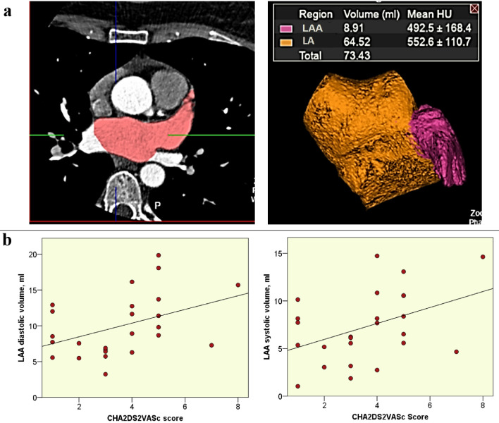
All patients undergoing cardiac computed tomography (CT) in La Princesa University Hospital of Madrid, between April and November 2019, with retrospective electrocardiogram (ECG)-gated acquisition (Siemens 64-detectors CT) including complete volume of LA were recruited. Patients with low quality LA and LAA imaging or not in sinus rhythm were excluded. The systolic and diastolic volumes of LA and LAA were measured with a Vitrea Workstation. Systolic and diastolic atrial segmentation was manually traced on axial plane images with subsequent representation as volume rendering (Fig. 1a). Volumes were collected as whole LA and LAA alone. Risk of stroke was estimated using the CHA2DS2-VASc score. The relationship of the CHA2DS2-VASc score with morphological and functional variables was analyzed by Pearson’s correlation. This study was approved by the Institutional Review Board, and was conducted in compliance with the ethical standards of the responsible institution on human subjects as well as with the Helsinki Declaration.
Figure 1.
(a) LA and LAA segmentation with 3D volume reconstruction are showed. (b) Correlation plots of LAA diastolic and systolic volumes with CHA2DS2-VASc score. LA: left atrium; LAA: left atrial appendage; 3D: three-dimensional; CHA2DS2-VASc: congestive heart failure; hypertension; ages ≥ 74 years (2 points); diabetes; stroke, transient ischemic attack, or systemic embolism (2 points); vascular disease; ages 65 - 74 years; sex (female).
Results
In the recruitment 20 patients were excluded for low quality of LA and LAA imaging. A total of 25 patients were finally included in the analysis; 56% were female and mean age was 75 ± 13 years; 80% presented with a CHA2DS2-VASc ≥ 2. Heart failure, hypertension, diabetes, previous stroke and vascular disease were present in the 48%, 60%, 16%, 32%, and 32% of the patients, respectively. According to the study indication, 10 were for transcatheter aortic valve implantation (TAVI) evaluation protocol, 14 for coronary artery disease assessment and one for pulmonary venous drainage assessment. None patient had mitral valve disease. Only one patient had a previous medical history of paroxysmal AF.
A positive correlation was documented between the CHA2DS2-VASc score and systolic (r = 0.419, P = 0.037) and diastolic (r = 0.415, P = 0.039) LAA volumes (Fig. 1b). Atrial volumes and left atrial ejection fraction showed no significant correlations with CHA2DS2-VASc.
Discussion
This is the first study using cardiac CT to correlate LA and LAA remodeling with the CHA2DS2-VASc score. Cardiac CT has a high spatial and temporal resolution that allows an accurate measurement of cardiac chambers. LAA volume measurement with other techniques as echocardiography or cardiac magnetic resonance is not as accurate.
After measuring volumes in both phases of the cardiac cycle and evaluating the clinical characteristics of each patient, this study showed a significant correlation between the remodeling of the LAA and the CHA2DS2-VASc. Specifically, it was observed that higher scores correspond to higher volumes of the LAA without altering the LA volumes. Our results offers a partial explication to justify the increased risk of stroke in patient with a higher CHA2DS2-VASc score as higher volumes of LAA might be related to a more intense blood stasis during AF. Components of CHA2DS2-VASc score as age, hypertension and diabetes may alter the LAA wall producing the observed higher volumes. Recent studies assessing LAA by cardiac CT focused on LAA morphology [3], however, none of them provided data about LAA volume. Interestingly, no volumetric differences have been reported among the different LAA morphologies [4]. Moreover, previous CT studies for LAA closure planning focused on orifice diameters and LAA depth, not on volumes [5]. Our exploratory results based on a small cohort of patients should be confirmed in further studies with a larger population including multivariate analysis.
Conclusions
In conclusion, this study shows, for the first time, a positive correlation between CHA2DS2-VASc score and LAA remodeling. These findings might offer a new marker for stratification of embolic risk based on LAA remodeling.
Acknowledgments
The investigators acknowledge the staff that participated in the research project.
Financial Disclosure
No funding should be reported.
Conflict of Interest
The authors have no conflict of interest to disclose
Informed Consent
All patients provided informed consents.
Author Contributions
ASR and AC designed the study, oversaw data collection, reviewed the literature, analyzed and interpreted the data, and drafted the manuscript. MJO, AV, SH, PC, LJJB engaged in data collection, data interpretation, and contributed to drafts of the manuscript. FA contributed to the literature review, data interpretation, and provided critical reviews of the manuscript.
Data Availability
The authors declare that data supporting the findings of this study are available from the corresponding author on request.
References
- 1.Beigel R, Wunderlich NC, Ho SY, Arsanjani R, Siegel RJ. The left atrial appendage: anatomy, function, and noninvasive evaluation. JACC Cardiovasc Imaging. 2014;7(12):1251–1265. doi: 10.1016/j.jcmg.2014.08.009. [DOI] [PubMed] [Google Scholar]
- 2.Gomez-Doblas JJ, Muniz J, Martin JJ, Rodriguez-Roca G, Lobos JM, Awamleh P, Permanyer-Miralda G. et al. Prevalence of atrial fibrillation in Spain. OFRECE study results. Rev Esp Cardiol (Engl Ed) 2014;67(4):259–269. doi: 10.1016/j.rec.2013.07.014. [DOI] [PubMed] [Google Scholar]
- 3.Hirata Y, Kusunose K, Yamada H, Shimizu R, Torii Y, Nishio S, Saijo Y. et al. Age-related changes in morphology of left atrial appendage in patients with atrial fibrillation. Int J Cardiovasc Imaging. 2018;34(2):321–328. doi: 10.1007/s10554-017-1232-x. [DOI] [PubMed] [Google Scholar]
- 4.Shimada M, Akaishi M, Kobayashi T. Left atrial appendage morphology and cardiac function in patients with sinus rhythm. J Echocardiogr. 2020;18(2):117–124. doi: 10.1007/s12574-020-00462-0. [DOI] [PubMed] [Google Scholar]
- 5.Rajwani A, Nelson AJ, Shirazi MG, Disney PJS, Teo KSL, Wong DTL, Young GD. et al. CT sizing for left atrial appendage closure is associated with favourable outcomes for procedural safety. Eur Heart J Cardiovasc Imaging. 2017;18(12):1361–1368. doi: 10.1093/ehjci/jew212. [DOI] [PubMed] [Google Scholar]
Associated Data
This section collects any data citations, data availability statements, or supplementary materials included in this article.
Data Availability Statement
The authors declare that data supporting the findings of this study are available from the corresponding author on request.


