Abstract
Purpose
Functional lung mapping from Ga68-ventilation/perfusion (V/Q) PET/CT, which has been shown to correlate with pulmonary function tests (PFTs), may be beneficial in a number of clinical applications where sparing regions of high lung function is of interest. Regions of clumping in the proximal airways in patients with airways disease can result in areas of focal intense activity and artefact in ventilation imaging. These artefacts may even shine through to subsequent perfusion images and create a challenge for quantitative analysis of PET imaging. We aimed to develop an automated algorithm that interprets the uptake histogram of PET images to calculate a peak uptake value more representative of the global lung volume.
Methods
Sixty-six patients recruited from a prospective clinical trial underwent both V/Q PET/CT imaging and PFT analysis before treatment. PET images were normalised using an iterative histogram analysis technique to account for tracer hotspots prior to the threshold-based delineation of varying values. Pearson’s correlation between fractional lung function and PFT score was calculated for ventilation, perfusion, and matched imaging volumes at varying threshold values.
Results
For all functional imaging thresholds, only FEV1/FVC PFT yielded reasonable correlations to image-based functional volume. For ventilation, a range of 10–30% of adapted peak uptake value provided a reasonable threshold to define a volume that correlated with FEV1/FVC (r = 0.54–0.61). For perfusion imaging, a similar correlation was observed (r = 0.51–0.56) in the range of 20–60% adapted peak threshold. Matched volumes were closely linked to ventilation with a threshold range of 15–35% yielding a similar correlation (r = 0.55–0.58).
Conclusions
Histogram normalisation may be implemented to determine the presence of tracer clumping hotspots in Ga-68 V/Q PET imaging allowing for automated delineation of functional lung and standardisation of functional volume reporting.
Keywords: V/Q PET/CT, Gallium 68, Regional lung function, Delineation
Introduction
Functional lung volume obtained from Ga68-ventilation/perfusion (V/Q) PET/CT imaging, a fractional measure of the total lung that exhibits appreciable uptake of inhaled or perfused tracer, has been shown to correlate with pulmonary function tests (PFTs) [1, 2]. This shows promise in utilising volumetric imaging to determine areas which may warrant avoidance in external beam radiotherapy [3, 4], assessment of radiation injury to the lung [5], and estimating the loss of function after surgery in the context of resection of lung cancers [6–8]. In previous work, functional volumes were assessed visually to determine appropriate uptake thresholds and manually corrected where necessary. This process is laborious, requires clinician input, and is subject to interobserver variability. Previously, an appropriate threshold to define a functional volume was adapted to each case, and in some instances, manual removal of non-physiological uptake was required [2].
As with Technegas, clumping of radiotracer is sometimes observed in the proximal airways with Galligas. More artefact is typical in patients with severe airway disease, and areas of uptake have focal intense uptake which interferes with quantitative analysis. As the perfusion acquisition is obtained shortly after the ventilation acquisition, “shine through” of these high count areas may occur, also resulting in artefact on the perfusion images. The aim of this work was to identify an automated, objective means to improve on the previous process by implementing an iterative normalisation of the maximum uptake voxel intensity prior to thresholding—an initialisation step to remove the requirement for manual pre-processing by the reviewer.
Methods
Patients
Sixty-six patients (43 males, 23 females; mean age 66 years, range 34–89) with stage III non-small cell lung cancer planned to receive radiation therapy with curative intent were analysed. Patients had ventilation/perfusion PET/CT and pulmonary function tests, as part of a study assessing lung function from radiotherapy (Australian New Zealand Clinical Trial Registry Trial ID 12613000061730). The study was approved by the Peter MacCallum Clinical Governance and Ethics Committee. The study design and patient cohort have been comprehensively described in a previous paper [9].
Pulmonary function tests
All patients underwent PFT assessment prior to radiation therapy according to forced vital capacity (FVC), forced expiratory volume (FEV), FEV/FVC ratio, and carbon monoxide diffusing capacity (DLCO). Mean baseline PFT assessments (actual/predicted) FVC, FEV, FEV/FVC ratio, and DLCO were 91%, 76%, 65%, and 64%, respectively. PFTs ranged from 47 to 134%, 31 to 121%, 30 to 90%, and 23 to 102% for FVC, FEV, FEV/FVC, and DLCO, respectively.
68Ga-V/Q PET/CT protocol
Ventilation PET imaging was acquired via inhalation of 68Ga carbon nanoparticles (Galligas) [10] followed by an injection of approximately 50 MBq of 68Ga-labelled macroaggregated albumin (MAA) for perfusion imaging. In 14 of 66 cases, only a perfusion study was performed due to tracer availability. PET studies were performed over two bed positions where each bed position was acquired for 5 min. The protocol for 68Ga-V/Q imaging has previously been described previously [11, 12].
Functional volume delineation
Functional volume delineation can be summarised in three steps: whole lung uptake characterisation, iterative reduction in outlier uptake values via histogram normalisation, and subsequent thresholding based on the converged peak intensity uptake value. To characterise whole lung uptake, PET voxels were limited to a lung contour that was defined based on CT margins. The converged peak intensity value is an iteratively reduced uptake value based on a histogram analysis of uptake inside the lung contour. The purpose of this step is to anchor threshold values to a more accurate value compared to an overall maximum and addresses non-physiological artefacts in both ventilation and perfusion imaging such as Galligas clumping in the airways.
Histogram normalisation using iterative convergence
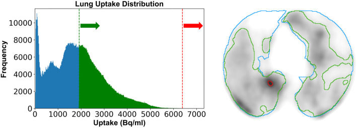
In order to remove the requirement for manual corrections of artefacts in ventilation imaging via airway clumping and perfusion imaging via MAA clumping, a reduction in the maximum uptake voxel was computationally implemented. The maximum intensity measured in the whole lung uptake was iteratively reduced to a new value (mean + 4 standard deviations) until a difference between the calculated value and previous maximum was less than 1%. In situations where the initial iteration calculates a value within 1% of the original maximum or greater than the original maximum, no reduction in maximum intensity was applied. An example of the reduced maximum intensity anchoring threshold is given in Fig. 1. This accounts for very high outlier values which otherwise skew the uptake distribution histogram of intensities within the lung and yields a peak intensity value which is in closer accordance with the uptake distribution within high functioning lung tissue. This iterative technique is similar to that used by Kipritidis et al. for clumping hotspot removal [13] and was implemented using Python 3.7 with libraries SimpleITK and NumPy.
Fig. 1.
Example of functional volume delineation in the presence of an airway artifact. Whole lung volume is shown in blue. The aggregated, non-physiological uptake (red) is detected by histogram analysis, and a more representative peak uptake value is designated. The resultant functional volume from 30% threshold is shown in green
Iterative maximum intensity reduction was considered to be converged when the convergence parameter (z) was greater than 99% (i.e. the step-wise value less than 1% of the previous). The first iteration step (i = 1) determined if the maximum intensity reduction was required at all. For example, if the initial calculated mean + 4 standard deviations inside the lung were within 1% of the original maximum, no reduction was applied and the original maximum was considered to be a representative maximum. The resolved peak intensity is a function of the mean (x̄) and standard deviation (σ) uptake inside the lung. Volumes above the peak at each step are ignored in successive iterations but are reintroduced into the final volume, reduced to the new peak intensity.
Thresholding
A range of threshold values (5–70% of the peak intensity, in 5% increments) were applied to each ventilation and perfusion image after the normalisation process to investigate an optimal functional correlation across the cohort. A fractional value was given to each image at each threshold, defining the fraction of functional lung volume delineated relative to total lung volume. Where both ventilation and perfusion imaging data was available (n = 52/66), a matched functional volume was generated using the intersection of functional volumes in both images at the same threshold. Functional volumes from all of the thresholds were then correlated against pulmonary function tests: FVC (%Predicted), FEV (%Predicted), FEV/FVC, and DLCO (%Predicted)—using Pearson correlation.
Results
Peak uptake normalisation was more frequently required in ventilation imaging (48/52) than perfusion imaging (21/66). Relative volumes normalised for ventilation and perfusion imaging were 0.23% and 0.06%, respectively. For all studies, across both imaging techniques, the maximum volume normalised was 4.88% in a perfusion image—as shown in Table 1.
Table 1.
Median, mean, and max volumes relative to the total lung that were normalised
| Imaging technique | Volume normalised (% relative to total lung) | ||
|---|---|---|---|
| Median | Mean | Max | |
| Ventilation (n = 48/52) | 0.23% | 0.53% | 3.34% |
| Perfusion (n = 21/66) | 0.06% | 0.51% | 4.88% |
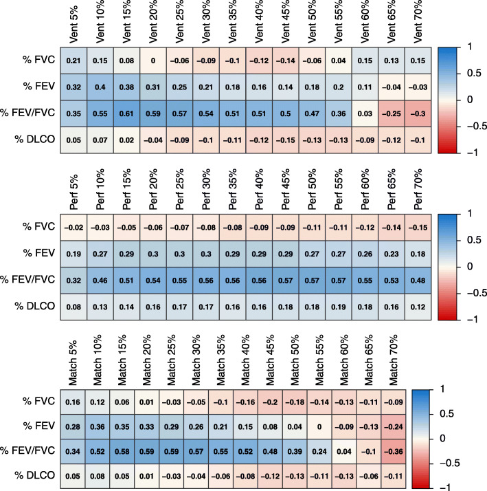
Correlograms illustrating the correlation of lung function measures and resolved peak intensity threshold for each imaging technique are given in Fig. 2. The range of correlations plateaus between threshold minimum and maximum boundaries for each volume. For all PFTs correlated against functional volumes, FEV/FVC consistently yielded the greatest correlation to PET-defined volumes. A moderate correlation between FEV/FVC and functional volume was achieved by selecting an appropriate threshold for ventilation, perfusion, and matched function from both image sets. For ventilation imaging, the highest correlations were observed with FEV/FVC using 10–30% thresholds (r = 0.54–0.61). For perfusion imaging, similar correlation coefficients (r = 0.56–0.57) between FEV/FVC and functional volume were observed from 30 to 55% threshold values. For the matched V/Q volumes, the greatest correlations between FEV/FVC and functional volume observed was of the range 15–30% (r = 0.57–0.59). A weak correlation was observed with other lung function measures FEV, DLCO, and functional volume.
Fig. 2.
Correlogram of baseline volumes vs. baseline PFT
Discussion
Radiopharmaceutical uptake globally throughout the lungs in ventilation/perfusion PET imaging is associated with overall lung function as measured by common PFT metrics. Whereas PFT can only define global lung function, assessment of regional lung function within anatomical subunits of the lung requires advanced imaging techniques. There is potential benefit in the ability to localise high- and low-functioning regions which may assist with surgical or radiotherapy planning. Previous work has investigated the correlation between FEV/FVC and an expert-defined manual contour of the functional lung as well as a semi-automated method [1, 2]. While the results of the automated method developed in this work are not as strongly correlated as the fully manual method (r = 0.78 and 0.81 for ventilation and perfusion, respectively), this study’s results were an improvement over the semi-automated method (r = 0.48 and 0.46) which involved defining a threshold at 15% of the maximum uptake inside the lung and manually removing regions of suspected clumping.
In principle, functioning lung tissue should exhibit both ventilation representing localised air flow and perfusion representing the ability to perform gas exchange to the blood. The matched functional volume, which includes the intersection of highly ventilated and perfused threshold regions, should be best correlated with pulmonary function tests. In this patient cohort, a range of threshold values applied to the reduced peak intensity shared similar correlations. Ventilation imaging alone had a limited range in which the correlation was relatively strong, this suggests that this ventilation technique may be more susceptible to variations in uptake distribution.
As a guide for potential threshold values in future applications, our observations suggest a range of 15–30% of adjusted peak uptake value for matched and ventilation imaging. For perfusion imaging, a broader range of thresholds (15–65%) offered a comparable correlation to lung function. Implementation of this methodology is proposed to be an addition to the typical workflow outlined: image acquisition, clinician assessment with raw imaging to determine the adequacy of the acquisition, and lastly automatically define regions of functional lung to provide volumes as supplementary information.
Ventilation imaging more frequently required a reduction in uptake maximum than perfusion imaging (48/52 and 21/66, respectively). A reduction in the uptake maximum is not a direct indication for the presence of tracer clumping—its intent is to provide a more representative peak value for the lungs that exhibit large heterogeneity of uptake. Additionally, it should be noted that when normalised volumes (0–3.34% ventilation, 0–4.88% perfusion) were excluded from the calculation of fractional lung function as opposed to being included, the resulting Pearson correlation remained relatively similar due to the relatively small magnitude of volume being normalised.
This methodology may be applied in future applications such as estimation of global lung function as a surrogate for conventional pulmonary function tests with added information on the regional distribution of function, lung avoidance in radiotherapy by providing contours as DICOM structures on the radiotherapy planning software, and planning of anatomical lung resections which may aid the surgeon in defining estimated loss of function after lobectomy, segmentectomy, and pneumonectomy in the context of resection of lung cancers. A core limitation of this study arises from its reliance on homogenous uptake in the global lung volume; perfusion imaging can exhibit an uptake gradient under influence of gravity [14], resulting in functional regions being excluded via this automated process. Although this gravitational effect may affect tracer pooling and distribution, the relationship between PFTs as a global measure and the automated estimation should be maintained. Additionally, the apparent pattern of tracer uptake is constrained by the resolving power of the PET imaging system which limits the ability to perceive potential regions of heterogeneous function due to the partial volume effect and respiratory motion. Future work may advance further by applying the suggested methodology to segmental lung regions rather than a global volume.
Conclusion
The method outlined describes an automated functional lung volume delineation technique that advances on previous studies by automating an initialisation step to manage the potential presence of hotspots prior to thresholding. Implementation of this methodology has the potential to provide consistency for lung function assessment in serial imaging, more accurate inter and intra-patient comparisons, and an avoidance map for clinical interventions.
Acknowledgements
The authors wish to thank Lisa Selbie for the coordination of the study and assistance with data curation.
Authors’ contributions
LM: study design, investigation, algorithm implementation, data analysis, data curation, and writing. PJ: study design, investigation, project administration, algorithm implementation, data analysis, and writing. NH: study design, investigation, project administration, data analysis, and writing. MB: investigation, data analysis, and writing. TK: study design, investigation, and writing. JC: data curation, investigation, data analysis, and writing. DS: study design, investigation, and writing. NB: study design, investigation, and writing. MH: study design, supervision, investigation, project administration, data analysis, and writing. SS: study design, supervision, investigation, project administration, data analysis, and writing. The authors read and approved the final manuscript.
Funding
Shankar Siva has received National Health and Medical Research Council scholarship funding APP1038399.
Availability of data and materials
Data is not appropriate to be shared.
Declarations
Ethics approval and consent to participate
The study was approved by the Peter MacCallum Clinical Governance and Ethics Committee as part of a clinical trial (Australian New Zealand Clinical Trial Registry Trial ID 12613000061730).
Date of registration: 16 January 2013, retrospectively registered
URL of trial registry record: ttps://www.anzctr.org.au/Trial/Registration/TrialReview.aspx?id=363411&isReview=true
Consent for publication
Not applicable
Competing interests
The authors declare that they have no competing interests.
Footnotes
Publisher’s Note
Springer Nature remains neutral with regard to jurisdictional claims in published maps and institutional affiliations.
References
- 1.Le Roux PY, Siva S, Steinfort DP, Callahan J, Eu P, Irving LB, et al. Correlation of 68Gallium ventilation-perfusion PET/CT with pulmonary function test indices for assessing lung function. J Nucl Med. 2015;56(11):1718–1723. doi: 10.2967/jnumed.115.162586. [DOI] [PubMed] [Google Scholar]
- 2.Le Roux PY, Siva S, Callahan J, et al. Automatic delineation of functional lung volumes with 68Ga-ventilation/perfusion PET/CT. EJNMMI Res. 2017;7(1):82. doi: 10.1186/s13550-017-0332-x. [DOI] [PMC free article] [PubMed] [Google Scholar]
- 3.Hardcastle N, Hofman MS, Hicks RJ, Callahan J, Kron T, MacManus MP, et al. Accuracy and utility of deformable image registration in 68Ga 4D PET/CT assessment of pulmonary perfusion changes during and after lung radiation therapy. Int J Radiat Oncol Biol Phys. 2015;93(1):196–204. doi: 10.1016/j.ijrobp.2015.05.011. [DOI] [PubMed] [Google Scholar]
- 4.Siva S, Thomas R, Callahan J, Hardcastle N, Pham D, Kron T, et al. High-resolution pulmonary ventilation and perfusion PET/CT allows for functionally adapted intensity modulated radiotherapy in lung cancer. Radiother Oncol. 2015;115(2):157–162. doi: 10.1016/j.radonc.2015.04.013. [DOI] [PubMed] [Google Scholar]
- 5.Siva S, Hardcastle N, Kron T, Bressel M, Callahan J, MacManus MP, et al. Ventilation/perfusion positron emission tomography-based assessment of radiation injury to lung. Int J Radiat Oncol Biol Phys. 2015;93(2):408–417. doi: 10.1016/j.ijrobp.2015.06.005. [DOI] [PubMed] [Google Scholar]
- 6.Argula RG, Strange C, Ramakrishnan V, Goldin J. Baseline regional perfusion impacts exercise response to endobronchial valve therapy in advanced pulmonary emphysema. Chest. 2013;144(5):1578–1586. doi: 10.1378/chest.12-2826. [DOI] [PubMed] [Google Scholar]
- 7.Leong P, Le Roux PY, Callahan J, Siva S, Hofman MS, Steinfort DP. Reduced ventilation-perfusion (V/Q) mismatch following endobronchial valve insertion demonstrated by gallium-68 V/Q photon emission tomography/computed tomography. Respirol Case Rep. 2017;5(5):e00253. doi: 10.1002/rcr2.253. [DOI] [PMC free article] [PubMed] [Google Scholar]
- 8.Le Roux PY, Leong TL, Barnett SA, Hicks RJ, Callahan J, Eu P, et al. Gallium-68 perfusion positron emission tomography/computed tomography to assess pulmonary function in lung cancer patients undergoing surgery. Cancer Imaging. 2016;16(1):24. doi: 10.1186/s40644-016-0081-5. [DOI] [PMC free article] [PubMed] [Google Scholar]
- 9.Siva S, Callahan J, Kron T, et al. A prospective observational study of gallium-68 ventilation and perfusion PET/CT during and after radiotherapy in patients with non-small cell lung cancer. BMC Cancer. 2014;14(1):1–8. doi: 10.1186/1471-2407-14-740. [DOI] [PMC free article] [PubMed] [Google Scholar]
- 10.Kotzerke J, Andreeff M, Wunderlich G. PET aerosol lung scintigraphy using Galligas. Eur J Nucl Med Mol Imaging. 2010;37(1):175–177. doi: 10.1007/s00259-009-1304-9. [DOI] [PubMed] [Google Scholar]
- 11.Callahan J, Hofman MS, Siva S, Kron T, Schneider ME, Binns D, et al. High-resolution imaging of pulmonary ventilation and perfusion with 68Ga-VQ respiratory gated (4-D) PET/CT. Eur J Nucl Med Mol Imaging. 2014;41(2):343–349. doi: 10.1007/s00259-013-2607-4. [DOI] [PubMed] [Google Scholar]
- 12.Hofman MS, Beauregard JM, Barber TW, Neels OC, Eu P, Hicks RJ. 68Ga PET/CT ventilation-perfusion imaging for pulmonary embolism: a pilot study with comparison to conventional scintigraphy. J Nucl Med. 2011;52(10):1513–1519. doi: 10.2967/jnumed.111.093344. [DOI] [PubMed] [Google Scholar]
- 13.Kipritidis J, Siva S, Hofman MS, Callahan J, Hicks RJ, Keall PJ. Validating and improving CT ventilation imaging by correlating with ventilation 4D-PET/CT using 68Ga-labeled nanoparticles. Med Phys. 2014;41(1):011910. doi: 10.1118/1.4856055. [DOI] [PubMed] [Google Scholar]
- 14.Hopkins SR, Wielpütz MO, Kauczor HU. Imaging lung perfusion. J Appl Physiol (1985) 2012;113(2):328–339. doi: 10.1152/japplphysiol.00320.2012. [DOI] [PMC free article] [PubMed] [Google Scholar]
Associated Data
This section collects any data citations, data availability statements, or supplementary materials included in this article.
Data Availability Statement
Data is not appropriate to be shared.


