



Abstract
Introduction
Venous thromboembolism (VTE) has a long‐term risk of recurrence, dependent on the presence or absence of provoking risk factors at the time of the event.
Objective
To compare clinical characteristics, anticoagulant patterns, and 12‐month outcomes in patients with transient provoking factors, active cancer, and unprovoked VTE.
Methods
The Global Anticoagulant Registry in the FIELD (GARFIELD)‐VTE is a prospective, observational study that enrolled 10 207 patients with objectively diagnosed VTE from 415 sites in 28 countries.
Results
Patients with transient provoking factors were younger (53.0 years) and more frequently women (61.2%) than patients with unprovoked VTE (60.3 years; 43.0% women) or active cancer (63.6 years; 51.7% women). After 6 months, 59.1% of patients with transient provoking factors remained on anticoagulation, compared to 71.3% with unprovoked VTE and 47.3% with active cancer. At 12 months, this decreased to 36.7%, 51.5%, and 25.4%, respectively. The risk of mortality (hazard ratio [HR], 1.21; 95% confidence interval [CI], 0.90‐1.62), recurrent VTE (HR, 0.84; 95% CI, 0.62‐1.14), and major bleeding (HR, 1.26; 95% CI, 0.86‐1.85) was comparable in patients with transient provoking factors and unprovoked VTE. Patients with minor and major transient provoking factors had a similar risk of recurrent VTE (HR, 0.99; 95% CI, 0.59‐1.66), but those with major transient risk factors had a lower risk of death (HR, 0.61; 95% CI, 0.38‐0.98).
Conclusion
At 1 year, nearly 40% of patients with transient provoking factors and slightly over half of patients with unprovoked VTE were on anticoagulant treatment. Event rates were comparable between the two groups. Risk of death was higher in patients with minor transient factors than in those with major transient factors.
Keywords: anticoagulants, deep vein thrombosis, pulmonary embolism, risk factors, venous thromboembolism
Essentials.
GARFIELD‐VTE records presenting risk factors and treatment patterns for patients with VTE worldwide.
Presenting persistent risk factors, transient risk factors, and unprovoked VTE are heterogeneous.
Long‐term outcomes are influenced by the presence and type of presenting risk factors.
Presenting risk factors impact the pattern of anticoagulation administered to patients with VTE.
1. INTRODUCTION
Venous thromboembolism (VTE) has a long‐term risk of recurrence, partially dependent on the presence of specific types of risk factors at the time of the initial VTE event. 1 , 2 , 3 Certain transient provoking risk factors such as major surgery or major trauma typically have a low risk of recurrence (<3% per year) after cessation of anticoagulation. 4 , 5 Patients hospitalized with acute medical illness such as heart failure or pneumonia have an intermediate risk of recurrence (3%–8% per year), as do patients with no identifiable risk factor (unprovoked), after completing time‐limited anticoagulation 5 . Patients with active cancer, a persistent provoking factor, have the highest risk of recurrent VTE after termination of anticoagulation. 4 Therefore, the presence or absence of risk factors influence both the treatment duration and the prognosis. 6 , 7
Current guidelines recommend anticoagulation treatment for 3 months after VTE caused by transient risk factors, and longer if caused by persistent risk factors. 2 However, evidence suggests that clinicians often treat VTE for >3 months. 8 , 9 , 10 , 11 This may reflect the fact that the duration of anticoagulation therapy is influenced by other factors, such as the site of VTE, congestive heart failure, and age. 8 Further study is warranted to determine whether the risk of recurrence outweighs the risk of bleeding in the presence or absence of provoking specific risk factors. 12 , 13 The Global Anticoagulant Registry in the FIELD–Venous Thromboembolic Events (GARFIELD‐VTE) is an ongoing, prospective, noninterventional registry designed to observe initial and extended therapeutic strategies and clinical outcomes for 3 years in patients with VTE worldwide, treated according to local standard practices. This article aims to compare clinical characteristics, anticoagulant treatment patterns, and 12‐month outcomes among patients with transient provoking risk factors, persistent provoking risk factors, and unprovoked VTE.
2. METHODS
2.1. Study design and participants
The design of the GARFIELD‐VTE registry has been described previously. 14 GARFIELD–VTE (ClinicalTrials.gov identifier: NCT02155491) has enrolled 10 684 patients with objectively diagnosed VTE from 415 sites in 28 countries.
Men and women aged ≥ 18 years with an objectively confirmed diagnosis of VTE within 30 days of entry into the registry were eligible for inclusion. Patients with recurrent VTE must have completed treatment for the previous event. The study excluded patients with superficial vein thrombosis, those participating in an interventional study that dictated treatments, or those for whom long‐term follow up was not possible. No specific treatments, tests, or procedures are mandated by the study protocol. Decisions to initiate, continue, or change treatment were solely at the discretion of the treating physicians and their patients. Thus, assignment of anticoagulation type at baseline was managed by the physician.
2.2. Selection of study sites
The national coordinating investigator identified the care settings they believed most accurately represented the management of patients with VTE in their country. The contract research organization provided a list of sites that reflected these care settings before contacting a random sample of sites for each care setting from the list. Sites that agreed to participate were recruited after a qualification telephone call, and all investigators completed an educational program providing guidance on patient screening, enrollment, and follow‐up in the registry.
2.3. Data collection
Data are captured using an electronic case report form (eCRF) designed by eClinicalHealth Services (Stirling, UK) and submitted electronically via a secure website to the registry coordinating center at the Thrombosis Research Institute, which was responsible for checking the completeness and accuracy of data collected from medical records. The GARFIELD‐VTE protocol mandates (i) centralized auditing of 10% of all eCRFs by comparison with source documentation, (ii) provision of electronic audit trails for all data modifications, and (iii) subjecting critical variables to additional audit. This study reports prospectively collected data from patients enrolled from May 12, 2014, to January 4, 2017. The data were extracted from the study database on December 8, 2018.
2.4. Clinical outcomes
The primary clinical outcomes were all‐cause mortality, recurrent VTE, and major bleeding. Major bleeding was defined as clinically overt bleeding associated with a critical site (eg, intracranial, intraspinal, intraocular), decrease in hemoglobin of ≥ 2 g/dL, transfusion of ≥ 2 units of packed red blood cells, or a fatal outcome. Nonmajor bleeding was defined as any overt bleeding not meeting the criteria for major bleeding. The rates of hospitalization, bleeding, cancer, stroke/transient ischemic attack, and myocardial infarction (MI) were also recorded. Cancer events that were diagnosed >30 days after VTE diagnosis date were considered as cancer end points. Patients with cancer ≤ 30 days from the VTE diagnosis date were considered to have either active cancer or a history of cancer. For all other outcomes, events that occurred from the day of diagnosis onward were considered outcomes. Only the first occurrence of each event type was considered.
2.5. Ethics statement
The registry is conducted in accordance with the Declaration of Helsinki and guidelines from the International Conference on Harmonisation on Good Clinical Practice and Good Pharmaco‐epidemiological Practice and adheres to all applicable national laws and regulations. An independent ethics committee for each participating country and the hospital‐based institutional review board approved the design of the registry. All patients provided written informed consent to participate. Confidentiality and anonymity of patients recruited into this registry are maintained.
2.6. Definitions
Categorization of patients with persistent provoking factors, transient provoking factors (major and minor), or unprovoked VTE was based on the guidance document published by the Scientific and Standardization Committees on Control of Anticoagulation and on Predictive Variables of the International Society of Thrombosis and Haemostasis. 15 Active cancer was considered a persistent provoking risk factor. Major transient provoking factors included surgery or trauma up to 3 months before enrollment. Minor transient provoking factors included hospitalization, pregnancy, hormone replacement therapy, oral contraception, and acute medical illness up to 3 months before enrollment.
2.7. Statistical analysis
Continuous variables are summarized as mean and standard deviation (SD), and categorical variables are presented as frequency and percentage. Event rates and the associated 95% confidence interval (CI) were estimated using Poisson regression and are expressed per 100 person‐years.
Time‐to‐event analyses of outcomes were performed using Cox proportional hazard models. The as‐treated population was used for the analyses; thus, patients were assessed according to their ongoing treatment regimens and analysis was restricted to patients with VTE who were receiving anticoagulation. Patients were analyzed according to anticoagulation type: parenteral therapy only, vitamin K antagonists (VKAs; with or without parenteral therapy), and direct oral anticoagulants (DOACs; with or without parenteral therapy). Day 1 of treatment was defined as the first day of anticoagulation stabilization, having continued without interruption before day 30. Constant exposure to treatment was assumed from day 1 until treatment completion or discontinuation. Eligible patients could not switch anticoagulation type. Follow‐up data was right censored when treatment was completed or discontinued. Temporary discontinuation of anticoagulation for ≤7 days was ignored. During the 30 days from VTE diagnosis, as patients can exhibit irregular treatment patterns due to this transition period, the temporary discontinuation rule did not apply during this phase. Discontinuation of anticoagulation for >7 days was considered permanent discontinuation. Intention‐to‐treat analysis was also carried out for the GARFIELD‐VTE registry, whereby patients were assessed according to their initial treatment assigned at baseline (see Appendix S1).
Hazard ratios (HRs) were adjusted to account for the following variables: age, sex, ethnicity, body mass index (BMI), type of VTE, recent bleeding or anemia, chronic heart failure, chronic immobilization, family history of VTE, history of cancer, known thrombophilia, prior episode of VTE, chronic kidney disease stage, and treatment at baseline. Missing values were imputed using multivariate imputation by the chained equations method. 16 Forests plots are used to present adjusted HRs and their 95% CIs. Model assumptions were tested to evaluate the adequacy of observed data. Cumulative incidence plots were estimated to account for the competing risk of mortality on recurrent VTE episodes and major bleeding events. All statistical analyses were performed using R statistical software 17 and SAS software version 9.4 (SAS Institute, Cary, NC, USA). The threshold for assessing statistical significance for two‐sided tests was P = 0.05.
3. RESULTS
3.1. Baseline characteristics
Of the total 10 868 patients enrolled, 10 207 (93.9%) were eligible for this analysis. A total of 1026 (10.1%) had a persistent provoking risk factor, 3134 (30.7%) had a transient provoking risk factor, and 6047 (59.2%) had unprovoked VTE (Figure 1). Baseline characteristics are provided in Table 1. Distribution of patient recruitment according to region, country, care setting, and treatment funding source is detailed in Appendix S1: Table S1.
FIGURE 1.

Study population flowchart. AC, anticoagulant; DOAC, direct oral anticoagulant; PAR, parenteral; VKA, vitamin K antagonist
TABLE 1.
Baseline characteristics
| Persistent provoking risk factor (N = 1026) | Transient provoking risk factor (N = 3134) | Unprovoked (N = 6047) | |
|---|---|---|---|
| Female, n (%) | 530 (51.7) | 1918 (61.2) | 2598 (43.0) |
| Age, y, mean (SD) | 63.6 (13.4) | 53.0 (18.1) | 60.3 (16.1) |
| Age groups, y, n (%) | |||
| <50 | 153 (14.9) | 1458 (46.5) | 1571 (26.0) |
| 50‐64 | 360 (35.1) | 762 (24.3) | 1895 (31.3) |
| 65‐74 | 315 (30.7) | 480 (15.3) | 1372 (22.7) |
| 75‐84 | 165 (16.1) | 335 (20.7) | 962 (15.9) |
| ≥85 | 33 (3.2) | 99 (3.2) | 247 (4.1) |
| Body mass index, kg/m2, mean (SD) | 26.6 (6.4) | 28.2 (6.6) | 28.9 (6.5) |
| Body mass index group, kg/m2, n (%) | |||
| Underweight (<18.5) | 58 (6.2) | 59 (2.1) | 84 (1.6) |
| Normal (18.5‐24.9) | 364 (38.9) | 945 (33.0) | 1455 (26.9) |
| Overweight (25‐29.9) | 300 (32.1) | 928 (32.4) | 2005 (37.1) |
| Obese (≥30) | 214 (22.9) | 935 (32.6) | 1866 (34.5) |
| Missing | 90 | 267 | 637 |
| Responsible physician, n (%) | |||
| Vascular medicine | 376 (36.6) | 1497 (47.8) | 2707 (44.8) |
| General practitioner | 34 (3.3) | 119 (3.8) | 214 (3.5) |
| Internal medicine (hematology and intensive care) | 566 (55.2) | 1288 (41.1) | 2596 (43.0) |
| Emergency medicine | 28 (2.7) | 71 (2.3) | 166 (2.7) |
| Cardiology | 22 (2.1) | 159 (5.1) | 359 (5.9) |
| Missing | 0 | 0 | 5 |
| Current/previous smoker, n (%) | 387 (39.1) | 1034 (34.0) | 2393 (41.3) |
| Missing | 36 | 96 | 246 |
| Chronic kidney disease stage (eGFR), n (%) | |||
| I – Normal (≥90) | 356 (37.8) | 1067 (40.6) | 1450 (28.7) |
| II – Mild (60‐89) | 354 (37.6) | 1067 (40.6) | 2388 (47.2) |
| III – Moderate (30‐59) | 191 (20.3) | 366 (13.9) | 1017 (20.1) |
| IV – Severe (15‐29) | 26 (2.8) | 48 (1.8) | 99 (2.0) |
| V – Failure (<15) | 14 (1.5) | 90 (3.4) | 103 (2.0) |
| Missing | 85 | 507 | 990 |
| Hemoglobin categories, n (%) | |||
| Low | 659 (71.6) | 1266 (47.6) | 1568 (30.8) |
| Medium | 253 (27.5) | 1367 (51.4) | 3409 (66.9) |
| High | 8 (0.9) | 28 (1.1) | 117 (2.3) |
| Missing | 106 | 473 | 953 |
| Platelet count, n (%) | |||
| Low | 171 (18.0) | 211 (7.8) | 553 (10.8) |
| Normal | 716 (75.2) | 2323 (86.2) | 4430 (86.4) |
| High | 65 (6.8) | 161 (6.0) | 145 (2.8) |
| Missing | 74 | 439 | 919 |
Hemoglobin was categorized as low (<13.5 g/dL for men, <12 g/dL for women), normal (13.5‐17.5 g/dL for men, 12‐15.5 g/dL for women), or high (>17.5 g/dL for men, >15.5 g/dL for women). High and low platelet counts were defined as > 450 × 109/L and < 150 × 109/L, respectively. Data available for all patients unless stated.
Abbreviations: eGFR, estimated glomerular filtration rate; SD, standard deviation.
Patients with transient provoking factors were younger than patients with active cancer or unprovoked VTE (53.0 ± 18.1 years, 63.6 ± 13.4 years, and 60.3 ± 16.1 years, respectively) and more frequently women (61.2%, 51.7%, and 43.0%, respectively). Patients with active cancer were more likely to be underweight with low BMI, than those with transient provoked or unprovoked VTE (6.2% vs 2.1% and 1.6%, respectively) and less likely to be obese (22.9% vs 32.6% and 34.5%, respectively). Patients with transient provoking factors or unprovoked VTE were equally likely to be treated by a specialist in vascular medicine (47.8% and 44.8%, respectively) or internal medicine (41.1% and 43.0%, respectively), whereas patients with cancer were more frequently seen by a specialist in internal medicine (55.2%). Patients with cancer more frequently had low hemoglobin levels (71.6%) and thrombocytopenia (18.0%) compared with patients with transient provoking factors or unprovoked VTE (Table 1).
3.2. Site of VTE
In each patient group, the majority of VTE events were deep vein thrombosis (DVT) alone, with the highest percentage of DVT events in patients with transient provoking factors (64.8%). Of these DVT events, the majority were in the lower limb (73.6%), with a minority occurring in the upper limb (4.2%) or vena cava (0.9%). Bilateral DVT occurred more frequently in patients with cancer (10.8%) than in those with transient provoking factors (6.3%) or no provoking factors (5.7%). Lower limb distal DVT was more frequent in patients with transient provoking factors (39.7%) and unprovoked VTE (33.2%) than those with active cancer (28.8%). Conversely, patients with active cancer were more likely to present with lower limb proximal DVT (44.9%) than patients with transient provoking factors or unprovoked proximal DVT (33.7% and 37.6%, respectively). Patients with both distal and proximal DVT were comparable among groups. Pulmonary embolism (PE) within the pulmonary arterial branch was closely distributed between the main, lobar, and segmental branches in patients with active cancer (28.1%, 29.1%, and 35.2%, respectively), transient provoked VTE (28.5, 28.4, and 31.4%, respectively), and unprovoked VTE (30.4, 30.0, and 30.8%, respectively). Subsegmental pulmonary arterial PE was less common in all three groups (7.6%, 11.7%, and 8.8%) (Table 2).
TABLE 2.
Site of VTE
| Persistent provoking risk factor (N = 1026) | Transient provoking risk factor (N = 3134) | Unprovoked (N = 6047) | |
|---|---|---|---|
| Site of VTE, n (%) | |||
| DVT only | 618 (60.2) | 2032 (64.8) | 3599 (59.5) |
| PE only | 278 (27.1) | 666 (21.3) | 1461 (24.2) |
| PE and DVT | 130 (12.7) | 436 (13.9) | 987 (16.3) |
| Site of DVT, n (%) | |||
| Upper limb | 67 (6.5) | 132 (4.2) | 210 (3.5) |
| Lower limb | 643 (62.7) | 2307 (73.6) | 4295 (71.0) |
| Caval vein (inferior) | 24 (2.3) | 18 (0.6) | 51 (0.8) |
| Caval vein (superior) | 14 (1.4) | 8 (0.3) | 25 (0.4) |
| No DVT | 278 (27.1) | 669 (21.3) | 1466 (24.2) |
| Unilateral or bilateral DVT, n (%) | |||
| Left | 394 (52.7) | 1345 (54.6) | 2432 (53.1) |
| Right | 273 (36.5) | 964 (39.1) | 1885 (41.2) |
| Both | 81 (10.8) | 155 (6.3) | 261 (5.7) |
| Missing | 278 | 670 | 1469 |
| Type of lower limb DVT, n (%) | |||
| Distal | 184 (28.8) | 905 (39.7) | 1409 (33.2) |
| Proximal | 287 (44.9) | 769 (33.7) | 1595 (37.6) |
| Both | 168 (26.3) | 608 (26.6) | 1243 (29.3) |
| Missing | 387 | 852 | 1800 |
| Pulmonary arterial branch involved, n (%) | |||
| Main | 114 (28.1) | 312 (28.5) | 739 (30.4) |
| Lobar | 118 (29.1) | 311 (28.4) | 728 (30.0) |
| Segmental | 143 (35.2) | 343 (31.4) | 748 (30.8) |
| Subsegmental | 31 (7.6) | 128 (11.7) | 214 (8.8) |
| Missing | 620 | 2040 | 3618 |
Abbreviations: DVT, deep vein thrombosis; PE, pulmonary embolism; VTE, venous thromboembolism.
3.3. Treatment
At day 1 of treatment, patients with active cancer were more likely to receive parenteral therapy (60.0%) and less likely to receive VKAs (13.5%) or DOACs (26.4%) compared with patients with transient (16.1%, 32.4%, and 51.5%, respectively) or unprovoked VTE (11.4%, 33.8%, and 54.9%, respectively) (Figure 2). The choice of treatment was comparable between patients with transient provoking factors and with unprovoked VTE; in both groups, a similar proportion received DOACs or VKAs.
FIGURE 2.

Anticoagulation treatment patterns for (A) persistent provoked VTE (B) transient provoked VTE, and (C) unprovoked VTE over time. AC, anticoagulant; DOAC, direct oral anticoagulant; LTFU, lost to follow‐up; PAR, parenteral therapy, VKA, vitamin K antagonist
After 3 months, 79.7% of patients with transient provoking risk factors remained on anticoagulant therapy, compared with 86.3% of those with unprovoked VTE and 66.7% of patients with active cancer. At 6 months, this decreased to 59.1% and 71.3% for patients with transient provoking risk factors and unprovoked VTE, respectively, with approximately half of these patients receiving DOACs. Patients with active cancer remained on anticoagulant therapy in 47.3% of cases with 27.5% receiving parenteral therapy alone. After 12 months of follow‐up, patients with unprovoked VTE were more likely to remain on anticoagulation (51.5%) compared with patients with transient (36.7%) or active cancer (25.4%) (Figure 2). Patients with active cancer were more likely to remain on parenteral therapy (13.5%) than VKAs (3.4%) or DOACs (33.7%). Patients with transient and unprovoked VTE were more likely to remain on DOACs (18.3% and 30.1%). Parenteral therapy and VKA usage was comparable between transient provoked and unprovoked VTE during the 12 months of follow‐up (parenteral, 4.0% and 3.3%; VKA, 14.4% and 18.1%). Similar trends were observed among the three patient groups when analyzed by intention to treat (Appendix S1: Figure S1).
3.4. Clinical outcomes
During 12 months of follow‐up, the rate of all‐cause mortality, recurrent VTE, and major bleeding was comparable between patients with transient provoking factors and unprovoked VTE (4.4 vs 2.9 per 100 person‐years, 3.7 vs 3.4 per 100 person‐years, and 2.5 vs 1.7 per 100 person‐years, respectively). The rate of all primary outcomes was higher in patients with active cancer. Stroke/transient ischemic attack was more frequent in patients with cancer (2.1 per 100 person‐years) than in patients with transient provoking factors or unprovoked VTE (0.6 and 0.4 per 100 person‐years). The rate of MI was comparable among all groups (0.5‐1.0 per 100 person‐years) (Table 3). The cumulative incidences of primary outcomes are shown in Figure 3.
TABLE 3.
Unadjusted 12‐month event rates
| Events | Persistent provoking risk factor (N = 1072) | Transient provoking risk factor (N = 3268) | Unprovoked (N = 6302) | |||
|---|---|---|---|---|---|---|
| n | Rate (95% CI) | n | Rate (95% CI) | n | Rate (95% CI) | |
| All‐cause mortality | 251 | 48.6 (42.9‐55.0) | 84 | 4.4 (3.6‐5.5) | 123 | 2.9 (2.4‐3.4) |
| Recurrent VTE | 37 | 7.3 (5.3‐10.1) | 70 | 3.7 (3.0‐4.7) | 144 | 3.4 (2.9‐4.0) |
| Major bleed | 55 | 10.9 (8.3‐14.1) | 48 | 2.5 (1.9‐3.4) | 74 | 1.7 (1.4‐2.2) |
| Any bleed | 128 | 26.5 (22.3‐31.5) | 246 | 13.6 (12.0‐15.4) | 422 | 10.2 (9.3‐11.2) |
| New cancer | 39 | 7.7 (5.7‐10.6) | 24 | 1.26 (0.9‐1.9) | 87 | 2.0 (1.7‐2.5) |
| Stroke/TIA | 11 | 2.1 (1.2‐3.9) | 12 | 0.6 (0.4‐1.1) | 17 | 0.4 (0.3‐0.6) |
| Myocardial infarction | 5 | 1.0 (0.4‐2.3) | 9 | 0.5 (0.3‐0.9) | 22 | 0.5 (0.3‐0.8) |
Event rates are shown per 100 person‐years (95% CI). New cancer was defined as cancer diagnosed > 30 days after VTE diagnosis.
Abbreviations: CI, confidence interval; TIA, transient ischemic attack; VTE, venous thromboembolism.
FIGURE 3.

Cumulative incidence curves for primary outcomes over 12 months of follow‐up in patients with persistent or transient provoking risk factors or unprovoked VTE. (A) All‐cause mortality; (B) recurrent VTE; (C) major bleeding. Data are shown as percentage of patients with event and 95% confidence intervals. VTE, venous thromboembolism
After adjustment, the incidence of all primary outcomes was comparable between patients with transient provoking factors and unprovoked VTE (all‐cause mortality: HR, 1.21; 95% CI, 0.90‐1.62; recurrent VTE: HR, 0.84; 95% CI, 0.62‐1.14; major bleeding: HR, 1.26; 95% CI, 0.86‐1.85, respectively) (Figure 4). Patients with active cancer had an increased incidence of all‐cause mortality (HR, 5.79; 95% CI, 4.21‐7.96) and major bleeding (HR, 3.24; 95% CI, 1.98‐5.32) compared with patients with unprovoked VTE (Figure 4). The risk of recurrent VTE was comparable (HR, 1.48; 95% CI, 0.90‐2.43). Using intention‐to‐treat analysis, the risk of recurrent VTE was slightly higher in patients with active cancer compared with those with unprovoked VTE (Appendix S1: Results). All other results and trends were mirrored by intention‐to‐treat analysis of this population. Full analysis is available upon request.
FIGURE 4.

Adjusted hazard ratios for mortality with 95% confidence intervals for 12‐month outcomes after VTE diagnosis between patients with persistent provoked VTE vs unprovoked VTE (reference group) and transient provoked VTE vs unprovoked VTE (reference group). Hazard ratios were adjusted for age, gender, ethnicity, body mass index, type of VTE, recent bleeding or anaemia, chronic heart failure, chronic immobilization, family history of VTE, history of cancer, known thrombophilia, prior episode of VTE, chronic kidney disease stage, and treatment at baseline. VTE, venous thromboembolism
3.5. Transient provoking risk factors
Of the 3143 patients with transient provoking risk factors, 1722 (54.8%) were considered to have major risk factors, and 1412 (44.9%) were considered to have minor risk factors. Patients with minor transient provoking risk factors were more often women compared with patients with major transient provoking factors (73.8% vs 50.9%). The mean age and BMI were comparable between groups (50.3 ± 19.1 years vs 55.2 ± 16.8 years, 28.2 ± 7.3 kg/m2 vs 28.2 ± 5.9 kg/m2, respectively). A full comparison of baseline characteristics for patients with major and minor transient risk factors is presented in Appendix S1: Table S2. The distribution of site of DVT and predisposing risk factors are shown in Table 4.
TABLE 4.
Characteristics of patients with major or minor transient provoking factors
| Major transient provoking risk factor (N = 1722) | Minor transient provoking risk factor (N = 1412) | |
|---|---|---|
| Female, n (%) | 876 (50.9) | 1042 (73.8) |
| Age, y, mean (SD) | 55.2 (16.8) | 50.3 (19.1) |
| Body mass index, kg/m2, mean (SD) | 28.2 (5.9) | 28.2 (7.3) |
| Missing | 152 | 115 |
| Site of VTE, n (%) | ||
| DVT only | 1166 (67.7) | 866 (61.3) |
| PE only | 325 (18.9) | 341 (24.2) |
| DVT and PE | 231 (13.4) | 205 (14.5) |
| Site of DVT, n (%) | ||
| Upper limb | 69 (4.9) | 63 (5.9) |
| Lower limb | 1315 (94.3) | 992 (92.6) |
| Caval vein | 10 (0.7) | 16 (1.1) |
| Missing | 328 | 341 |
| Predisposing risk factors, n (%) | ||
| Chronic heart failure | 33 (1.9) | 89 (6.3) |
| Chronic immobilization | 177 (10.3) | 135 (9.6) |
| Known thrombophilia | 38 (2.2) | 41 (2.9) |
| Family history of VTE | 102 (5.9) | 104 (7.4) |
| Prior episode of VTE | 182 (10.6) | 144 (10.2) |
| Renal insufficiency | 50 (2.9) | 83 (5.9) |
Abbreviations: DVT, deep vein thrombosis; SD, standard deviation; VTE, venous thromboembolism.
At day 1 of treatment, patients with major transient provoking factors were more likely to receive a DOAC compared with patients with minor transient provoking factors (Figure 5). Patients with major transient provoking factors were more likely to discontinue anticoagulation over time. Major transient provoking factors were associated with a greater reduction in DOAC usage at 3 months, compared to minor transient factors. Conversely, patients with minor transient factors were more likely to cease DOAC usage at 6 months. Discontinuation of VKAs and parenteral therapy was comparable between groups. A slightly higher proportion of patients with minor transient provoking factors remained on anticoagulation therapy at 12 months.
FIGURE 5.
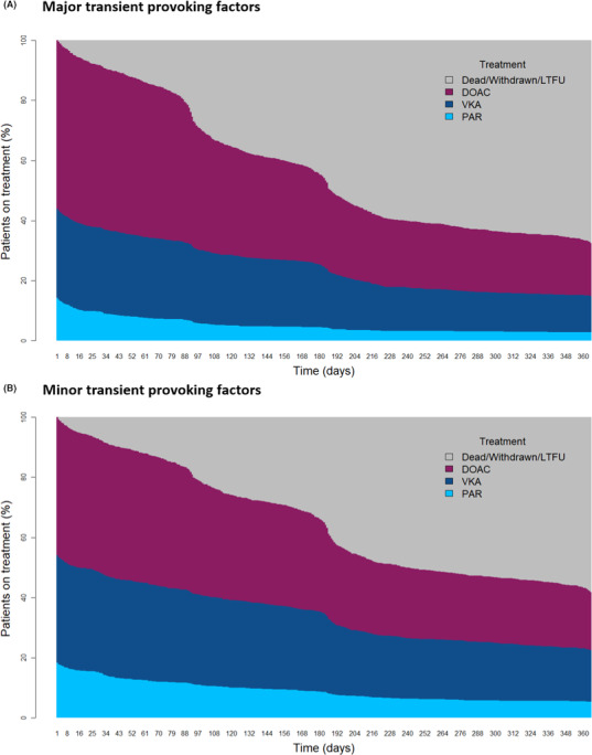
Anticoagulant treatment patterns over 12 months of follow‐up in patients with major or minor transient provoking risk factors. DOAC, direct oral anticoagulant; LTFU, lost to follow‐up; PAR, parenteral therapy; VKA, vitamin K antagonist
The rate of all‐cause mortality was significantly higher in patients with minor transient risk factors than in patients with major transient risk factors (6.1 vs 2.8 per 100 person‐years, respectively). This was predominately due to patients with acute medical illness and/or hospitalization (14.21 per 100 person‐years) as opposed to patients with estrogen or pregnancy‐related minor transient risk factors (2.47 per 100 person‐years). The rates of recurrent VTE (4.1 vs 4.3 per 100 person‐years) and major bleeding (3.2 vs 1.9 per 100 person‐years) were comparable (Appendix S1: Table S3).
After adjustment, patients with major transient provoking risk factors had a decreased incidence of all‐cause mortality compared with patients with minor transient risk factors (HR, 0.61; 95% CI, 0.38‐0.98). The incidences of both recurrent VTE (HR, 0.99; 95% CI, 0.59‐1.66) and major bleeding (HR, 0.79; 95% CI, 0.43‐1.45) were comparable. There was no difference in the incidence of other outcomes between patients with major or minor transient provoking factors (Appendix S1: Table S4). Figure 6 shows the cumulative incidence of primary outcomes in patients with major or minor transient provoking factors. These results and trends were mirrored in the intention‐to‐treat analysis of this population (Appendix S1: Results)
FIGURE 6.

Cumulative incidence curves for primary outcomes in patients with major or minor transient provoking risk factors. (A) All‐cause mortality; (B) recurrent VTE; (C) major bleeding. Data are shown as percentage of patients with event. VTE, venous thromboembolism
4. DISCUSSION
In this global VTE registry, both anticoagulation patterns and 12‐month outcomes were comparable between patients with transient provoking risk factors and unprovoked VTE, which made up 30% and 10% of the population, respectively. Active cancer was associated with a higher use of parenteral therapy alone, as well as an increased risk of all‐cause mortality and major bleeding. Subanalysis of transient provoking factors revealed that patients with minor transient provoking factors were more often female, more likely to discontinue DOAC use, and had a higher risk of all‐cause mortality compared with those with major transient provoking factors.
Patients with transient provoking risk factors were more often female, likely due to the inclusion of estrogen use and pregnancy in this category. This is further reflected in the substantially higher proportion of female patients within the minor transient factor category, which again includes estrogen use and pregnancy. Patients with transient risk factors were younger than patients with active cancer or unprovoked VTE, perhaps because older age increases the risk of both unprovoked VTE 8 and incidence of cancer. 18 These observations are consistent with a meta‐analysis of randomized controlled trials, which showed a higher proportion of men and a higher mean age in patients with unprovoked VTE than in those with provoked VTE. 19
The choice of anticoagulant treatment was comparable between patients with transient provoking factors and unprovoked VTE, with approximately half of these patients receiving a DOAC, and one‐third receiving a VKA at day 1 of treatment. This reflects a recent study of initial anticoagulation strategies within GARFIELD‐VTE, revealing that approximately 50% of patients with either DVT or PE ± DVT were prescribed a DOAC, and approximately one‐third received a VKA throughout the registry. 20 In the current study, the rate of anticoagulation discontinuation over time was greater in patients with transient provoked VTE compared with patients with unprovoked VTE, suggesting a greater concern of major bleeding in the former group. Patients with VTE and active cancer were most likely to be prescribed parenteral therapy, in accordance with the guidelines for treating cancer‐associated thrombosis at the time the study was conducted. 6 , 21 Current American College of Chest Physicians guidelines recommend anticoagulation treatment for 3 months after a diagnosis of VTE caused by transient risk factors, and longer if caused by active cancer. Within GARFIELD‐VTE, however, almost 40% of patients with a transient provoking risk factor remained on anticoagulation after 12 months. Extended duration of anticoagulation in patients with transient risk factors was also observed in the RIETE (Registro Informatizado de la Enfermedad TromboEmbolica) registry, 8 suggesting that physicians prioritize the risk of recurrent VTE after discontinuation of anticoagulation over the risk of bleeding. This trend was also confirmed when major and minor transient risk factors were separately analyzed, with minimal difference in the proportion of patients remaining on anticoagulant therapy throughout the 12 months of follow‐up.
The optimal duration of treatment for secondary VTE prevention remains uncertain and should be determined by balancing the risk of recurrent VTE and other outcomes with the risk of bleeding. 22 , 23 Although several research efforts have piloted risk modeling to predict VTE recurrence, such scoring systems are rarely used. 24 , 25 , 26 , 27 , 28 , 29 , 30 The prediction of bleeding risk is challenging, and none of the bleeding risk scores are adequately validated in patients with VTE. 31 , 32 , 33 , 34 , 35 The 2019 European Society of Cardiology (ESC) PE guidelines have urged clinicians to stop dichotomizing VTE into provoked and unprovoked, and instead to use patients’ individual risk factors to estimate the likelihood of recurrent VTE after stopping anticoagulation. 22 , 36 Data from GARFIELD‐VTE support this recommendation.
Despite the reported increased risk of recurrence in patients with unprovoked VTE, after adjustment for baseline characteristics, we failed to observe a difference between patients with transient provoking factors and unprovoked VTE, possibly because of the greater proportion of patients with unprovoked VTE on treatment at 12 months. Indeed, the risk of all primary outcomes was comparable between these patient groups. The overlap of prolonged anticoagulation between these groups may be indicative of parallel concerns for long‐term outcomes of these patients. Although extended anticoagulation is recommended for unprovoked VTE, evidence suggests that patients with minor transient provoked VTE also have a high risk of recurrent VTE and require long‐term anticoagulation therapy. 36 Indeed, many patients remain on anticoagulation therapy indefinitely, suggesting a greater concern for recurrent VTE than major bleeding. 8 These findings are in agreement with the latest guidance issued by the ESC, which suggest patients with VTE of unknown etiology and those with minor transient risk factors are at an equivalent risk of long‐term recurrence (3%–8% per year). 37 As expected, patients with VTE and active cancer had an increased risk of all‐cause mortality and major bleeding compared with those with unprovoked VTE.
Among patients with transient risk factors, patients with minor transient risk factors were at an increased risk of death compared with those with major transient risk factors (surgery and trauma). Overall, patients with minor transient provoking factors had a higher prevalence of chronic heart failure and renal insufficiency. Previous studies have shown that patients with VTE with minor transient factors have an increased risk of recurrent VTE compared with patients with major transient risk factors. 4 , 5 , 15 However, in this registry, there was no difference in the risk of recurrent VTE between these patient groups, an observation that may be partially explained by a slightly longer duration of anticoagulation in patients with minor transient risk factors. The rate of major bleeding was also comparable, despite a marginally higher proportion of patients with minor transient risk factors remaining on anticoagulation at 12 months.
Provoking factors were variable among patients with different DVTs. In agreement with previous studies, patients with transient provoking factors most frequently experienced lower limb distal DVT, whereas patients with active cancer most frequently experienced lower limb proximal DVT. The impact of DVT type on anticoagulation patterns, outcomes, and their associated risk factors over 12 months has been previously investigated in the GARFIELD‐VTE registry. Patients with isolated distal DVT (IDDVT) were found to comprise one‐third of patients with DVT within the registry; were more often associated with provoking risk factors; and had a lower risk of all‐cause mortality, major bleeding, and cancer compared with patients with proximal DVT or PE. 38 Patients with IDDVT were also less likely to remain on anticoagulation at 12 months.
This study has several limitations, including the collection of nonrandomized data, the absence of central adjudication of outcome events, and lack of data for patient anticoagulation preferences. Varied clinical experience among the chosen sites of study and missing data are additional limitations. Furthermore, this analysis was not designed to assess the impact of duration or discontinuation of anticoagulation therapy on outcomes. The strength of this study is that GARFIELD‐VTE is a large prospective study that was well powered to provide insights into the distribution of VTE risk factors and associated anticoagulation patterns and outcomes.
In summary, GARFIELD‐VTE provides data for real‐world treatment patterns and clinical outcomes in patients with cancer‐associated VTE, transient provoked VTE, or unprovoked VTE. Although outcome event rates were similar between patients with transient provoking factors and unprovoked VTE, duration of anticoagulation differed. Anticoagulation duration was not in line with current recommendations from international clinical guidelines, suggesting that real‐world duration of therapy is influence by the presence and type of provoking factors. The higher rate of anticoagulation continuation in patients with unprovoked VTE suggests a greater concern for recurrent VTE than major bleeding in this patient cohort compared to transient provoked or active cancer.
AUTHOR CONTRIBUTIONS
W.A., A.F., S.H., J.W, S.G., A.T., S.G., P.A., J.D.N., G.K., S.S., H.B., L.M., P.P., and A.K. all contributed to the concept, design, and conduct of the study. A.F. conducted the statistical analysis. All authors contributed to data interpretation. W.A. wrote the manuscript. All authors critically reviewed the manuscript. A.K. and G.K. handled funding and supervised the registry.
RELATIONSHIP DISCLOSURE
W.A. received honoraria from Boehringer Ingelheim, Bayer Pharma AG, Bristol Myers Squibb, Pfizer, Daiichi‐Sankyo, Portola, Aspen, and Sanofi. S.H. received honoraria from Aspen, Bayer Pharma AG, Bristol Myers Squibb, Daiichi‐Sankyo, Pfizer, Portola, and Sanofi. J.I.W, received research support from the Canadian Institutes of Health Research, Heart and Stroke Foundation, and the Canadian Fund for Innovation; and honoraria from Bayer Pharma AG, Boehringer‐Ingelheim, Bristol Myers Squibb, Daiichi‐Sankyo, Ionis, Janssen, Merck, Portola, Pfizer, Servier, Novartis, Anthos, and Tetherex. S.Z.G. received research support from Bayer Pharma AG, Boehringer‐Ingelheim, BMS, BTG EKOS, Daiichi, Janssen, NHLBI, and Thrombosis Research Institute; and consultancy fees from Bayer Pharma AG and Boehringer‐Ingelheim. A.G.G.T. received personal fees from Bayer Pharma AG and Janssen. S.G. received research funding from Ono, Bristol Myers Squibb, Sanofi, and Pfizer; and personal fees from Thrombosis Research Institute and the American Heart Association. J.D.N. received honoraria from Bayer Pharma AG, Boehringer‐Ingelheim, Bristol Myers Squibb, Merck Sharp & Dohme, Leo Pharma, and Pfizer. S.S. received speaker fees from Bayer Pharma AG, Boehringer‐Ingelheim, Bristol Meyer Squibb, Daiichi‐Sankyo, Sanofi Aventis, and Pfizer; and consultancy fees from Bayer Pharma AG, Boehringer‐Ingelheim, Daiichi‐Sankyo, Sanofi Aventis, Aspen, and Pfizer. H.B. received honoraria from Bayer Pharma AG. L.M. received grants and personal fees from Bayer Pharma AG, Boehringer‐Ingelheim, Pfizer, and Daiichi‐Sankyo. P.P. received personal fees from Bayer Pharma AG, Pfizer, Daiichi‐Sankyo, and Sanofi. A.K.K. received research grants from Bayer Pharma AG and Sanofi and personal fees from Bayer Pharma AG, Sanofi SA, Janssen Pharma, Verseon, Pfizer, and Anthos Therapeutics. P.A., A.F., and G.K. declare no conflicts of interest.
Supporting information
Appendix S1
ACKNOWLEDGEMENTS
Nick Burnley‐Hall and Rebecca Watkin (Thrombosis Research Institute, London, UK) provided drafts and editorial assistance to the authors during preparation of this manuscript. Statistical support was provided by Henrik Fryk (Thrombosis Research Institute, London, UK). Programming support was provided by Madhusudana Rao (Thrombosis Research Institute, London, UK). The authors thank the physicians, nurses, and patients involved in the GARFIELD‐VTE registry.
Ageno W, Farjat A, Haas S, et al. Provoked versus unprovoked venous thromboembolism: Findings from GARFIELD‐VTE. Res Pract Thromb Haemost. 2021;5:326–341. 10.1002/rth2.12482
Handling Editor: Susan Kahn
REFERENCES
- 1. Hutten BA, Prins MH, Gent M, Ginsberg J, Tijssen JGP, Büller HR, et al. Incidence of recurrent thromboembolic and bleeding complications among patients with venous thromboembolism in relation to both malignancy and achieved international normalized ratio: a retrospective analysis. J Clin Oncol. 2000;18(17):3078–83. [DOI] [PubMed] [Google Scholar]
- 2. Kearon C, Akl EA, Comerota AJ, Prandoni P, Bounameaux H, Goldhaber SZ, et al. Antithrombotic therapy for VTE disease: Antithrombotic Therapy and Prevention of Thrombosis, 9th ed: American College of Chest Physicians Evidence‐Based Clinical Practice Guidelines. Chest. 2012;141(2 Suppl):e419S–e496S. [DOI] [PMC free article] [PubMed] [Google Scholar]
- 3. Tromeur C, Sanchez O, Presles E, Pernod G, Bertoletti L, Jego P, et al. Risk factors for recurrent venous thromboembolism after unprovoked pulmonary embolism: the PADIS‐PE randomised trial. Eur Respir J. 2018;51(1):1701202. [DOI] [PubMed] [Google Scholar]
- 4. Iorio A, Kearon C, Filippucci E, Marcucci M, Macura A, Pengo V, et al. Risk of recurrence after a first episode of symptomatic venous thromboembolism provoked by a transient risk factor: a systematic review. Arch Intern Med. 2010;170(19):1710–6. [DOI] [PubMed] [Google Scholar]
- 5. Konstantinides SV, Meyer G, Becattini C, Bueno H, Geersing G‐J, Harjola V‐P, et al. 2019 ESC Guidelines for the diagnosis and management of acute pulmonary embolism developed in collaboration with the European Respiratory Society (ERS): The Task Force for the diagnosis and management of acute pulmonary embolism of the European Society of Cardiology (ESC). Eur Respir J. 2019;54(3):1901647. [DOI] [PubMed] [Google Scholar]
- 6. Kearon C, Akl EA, Ornelas J, Blaivas A, Jimenez D, Bounameaux H, et al. Antithrombotic therapy for VTE disease: CHEST Guideline and Expert Panel Report. Chest. 2016;149(2):315–52. [DOI] [PubMed] [Google Scholar]
- 7. Fahrni J, Husmann M, Gretener SB, Keo H‐H. Assessing the risk of recurrent venous thromboembolism–a practical approach. Vasc Health Risk Manag. 2015;11:451–9. [DOI] [PMC free article] [PubMed] [Google Scholar]
- 8. Ageno W, Samperiz A, Caballero R, Dentali F, Di Micco P, Prandoni P, et al. Duration of anticoagulation after venous thromboembolism in real world clinical practice. Thromb Res. 2015;135(4):666–72. [DOI] [PubMed] [Google Scholar]
- 9. Khorana AA, McCrae KR, Milentijevic D, Fortier J, Nelson WW, Laliberté F, et al. Current practice patterns and patient persistence with anticoagulant treatments for cancer‐associated thrombosis. Res Pract Thromb Haemost. 2017;1(1):14–22. [DOI] [PMC free article] [PubMed] [Google Scholar]
- 10. Whitworth MM, Haase KK, Fike DS, Bharadwaj RM, Young RB, MacLaughlin EJ. Utilization and prescribing patterns of direct oral anticoagulants. Int J Gen Med. 2017;10:87–94. [DOI] [PMC free article] [PubMed] [Google Scholar]
- 11. Khorana AA, Yannicelli D, McCrae KR, Milentijevic D, Crivera C, Nelson WW. Evaluation of US prescription patterns: Are treatment guidelines for cancer‐associated venous thromboembolism being followed? Thromb Res. 2016;145:51–3. [DOI] [PubMed] [Google Scholar]
- 12. Baglin T, Douketis J, Tosetto A, Marcucci M, Cushman M, Kyrle P, et al. Does the clinical presentation and extent of venous thrombosis predict likelihood and type of recurrence? A patient‐level meta‐analysis. J Thromb Haemost. 2010;8(11):2436–42. [DOI] [PubMed] [Google Scholar]
- 13. Weitz JI, Lensing AWA, Prins MH, Bauersachs R, Beyer‐Westendorf J, Bounameaux H, et al. Rivaroxaban or aspirin for extended treatment of venous thromboembolism. N Engl J Med. 2017;376(13):1211–22. [DOI] [PubMed] [Google Scholar]
- 14. Weitz JI, Haas S, Ageno W, Angchaisuksiri P, Bounameaux H, Nielsen JD, et al. Global Anticoagulant Registry in the Field ‐ Venous Thromboembolism (GARFIELD‐VTE). Rationale and design. Thromb Haemost. 2016;116(6):1172–9. [DOI] [PubMed] [Google Scholar]
- 15. Kearon C, Ageno W, Cannegieter SC, Cosmi B, Geersing G‐J, Kyrle PA, et al. Categorization of patients as having provoked or unprovoked venous thromboembolism: guidance from the SSC of ISTH. J Thromb Haemost. 2016;14(7):1480–3. [DOI] [PubMed] [Google Scholar]
- 16. Stef van Buuren KG‐O, Groothuis‐Oudshoorn K. mice: Multivariate imputation by chained equations in R. J Stat Softw. 2011;45(3):1–67. [Google Scholar]
- 17. R Core Team . R: a language and environment for statistical computing. R Foundation for Statistical Computing, Vienna, Austria. 2018.
- 18. White MC, Holman DM, Boehm JE, Peipins LA, Grossman M, Jane Henley S, et al. Age and cancer risk: a potentially modifiable relationship. Am J Prev Med. 2014;46(3 Suppl 1):S7–S15. [DOI] [PMC free article] [PubMed] [Google Scholar]
- 19. Di Minno MND, Ambrosino P, Lupoli R, Di Minno A, Dentali F. Direct oral anticoagulants for the treatment of unprovoked venous thromboembolism: a meta‐analysis of randomised controlled trials. Blood Trans. 2015;13(3):391–5. [DOI] [PMC free article] [PubMed] [Google Scholar]
- 20. Haas S, Ageno W, Weitz JI, Goldhaber SZ, Turpie AGG, Goto S, et al. Anticoagulation therapy patterns for acute treatment of venous thromboembolism in GARFIELD‐VTE patients. J Thromb Haemost. 2019;17(10):1694–706. [DOI] [PubMed] [Google Scholar]
- 21. Lyman GH, Khorana AA, Kuderer NM, Lee AY, Arcelus JI, Balaban EP, et al. Venous thromboembolism prophylaxis and treatment in patients with cancer: American Society of Clinical Oncology clinical practice guideline update. J Clin Oncol. 2013;31(17):2189–204. [DOI] [PubMed] [Google Scholar]
- 22. Albertsen Ida E, Piazza G, Goldhaber SZ. Let’s stop dichotomizing venous thromboembolism as provoked or unprovoked. Circulation. 2018;138(23):2591–3. [DOI] [PubMed] [Google Scholar]
- 23. Klok FA, Huisman MV. How I assess and manage the risk of bleeding in patients treated for venous thromboembolism. Blood. 2020;135(10):724–34. [DOI] [PubMed] [Google Scholar]
- 24. Rodger MA, Kahn SR, Wells PS, Anderson DA, Chagnon I, Le Gal G, et al. Identifying unprovoked thromboembolism patients at low risk for recurrence who can discontinue anticoagulant therapy. CMAJ. 2008;179(5):417–26. [DOI] [PMC free article] [PubMed] [Google Scholar]
- 25. Tosetto A, Iorio A, Marcucci M, Baglin T, Cushman M, Eichinger S, et al. Predicting disease recurrence in patients with previous unprovoked venous thromboembolism: a proposed prediction score (DASH). J Thromb Haemost. 2012;10(6):1019–25. [DOI] [PubMed] [Google Scholar]
- 26. Eichinger S, Heinze G, Jandeck LM, Kyrle PA. Risk assessment of recurrence in patients with unprovoked deep vein thrombosis or pulmonary embolism: the Vienna prediction model. Circulation. 2010;121(14):1630–6. [DOI] [PubMed] [Google Scholar]
- 27. Ay C, Dunkler D, Marosi C, Chiriac A‐L, Vormittag R, Simanek R, et al. Prediction of venous thromboembolism in cancer patients. Blood. 2010;116(24):5377–82. [DOI] [PubMed] [Google Scholar]
- 28. Verso M, Agnelli G, Barni S, Gasparini G, LaBianca R. A modified Khorana risk assessment score for venous thromboembolism in cancer patients receiving chemotherapy: the Protecht score. Intern Emerg Med. 2012;7(3):291–2. [DOI] [PubMed] [Google Scholar]
- 29. Pelzer U, Sinn M, Stieler J, Riess H. Primary pharmacological prevention of thromboembolic events in ambulatory patients with advanced pancreatic cancer treated with chemotherapy? Dtsch Med Wochenschr. 2013;138(41):2084–8. [DOI] [PubMed] [Google Scholar]
- 30. Timp JF, Braekkan SK, Lijfering WM, van Hylckama Vlieg A, Hansen J‐B, Rosendaal FR, et al. Prediction of recurrent venous thrombosis in all patients with a first venous thrombotic event: the Leiden Thrombosis Recurrence Risk Prediction model (L‐TRRiP). PLoS Medicine. 2019;16(10):e1002883. [DOI] [PMC free article] [PubMed] [Google Scholar]
- 31. Ruíz‐Giménez N, Suárez C, González R, Nieto J, Todolí J, Samperiz Á, et al. Predictive variables for major bleeding events in patients presenting with documented acute venous thromboembolism. Findings from the RIETE registry. Thromb Haemost. 2008;100(1):26–31. [DOI] [PubMed] [Google Scholar]
- 32. Kooiman J, van Hagen N, Iglesias del Sol A, Planken EV, Lip GYH, van der Meer FJM, et al. The HAS‐BLED score identifies patients with acute venous thromboembolism at high risk of major bleeding complications during the first six months of anticoagulant treatment. PLoS One. 2015;10(4):e0122520. [DOI] [PMC free article] [PubMed] [Google Scholar]
- 33. Landefeld CS, Goldman L. Major bleeding in outpatients treated with warfarin: incidence and prediction by factors known at the start of outpatient therapy. Am J Med. 1989;87(2):144–52. [DOI] [PubMed] [Google Scholar]
- 34. Kuijer PMM, Hutten BA, Prins MH, Büller HR. Prediction of the risk of bleeding during anticoagulant treatment for venous thromboembolism. Arch Intern Med. 1999;159(5):457–60. [DOI] [PubMed] [Google Scholar]
- 35. Klok FA, Hösel V, Clemens A, Yollo WD, Tilke C, Schulman S, et al. Prediction of bleeding events in patients with venous thromboembolism on stable anticoagulation treatment. Eur Respir J. 2016;48(5):1369–76. [DOI] [PubMed] [Google Scholar]
- 36. Prins MH, Lensing AWA, Prandoni P, Wells PS, Verhamme P, Beyer‐Westendorf J, et al. Risk of recurrent venous thromboembolism according to baseline risk factor profiles. Blood Adv. 2018;2(7):788–96. [DOI] [PMC free article] [PubMed] [Google Scholar]
- 37. Konstantinides SV, Meyer G, Becattini C, Bueno H, Geersing G‐J, Harjola V‐P, et al. 2019 ESC Guidelines for the diagnosis and management of acute pulmonary embolism developed in collaboration with the European Respiratory Society (ERS): The Task Force for the diagnosis and management of acute pulmonary embolism of the European Society of Cardiology (ESC). Eur Heart J. 2019.41(4):543–603. [DOI] [PubMed] [Google Scholar]
- 38. Schellong SM, Goldhaber SZ, Weitz JI, Ageno W, Bounameaux H, Turpie AGG, et al. Isolated distal deep vein thrombosis: perspectives from the GARFIELD‐VTE registry. Thromb Haemost. 2019;19(10):1675–85. [DOI] [PubMed] [Google Scholar]
Associated Data
This section collects any data citations, data availability statements, or supplementary materials included in this article.
Supplementary Materials
Appendix S1