Abstract
Background
Toxoplasmosis is an infectious disease caused by a protozoan parasite named Toxoplasma gondii (T.gondii). Pregnant women are considered one of the risk groups. The objective of this retrospective study is to provide an updated estimate of the seroprevalence of anti-T. gondii antibodies among a group of Moroccan pregnant women monitored at the Parasitology Laboratory of the National Institute of Hygiene in Rabat in Morocco.
Methods
Serum samples were tested for the presence of specific anti-T. gondii immunoglobulin G (IgG) and immunoglobulin M (IgM) antibodies using indirect enzyme-linked immunosorbent assay (ELISA). Anti-Toxoplasma IgM- and IgG-positive cases were also evaluated with the anti-Toxoplasma IgG avidity test. All cases were evaluated according to the age, parity, and historical of abortion.
Results
Among 677 pregnant women, 94.1% (637/677) were serologically screened for the first time and therefore had no knowledge of their serological status, and only 5.9% (40/677) were screened for the second or third time. The overall anti-T. gondii IgG and IgM seropositivity among the 637 pregnant women included in the study analysis was 43% (274/637) and 3.9% (25/637), respectively. The use of the IgG avidity test allowed excluding recent infection among 83% of cases with IgG and IgM positive sera. The mean age was 29.4 ± 6.3 years. The result of the bivariate analysis revealed that the age influenced significantly the seroprevalence rate, while the parity and the existence of previous spontaneous abortion did not have any significant statistical correlation with seropositivity to T. gondii.
Conclusion
This study shows that 43% of pregnant women were positive and 57% of them had no antibody against the T. gondii infection. However, the pregnancy follow-up and the counseling of pregnant women remain essential for the prevention of congenital toxoplasmosis.
Keywords: Toxoplasmosis, Toxoplasma gondii, Pregnant women, Prevalence, Rabat, Morocco
Introduction
Toxoplasmosis is a parasitic disease caused by the protozoan Toxoplasma gondii (T. gondii) [1]. The Food and Agriculture Organization and the World Health Organization ranked toxoplasmosis fourth among the 24 most harmful food-borne pathogens [2]. In 80% of cases, the infection is asymptomatic in immunocompetent subjects [1]. However, the clinical aspects are serious in immunocompromised individuals and seronegative pregnant women (fetopathy) and newborns [3]. Human contamination occurs either by ingestion of oocysts (sporozoites) contained in water, vegetables, or fruits contaminated with cat excrement or by ingestion of cysts (bradyzoites) contained in raw or undercooked meat [4]. Other modes of transmission include transmission by blood transfusion, laboratory accidents, or organ transplantation. Vertical transmission, namely, maternal-fetal transmission, is likely to occur through tachyzoites in the maternal blood by transplacental passage and lead to congenital toxoplasmosis [5]. Toxoplasmosis is distributed worldwide in humans and warm-blooded animals [6]. One third of the human population is generally considered to be infected with T. gondii. The highest prevalence rates are found in the humid tropics [7, 8], and the lowest rates are found in the cold and Saharan regions [9]. Regional variations in seroprevalence are related to both climatic conditions, which may or may not favor the survival of oocysts, and factors such as the religious, cultural, and socioeconomic practices of each subpopulation [8].
The impact of toxoplasmosis on the health of the mother and the newborn should not be neglected. The surveillance, prevention, and control of toxoplasmosis are based mainly on research on IgG and IgM antibodies against T. gondii. The screening of the disease must be conducted early during pregnancy to allow the early detection of seroconversion that can lead to congenital toxoplasmosis. The clinical manifestations of congenital toxoplasmosis can be particularly severe when fetal contamination occurs during the first trimester of pregnancy, which is why it is recommended to perform toxoplasmosis serodiagnosis during the first months of pregnancy [10]. However, in the absence of a national toxoplasmosis surveillance program, it is common for a pregnant woman to arrive at the laboratory for toxoplasmic serology testing in the last trimester of pregnancy without knowledge of her previous serological status, even though the first and second trimesters are the most critical for fetal contamination.
In Morocco, a recent review reported there are only a few studies addressing the prevalence of toxoplasmosis [11]. Hence, we felt it was appropriate to update the data on the prevalence of toxoplasmosis among pregnant women in the region of Rabat and its surroundings and look for possible associations between seropositivity and the following factors: age, parity, and history of abortion.
Materials and methods
Study area
This descriptive retrospective study was conducted from April 2014 to September 2018 in Rabat that is located along the Atlantic Ocean, north-west of Morocco, and belonged at Rabat-Salé-Kénitra region (34° 02′ 00″ North, 6° 50′ 00″ West). Rabat is the capital of Morocco with an estimated population of 577,825 inhabitants (in 2014). The climate is Mediterranean with warm to hot dry summers and mild damp winters. Rabat belongs to the sub-humid bioclimatic zone with an average annual precipitation of 302.7 mm.
Population study
This retrospective study was held on 677 asymptomatic pregnant women who were screened at the National Institute of Hygiene in Rabat, Morocco, for antenatal follow-up anti-T. gondii. Information about them such as age, parity, and history of abortion was reported from medical records. No personal identifiers were included in data collection forms.
Inclusion/exclusion criteria
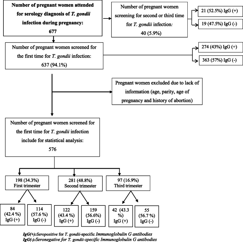
Among 677 pregnant women attended for serology diagnosis of T. gondii infection during pregnancy, only 576 who fulfilled the inclusion criteria were included in the analysis; All pregnant women who underwent pregnancy serological examination of toxoplasmosis for the first time during the studied time frame period were included. Additionally, the exclusion criteria were all pregnant women with uncompleted information (pregnant women without information of age, parity, and history of abortion) (Fig. 1).
Fig. 1.
Flowchart explaining the selection of data for the analysis with details of their distribution of pregnant women by serological status for Immunoglobulin G Toxoplasma gondii infection antibodies
Serological evaluation
Conventional ELISA
The blood samples were collected from each pregnant woman and tube-coated EDTA and were afterwards centrifuged to remove plasma and harvested sera stored at − 20 °C until tested. Serological diagnosis was done following the ELISA technique (enzyme-linked immunosorbent assay) using “Platelia Toxo” kits IgG and IgM (Biorad, France) for immunoglobulin G (IgG) and immunoglobulin M(IgM) anti-toxoplasmic research according to the manufacturer’s protocol. Briefly, the sera were diluted serially and then distributed in the microplate wells which were coated by the T. gondii antigen. After incubation of 1 h at 37 °C, a monoclonal antibody labeled with peroxidase was added to the microplate wells as the conjugate. During the second incubation of 1 h at 37 °C, the labeled antibody binds to the serum IgG captured by the T. gondii antigen. The presence of immune-complexes (T. gondii antigen, IgG antibodies to T. gondii) and anti-IgG conjugate was demonstrated by the addition in each well of an enzymatic development solution. The optical density reading obtained with a spectrophotometer set at 450/620 nm is proportional to the amount of IgG antibodies to T. gondii present in the sample and is converted into IU/ml using a standard curve calibrated against. The positive cut-off value of IgG antibodies was defined at the upper limit of 9IU/ml. For the search of specific IgM antibodies, the immunocapture technique was performed. Anti-human μ-chain antibodies are coated on wells of the microplate. A mixture of the T. gondii antigen and the monoclonal anti-T. gondii antigen antibody labeled with peroxidase was used as the conjugate. The optical densities (OD) were obtained with a spectrophotometer set at 450/620 nm after stopping the reaction with a sulfuric acid solution. The cut-off value (CO) corresponds to the mean value of the OD of the cut-off control duplicates. The IgM results were calculated qualitatively, and it is expressed by the ratio of samples OD and CO. The sample was considered positive if the ratio was ≥ 1.
Avidity ELISA
All IgG- and IgM-positive samples included in the study were analyzed for IgG avidity at the first evaluation at a gestational age ≤ 20 weeks by the kit “Platelia Toxo avidity, BioRad,” according to the manufacturer’s protocol. The principle of this method relies on the measurement of the avidity of the IgG antibodies to T. gondii. The use of an agent as urea dissociating the link antigen/antibody in parallel with the usual technique of IgG antibodies measurement allows comparison of the optical density (OD) obtained after dissociating agent action and OD obtained without dissociating agent action. The avidity is considered low when the antigen/antibody link is easily dissociated. The optical density reading obtained with a spectrophotometer set at 450/620 nm. The diagnostic value was defined as avidity index (AI). The avidity index was determined as the following criteria: A low index of avidity below 0.4 does not exclude a recent primary infection of less than 20 weeks while high index avidity or equal to 0.5 can be excluded. In case of intermediate index of avidity (0.4 ≤ AI < 0.5), an assay on a second sample was recommended.
Statistical analysis
The data input was done on Microsoft Office Excel 2010, and the analysis was performed using EpiInfo (ver. 2007 CDC, USA) Software. A descriptive analysis of the data was made to identify the characteristics of the different variables studied in EpiInfo. The chi-square test (bivariate test) from EpiInfo was used to determine the associated seroprevalence of T. gondii. Statistical significance was set to a value of p < 0.05.
Results
Demographic characteristics of pregnant women
In this retrospective study, among 637 pregnant women aged between 16 and 46 years were screened for the first time for T. gondii infection, only 576 were included in analysis, and they have complete information about age, parity, and history of abortion. The average age was 29.4 ± 6.3 years. The most of pregnant women were in the age groups 16–25 years and 26–30 years. We note that the majority of women screened were nulliparous 47.8% (275/576) followed by primiparous 45.1% (260/576) and multiparous women 41(7.1%). About 34.3% (198/576) of the pregnant women were in their first trimester, and 48.8% (281/576) and 16.9% (97/576) in the second and third trimesters, respectively (Table 1).
Table 1.
Sociodemographic characteristics of pregnant women
| Characteristic | Numbers | (%) | 95% CI* |
|---|---|---|---|
| Age ( years) mean: 29.4 ± 6.3 | |||
| 16–25 | 189 | 32.8 | 29.0–36.8 |
| 26–30 | 144 | 25.0 | 21.6–28.8 |
| 31–35 | 138 | 24.0 | 20.6–27.7 |
| 36–46 | 105 | 18.2 | 15.2–21.7 |
| Age of pregnancy | |||
| First trimester | 198 | 31.1 | 27.5–34.9 |
| Second trimester | 281 | 44.1 | 40.2–48.1 |
| Third trimester | 97 | 15.2 | 12.6–18.3 |
| Parity | |||
| Nulliparous | 275 | 47.8 | 47.7–51.9 |
| Primiparous (1 P**) | 260 | 45.1 | 92.9–29.7 |
| Multiparous (≥ 2P** | 41 | 7.1 | 100.0–30.2 |
| History of abortion | |||
| No abortion | 446 | 77.4 | 73.8–80.7 |
| One abortion | 95 | 16.5 | 13.6–19.8 |
| Two or more abortion | 35 | 6.1 | 4.3–8.4 |
CI confidence interval ** : Number of pregnancy .≥ 2P**: equal or more than two pregnancy
Serological status of pregnant women
According to the serological status for T. gondii infection, among 677 pregnant women, 94.1% (637 /677) [95%CI 92.0–95.7%] were serologically screened for the first time and therefore ignored their serological status, and 5.9% (40/677) [95%CI 4.0–9.0%] were screened for the second or third time (Fig. 1).
The results showed that the T. gondii seroprevalence of specific IgG was 43% (274/637) [95% CI 39.1–47.0%] while 57% (363/637) [95% CI 53.0–60.9%] were seronegative for anti-T. gondii-specific IgG antibodies. Furthermore, the percentage of T. gondii IgM-positive antibodies were 3.9% (25/637) [3.0–6.3%].
Among 25 pregnant women who had a positive IgG and IgM T. gondii infection during pregnancy, 18 cases are in their first month of pregnancy and are submitted to the avidity test. The results of the avidity test are shown in Table 2. Anti-T. gondii IgG avidity in the majority of patients studied 83.3% (15/18) had high avidity while a low avidity was detected in two patients (Table 2).
Table 2.
Comparison of results of IgM ELISA with IgG avidity test in 18 serum samples taken from pregnant women in Rabat region in Morocco
| IgG Avidity ELISA (%) | |||
|---|---|---|---|
| Low | Borderline | Higher | |
| IgG and IgM positive | 2 (11.1%) | 1 (5.6%) | 15 (83.3%) |
Risk factors associated with T. gondii infection
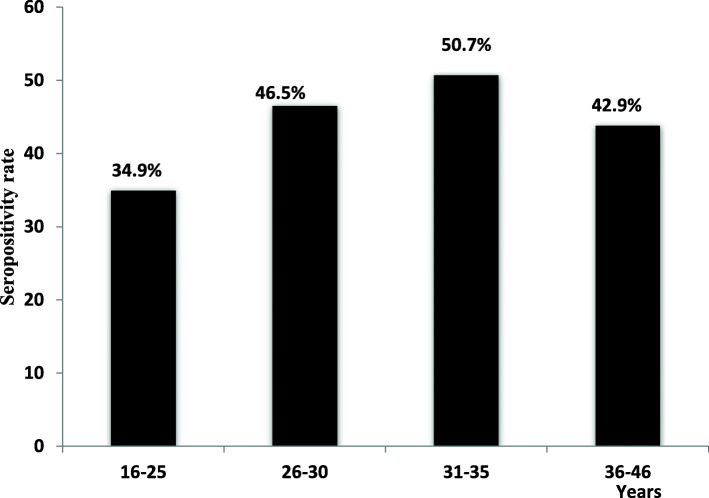
Seroprevalence of the parasite in the pregnant women based on various age groups, parity, and history of abortion is shown in Table 3. The results of bivariate analysis of age associated with seropositivity showed a significant difference in both the parasitic infection was more prevalent in the age group 31–35 years (50.7%) (70/138) followed by age group 26–30 years 46.5% (67/144) and 36–46 years 42.9% (56/105). The lowest percentage of T. gondii infection is found for women in the age group 16–25 years with about 35.7% (Fig. 2). This result is statically significant (p value = 0.0276).
Table 3.
Seroprevalence of toxoplasmosis according to age, parity, and history of abortion among pregnant women in Rabat region
| Variable | Toxoplasma seroprevalence | X2 | p value | |
|---|---|---|---|---|
| Seropositivity, n (%) | Seronegativity, n (%) | |||
| Age | ||||
| 16–25 | 66 (34.9%) | 123 (65.1%) | 9.1283 | 0.0276* |
| 26–30 | 67 (46.5%) | 77 (53.5%) | ||
| 31–35 | 70 (50.7%) | 68 (49.3%) | ||
| 36–46 | 45 (42.9%) | 60 (57.1%) | ||
| Age of pregnancy | ||||
| First trimester | 84 (42.4%) | 114 (57.6%) | .0495 | 0.9756 |
| Second trimester | 122 (43.4%) | 159 (56.6%) | ||
| Third trimester | 42 (43.3%) | 55 (56.7%) | ||
| Paritiy | ||||
| Nulliparous | 110 (40.0%) | 165 (60.0%) | 2.5627 | 0.2777 |
| Primparous (1 Pa) | 117 (45.0%) | 143 (55.0%) | ||
| Multiparous (≥ 2Pa | 21 (51.2%) | 20 (48.8%) | ||
| No abortion | 188 (42.2%) | 258 (57.8%) | .7715 | 0.6799 |
| History of abortion | ||||
| One abortion | 43 (45.3%) | 52 (54.7%) | ||
| Two or more abortion | 17 (48.6%) | 18 (51.4%) | ||
*NS statistically significant, aNumber of pregnancy
Fig. 2.
Distribution of pregnant women by serostatus for IgG T. gondii antibodies in various age group (years)
Multiparous women are the most immune for T. gondii infection with 51.2% (21/41), followed by primiparous 45% (117/260) and nulliparous 40% (110/275) (p value = 0.2777).
Regarding the history of abortion, among women who have had more than one abortion, 48.6% (17/35) are found to be positive for T. gondii infection compared to the women who had no abortion (42.2%) (188/446). Accordingly, even though a total IgG seropositivity was increased in pregnant women with a history of abortion, there are no significant associations observed between IgG seropositivity and the number of abortions (p value = 0.6799). On the other hand, a total IgG seropositivity rate was not more varied with the age of pregnancy (Table 3).
Discussion
In Morocco, there are very few documented data on T. gondii seroprevalence [11]. Adequate information on the prevalence and transmission routes would provide proper risk assessment options for pregnant women and would be helpful in the planning and implementation of control and preventive strategies for toxoplasmosis disease.
The T. gondii seroprevalence among pregnant women in the study area was 43%. Comparing the levels of seropositive IgG antibodies obtained in our study with those in previous studies conducted in 2007 and 2014, which were 51% and 47%, respectively, in the same region studied [11], we noticed a decrease in the seroprevalence during the last 5 years. A previous study showed that the majority of pregnant women with anti-toxoplasma antibodies have continuous contact with the soil in activities such as gardening, agricultural activities, or other activities [12]. Therefore, the noticeable decline in the seroprevalence may be due to the regression of contact with the soil. In addition, changes in lifestyle that are related to development, hygiene measures, and higher levels of education can contribute to reducing this prevalence.
When we compared our findings to those of previous studies reported in different cities in Morocco, we found that T. gondii seroprevalence in these regions continues to be higher than that in cities at lower altitudes and with higher humidity in Morocco, such as Fes, where the prevalence is 39.7% [13]. It is also higher than those reported in Nador, Tetouan, and Kenitra, where the seroprevalences are 34.3%, 42%, and 37.7%, respectively. However, the current finding indicates that the seroprevalence remains lower than that reported in Essaouira (48%) [14]. In addition, our results remain close to those reported in the two neighboring Maghreb countries: Tunisia (45.6%) [15] and Algeria (47.8%) [16]. This could be explained by the fact that Morocco, Tunisia, and Algeria share the same culinary habits, cultural habits, and climatic conditions. However, the results of other surveys show a lower and higher prevalence in different areas in Africa, in Europe, and in Asia: 27% in Sudan [17], 35.6% in Ethiopia [18], 44% in Tanzania [19], 47% in Benin [20], 13.8% in Italy [21], 31.5% in Austria [22], 55.8% in Romania [23], 31% in Turkey [24], 33% in Iran [25], 34.5% in Pakistan [26], 82.6% in Lebanon [27], and 35.8% in Peru [28]. This variation in the rate of T. gondii infection between countries and regions could be attributed to dietary habits, health standards, lack of awareness of disease transmission, and the socioeconomic level. Improvements in hygiene conditions and farming systems, together with increased socioeconomic levels, have led to a declining seroprevalence in most industrialized countries [10].
On the other hand, our findings indicate that 57% are seronegative and are therefore likely to be infected during pregnancy, so serological monitoring is required during each trimester with prophylaxis to follow throughout the pregnancy. However, we noted that only 5.9% of all pregnant women were undergoing T. gondii antibody screening, with 47.5% seronegativity, and among the seronegative cases, 15.2% were in their third trimester when they underwent screening for T. gondii antibodies for the first time. Hence, in Morocco, no systematic prenatal toxoplasmosis screening has been set up. The health system does not have a monitoring program for toxoplasmosis. Therefore, there is no follow-up of T. gondii seronegative pregnant women to properly control the risk of infection with congenital toxoplasmosis until delivery. Moreover, serological screening for T. gondii infection in our country is still considered a biological test not required by doctors. Thus, the serological diagnosis of T. gondii infection is not always requested at the first prenatal visit, and the follow-up of pregnant women is poorly conducted or is not conducted at all. Besides, the lack of knowledge of toxoplasmosis among pregnant women has been reported previously as a main risk factor for contracting disease. Only 5.9% were aware of toxoplasmosis. The lack of knowledge may put unaware women to not follow-up the serology and put them in a high-risk group, susceptible to contract T. gondii infection during pregnancy leading to an acute infection [12]. On the other hand, a recent study reported a moderate knowledge of toxoplasmosis among health professional including physicians and nurses on this parasitic infection and thus failed to provide sufficient information to the pregnant women [29]. Hence, healthcare professionals should advise patients to follow-up the serology specially for the seronegative pregnant women in order to avoid seroconversion during pregnancy.
Indeed, serological screening is considered the key element in the prevention of congenital toxoplasmosis. In fact, monthly serology for seronegative pregnancy is mandatory until delivery. The interpretation of the serological results must be carried out by a competent and experienced biologist, since from the conclusions of the serological analysis, all subsequent prenatal and postnatal behavior is ensured [30]. Even when primary infection occurs during pregnancy, early diagnosis and treatment can reduce the frequency and severity of the disease in neonates [31].
The current study reveals that among seropositive women, the majority were found to have a chronic or past infection based on serology of IgM antibodies. However, 18 (2.8%) of the 637 pregnant women consulting in their first trimester were susceptible to a recent infection, and only two (2) subjects were positive for IgM with a low avidity using the avidity IgG method. This test excluded 83.3% of pregnant women. Therefore, IgM positivity does not always predict an acute infection. IgG avidity tests in addition to IgM and IgG antibody testing should be performed in the first trimester of pregnancy. The avidity test seems necessary to confirm or exclude an infection of less than 4 months in pregnancy, which is likely to lead to congenital toxoplasmosis [32, 33].
In this study, the risk of contracting T. gondii increased with the age of the pregnant woman; increased odds of having T. gondii infection were observed in the groups of pregnant women aged ≥ 25 years compared to the group of pregnant women aged 16–25 years. This finding is consistent with the studies conducted in Fes [13] and in Rabat [34], where the prevalence did not increase linearly with age. In contrast, our results are not in agreement with those found in France [35], where a positive correlation between seroprevalence and age was found; our results are probably due to the low number of women in the sample of our study.
Conclusion
This descriptive study provides key and update baseline data about the T. gondii seroprevalence among pregnant women in the region of Rabat, Morocco. In this study, it is noted that the number of non-immunized pregnant women increases during the last years in the region studied. Therefore, the risk of fetal infection has increased because the possibility of seroconversion during pregnancy is higher among pregnant women. On the other hand, we report that few pregnant women have followed-up of toxoplasmosis during pregnancy especially among the number of non-immunized pregnant. Undoubtedly, a national program of screening and surveillance of seronegative pregnant women is required.
Limitations
Our study presents some limitations mainly due to the fact that these data are derived from a retrospective evaluation of pregnant women. For this reason, important data, such as possible risk factors for T. gondii infection (consumption of uncooked meat, contact with cats, or other animals…) are not available. Furthermore, some factors could explain the regression of T. gondii IgG exposure; however, these factors were not ascertained in this study and constitute one of the limitation of the survey. Hence, there is a need for a national sample survey estimating the real potential burden of this infection on maternal and its impact on fetal health. Despite these limitations, we have for the first time been able to report the number of pregnant women who follow-up the screening of toxoplasmosis.
Acknowledgements
The authors wish to thank all pregnant women who attended in parasitology laboratory in the National Institute of Hygiene in Rabat for toxoplasmosis.
Abbreviations
- AI
Avidity index
- CO
Cut-off value
- ELISA
Enzyme-linked immunosorbent assay
- EDTA
Ethylenediaminetetra-acetic acid
- IgG
Immunoglobulin G
- IgM
Immunoglobulin M
- OD
Optical density
- T. gondii
Toxoplasma gondii
Authors’ contributions
LM has conceptualized the study and significantly contributed to the data collection, analysis, and write up. SA and TZ supervised the study and have supported the implementation of this study and provided feedback during the write up. DO collected data. PF contributed to the critical review of the manuscript for intellectual content of the study. The authors reviewed and approved the final manuscript.
Funding
There is no funding for this research.
Availability of data and materials
The datasets used during the current study are available from the corresponding author on reasonable request.
Declarations
Ethics approval and consent to participate
This survey utilized a retrospective analysis routine reports which were anonymized. All data used in this study cannot be directly associated with any individual. Therefore, no consent or ethical clearance is applicable for this study. The confidentiality of the details of the subjects was assured for all pregnant women followed up in our laboratory.
Consent for publication
Not applicable
Competing interests
The authors declare that they have no competing interests.
Footnotes
Publisher’s Note
Springer Nature remains neutral with regard to jurisdictional claims in published maps and institutional affiliations.
References
- 1.Yera H, Paris L, Bastien P, Candolfi E. Diagnostic biologique de la toxoplasmose congénitale. Rev Francoph des Lab. 2015;470:65–72. [Google Scholar]
- 2.Food and Agriculture Organization of the United Nations/World Health . Multicriteria-based ranking for risk management of food-borne parasites. 2014. p. 302. [Google Scholar]
- 3.Berthélémy S. Toxoplasmose et grossesse. Actual Pharmacol. 2014;53(541):43–45. [Google Scholar]
- 4.Dalgıç N. Congenital toxoplasma gondii infection. Marmara Med J. 2008;21(1):89–101. [Google Scholar]
- 5.Helieh SOZ. Fetomaternal and ediatric toxoplasmosis. J Pediatr Infect Dis. 2017;12(4):1–7. doi: 10.1055/s-0037-1603942. [DOI] [PMC free article] [PubMed] [Google Scholar]
- 6.Halonen SK, Weiss LM. Toxoplasmosis. Handb Clin Neurol. 2013;114(08):125–145. doi: 10.1016/B978-0-444-53490-3.00008-X. [DOI] [PMC free article] [PubMed] [Google Scholar]
- 7.Dubey JP, Lago EG, Gennari SM, Su C, Jones JL. Toxoplasmosis in humans and animals in Brazil: high prevalence, high burden of disease, and epidemiology. Parasitology. 2012;139(11):1375–1424. doi: 10.1017/S0031182012000765. [DOI] [PubMed] [Google Scholar]
- 8.Pappas G, Roussos N, Falagas ME. Toxoplasmosis snapshots: global status of toxoplasma gondii seroprevalence and implications for pregnancy and congenital toxoplasmosis. Int J Parasitol. 2009;39(12):1385–1394. doi: 10.1016/j.ijpara.2009.04.003. [DOI] [PubMed] [Google Scholar]
- 9.Dar FK, Alkarmi T, Uduman S, Abdulrazzaq Y, Grundsell H, Hughes P. Gestational and neonatal toxoplasmosis: regional seroprevalence in the United Arab Emirates. Eur J Epidemiol. 1997;13(5):567–571. doi: 10.1023/A:1007392703037. [DOI] [PubMed] [Google Scholar]
- 10.Robert-Gangneux F, Dardé ML. Epidemiology of and diagnostic strategies for toxoplasmosis. Clin Microbiol Rev. 2012;25(2):264–296. doi: 10.1128/CMR.05013-11. [DOI] [PMC free article] [PubMed] [Google Scholar]
- 11.Laboudi M. Review of toxoplasmosis in Morocco: seroprevalence and risk factors for toxoplasma infection among pregnant women and HIV-infected patients. Pan Afr Med J. 2017;27(269):1–6. doi: 10.11604/pamj.2017.27.269.11822. [DOI] [PMC free article] [PubMed] [Google Scholar]
- 12.Laboudi M, El Mansouri B, Sebti F, Amarir F, Coppitiers Y, Rhajaoui M. Facteurs de risque d’une sérologie toxoplasmique positive chez la femme enceinte au Maroc. Parasite. 2009;16:1–2. doi: 10.1051/parasite/2009161071. [DOI] [PubMed] [Google Scholar]
- 13.Tlamcani Z, Yahyaoui G, Mahmoud M. Prevalence of immunity to toxoplasmosis among pregnant women in university hospital center Hassan II of FEZ city (Morocco) Acta Medica Int. 2017;4(1):43. doi: 10.5530/ami.2017.4.8. [DOI] [Google Scholar]
- 14.Errifaiy H. Evaluation des connaissances, des comportements et des statuts immunitaires des femmes enceintes par rapport à la toxoplasmose : Enquête épidémiologique dans la région Essaouira-Safi . Faculté de médecine et de pharmacie de Marrakech, Thèse N° X/2014. 2014.
- 15.Ben Abdallah R, Siala A, Bouafsoun A, Maatoug R, Souissi O, Aoun K. Dépistage de la toxoplasmose materno-foetale étude des cas suivis à l’Institut Pasteur de Tunis (2007–2010) Bull Soc Pathol Exot. 2013;106(2):108–112. doi: 10.1007/s13149-013-0287-8. [DOI] [PubMed] [Google Scholar]
- 16.Messerer L, Bouzbid S, Mokhtar B. Seroprevalence of toxoplasmosis in pregnant women in Annaba, Algeria. Rev Epidemiol Sante Publique. 2014;62:160–165. doi: 10.1016/j.respe.2013.11.072. [DOI] [PubMed] [Google Scholar]
- 17.Madinna M, Fathy F, Mirghan A, Mohamed MM, Muneer MS, Ahmed AE, et al. Prevalence and risk factors profile of seropositive toxoplasmosis gondii infection among apparently immunocompetent Sudanese women. BMC Res Notes. 2019;12(1):1–6. doi: 10.1186/s13104-018-4038-6. [DOI] [PMC free article] [PubMed] [Google Scholar]
- 18.Teweldemedhin M, Gebremichael A, Geberkirstos G, Hadush H, Gebrewahid T, Asgedom SW, et al. Seroprevalence and risk factors of toxoplasma gondii among pregnant women in Adwa district, northern Ethiopia. BMC Infect Dis. 2019;19(327):1–9. doi: 10.1186/s12879-019-3936-0. [DOI] [PMC free article] [PubMed] [Google Scholar]
- 19.Eliakimu P, Kiwelu I, Mmbaga B, Nazareth R, Sabuni E, Maro A, et al. Toxoplasma gondii seroprevalence among pregnant women attending antenatal clinic in northern Tanzania. Trop Med Health. 2018;46(39):1–8. doi: 10.1186/s41182-018-0122-9. [DOI] [PMC free article] [PubMed] [Google Scholar]
- 20.Tonouhewa ABN, Amagbégnon R, Atchadé SP, Hamidović A, Mercier A, Dambrun M, et al. Seroprevalence of toxoplasmosis among pregnant women in Benin: meta-analysis and meta-regression. Bull Soc Pathol Exot. 2019;112(2):79–89. doi: 10.3166/bspe-2019-0078. [DOI] [PubMed] [Google Scholar]
- 21.Fanigliulo D, Marchi S, Montomoli E, Trombetta CM. Toxoplasma gondii in women of childbearing age and during pregnancy : seroprevalence study in central and southern Italy from 2013 to 2017. Parasite. 2020;2:27–30. doi: 10.1051/parasite/2019080. [DOI] [PMC free article] [PubMed] [Google Scholar]
- 22.Berghold C, Herzog SA, Jakse H, Berghold A. Prevalence and incidence of toxoplasmosis : a retrospective analysis of mother-child examinations, Styria , Austria , 1995 to 2012. Eurosurveillance. 2016;21(33):1–7. doi: 10.2807/1560-7917.ES.2016.21.33.30317. [DOI] [PMC free article] [PubMed] [Google Scholar]
- 23.Olariu TR, Ursoniu S, Hotea I, Dumitrascu V, Anastasiu D, Lupu MA. Seroprevalence and risk factors of toxoplasma gondii infection in pregnant women from Western Romania. Vector-Born Zoonotic Dis. 2020:1–5. 10.1089/vbz.2019.2599. [DOI] [PubMed]
- 24.Tanrıverdi EÇ, Kadıoğlu BG, Alay H, Özkurt Z. Retrospective evaluation of anti-toxoplasma gondii antibody among first trimester pregnant women admitted to Nenehatun maternity hospital between 2013-2017 in Erzurum. Turkiye Parazitol Derg. 2018;42:101–105. doi: 10.5152/tpd.2018.5462. [DOI] [PubMed] [Google Scholar]
- 25.Mousavi-Hasanzadeh M, Sarmadian H, Ghasemikhah R, Didehdar M, Shahdoust M, Maleki M, and Taheri M. Evaluation of toxoplasma gondii infection in western Iran: seroepidemiology and risk factors analysis. Trop Med Health. 2020;48(35):1-7. [DOI] [PMC free article] [PubMed]
- 26.Shah F, Hasnain J, Muhammad H, Muhammad TK, Farman UJ, Tanveer I, et al. Seroprevalence of human toxoplasma gondii infection among pregnant women in Charsadda, KP, Pakistan. J Parasit Dis. 2018;42(4):554–8 [DOI] [PMC free article] [PubMed]
- 27.Nahouli H, El Arnaout N, Chalhoub E. Anastadiadis E, El Hajj H. Seroprevalence of anti-toxoplasma gondii antibodies among Lebanese pregnant women. Vector-Born Zoonotic Dis. 2017;17(12):785–90. [DOI] [PubMed]
- 28.Silva-Díaz H, Arriaga-deza EV, Failoc-Rojas VE, Alarcón-Flores YR, Rojas-Rojas SY, Becerra-Gutiérrez LK, et al. Seroprevalence of toxoplasmosis in pregnant women and its associated factors among hospital and community populations in Lambayeque, Peru. Rev Soc Bras Med Trop. 2020;53(e201901):1–6. doi: 10.1590/0037-8682-0164-2019. [DOI] [PMC free article] [PubMed] [Google Scholar]
- 29.Laboudi M, AitHamou S, Mansour I, Hilmi I, Sadak A. The first report of the evaluation of the knowledge regarding toxoplasmosis among health professionals in public health centers in Rabat, Morocco. Trop Med Health. 2020;48(17):1–8. doi: 10.1186/s41182-020-00208-9. [DOI] [PMC free article] [PubMed] [Google Scholar]
- 30.Villard O, Jung-Etienne J, Cimon B, Franck J, Fricker-Hidalgo H, Godineau N, et al. Le réseau du Centre national de Référence de la toxoplasmose. Sérodiagnostic la toxoplasmose en. 2011;2010:1–7. [Google Scholar]
- 31.Hernandez JR, Bonobio MV, Perea E. IgG avidity for toxoplas¬mosis detection by the liaison system. Clin Microbiol Infect. 2003;9:254. doi: 10.1046/j.1469-0691.2003.00740.x. [DOI] [Google Scholar]
- 32.Robert-Gangneaux F, Viieljeuf C, Tourte-Schaefer C, Depouy-Camet J. Apport de l’avidité des anticorps dans la datation d’une séroconversion toxoplasmique. Ann Biol Clin (Paris) 1998;56(5):586–589. [PubMed] [Google Scholar]
- 33.Laboudi M, Sadak A. Serodiagnosis of toxoplasmosis: the effect of measurement of IgG avidity in pregnant women in Rabat in Morocco. Acta Trop. 2017;172:139–142. doi: 10.1016/j.actatropica.2017.04.008. [DOI] [PubMed] [Google Scholar]
- 34.Laboudi M, El Mansouri B, Rhajaoui M. The role of the parity and the age in acquisition of toxoplasmosis among pregnant women in Rabat - Morocco. Int J Innov Appl Stud. 2014;6(3):488–492. [Google Scholar]
- 35.Berger F, Goulet V, Le Strat Y, Desenclos JC. Toxoplasmosis among pregnant women in France: risk factors and change of prevalence between 1995 and 2003. Rev Epidemiol Sante Publique. 2009;57(4):241–248. doi: 10.1016/j.respe.2009.03.006. [DOI] [PubMed] [Google Scholar]
Associated Data
This section collects any data citations, data availability statements, or supplementary materials included in this article.
Data Availability Statement
The datasets used during the current study are available from the corresponding author on reasonable request.


