Abstract
The parotid duct (Stenson's duct) can be damaged during traumatic injuries and surgical interventions. Early diagnosis and treatment of a duct injury is of great importance because complications such as sialocele and salivary gland fistula may develop if the duct is not surgically repaired. We think that the feeding tube is an ideal material in the parotid duct repair because of its technical characteristics, availability, and low cost. In this article, we described the use of a feeding tube for the treatment of a parotid duct rupture in a facial stab wound laceration, as it is a low-cost and easy-to-access material readily available in every operating room.
Keywords: Feeding tube, parotid duct, repair
INTRODUCTION
The parotid duct can be damaged in traumatic injuries and surgical interventions. Early diagnosis and treatment of a duct injury is of great importance because complications such as sialocele and salivary gland fistula may develop if the duct is not surgically repaired.[1] A guidance material is often needed to expose and repair the damaged duct. In the literature, a great variety of materials such as epidural catheter, urethral catheter, double-J catheter, catgut, and Vitallium wire have been used as an intraductal stent after repair.[2,3,4,5]
In this article, we described the use of a feeding tube for the treatment of parotid duct rupture in a facial stab wound laceration, as it is a low-cost and easy-to-access material in every operating room.
CASE STUDY/TECHNIQUE
A 24-year-old male patient with a sutured stab wound presented to the Department of Oral and Maxillofacial Surgery, Rajaie Trauma Center, Shiraz, Iran, with a left buccal swelling [Figure 1]. Physical examination of the patient revealed laceration of the buccal branch of the facial nerve as evidenced by the inability to blow the cheek. Aspiration examination of the swelling revealed that it was saliva [Figure 2].
Figure 1.
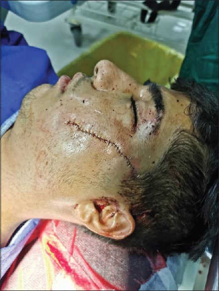
A 24-year-old male patient with stab wound laceration sutured, which saliva collection made swelling
Figure 2.
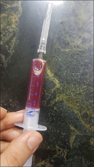
Saliva aspiration
Under hypotensive anesthesia, the sutures were removed [Figure 3] and the exposed area was explored and copiously irrigated. The distal part of the parotid duct was found in the left malar region, and the feeding tube of size 6 (green) was inserted into the parotid papilla at the level of the second molar tooth in the mouth [Figure 4].
Figure 3.
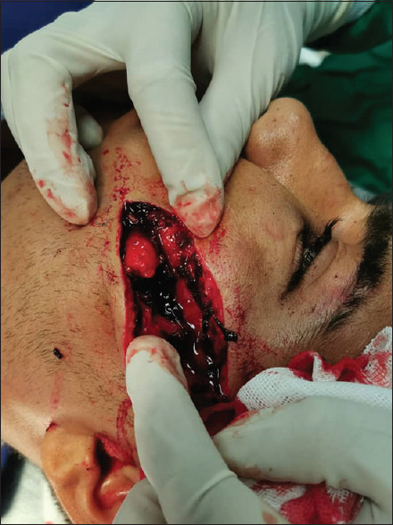
The sutured laceration reopened
Figure 4.
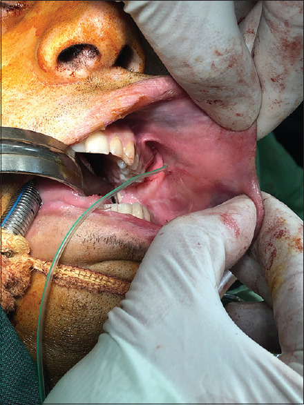
Distal part of the parotid duct was found intraorally, and the feeding tube was inserted
The end of the tube was seen to be coming out of the laceration line; the proximal end of the lacerated parotid duct was found both by meticulous exploration of the site and by milking the parotid gland to see where saliva exited. The proximal end of the lacerated parotid duct [Figure 5] was subsequently cannulated, using the tube as an intraductal stent.
Figure 5.
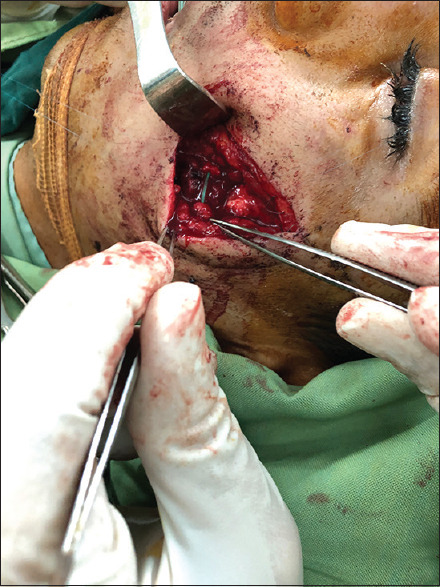
Proximal part of the parotid duct was found and connected to the feeding tube
The parotid duct was repaired with prolene 7.0 suture over the stent, in accordance with the surgical technique [Figure 6]. The excess part of the tube was cut out, leaving the end of the silicon tube in the mouth fixed to the buccal mucosa at two points, and the incisions in the skin were repaired and compressive dressing was applied to reduce the chance of hematoma formation. The patient was discharged 3 days postoperatively with instructions and antibiotics.
Figure 6.
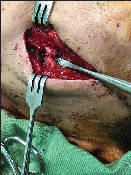
The parotid duct was sutured with prolene 7.0
Postoperatively, normal salivary flow was noted through the tube with no complications [Video 1]. By the third week, the patient was decannulated.
DISCUSSION
Although parotid duct injuries are often penetrating injuries, they may also develop as a consequence of tumor excisions and blunt traumas or as an iatrogenic complication.[6] The most common and difficult-to-treat complications observed in parotid duct injuries are sialocele and parotid gland fistula.[1] Many procedures intended for the prevention of complications include follow-up with aspiration and compressive dressing, primary saturation of the duct, parasympathetic denervation, ligation of the duct, fistulation of the duct into the oral cavity, superficial or total parotidectomy, and radiotherapy.[1,7,8,9] Because of the availability of advanced surgical techniques and suture materials, as well as the good results of surgical repair, the duct is recommended to be repaired in all cases where possible.
Stent use is preferred and has been widely accepted in direct repair because it not only prevents the suture from passing through the rear wall during repair but also prevents the duct from becoming collapsed and obstructed during recovery.[3,10] In literature, a great variety of materials such as epidural catheter, urethral catheter, double-J catheter, catgut, and Vitallium wire have been used as an intraductal stent after repair.[2,3,4,5] The feeding tube that we used had ideal technical characteristics, with its elasticity as well as noncollapsible structure. The space inside the tube allows for uninterrupted drainage of secretion from the salivary gland, during the postoperative period. In this way, it ensures the continuation of the salivary flow; thus, it supports the repair line.
CONCLUSIONS
In this case report, we described a successful use of an easy, yet efficient technique to manage the parotid duct injury using a feeding tube. We conclude that the use of a feeding tube in the repair of the parotid duct during traumatic facial and/or parotid injuries is a valuable technique to be used in surgical practice as it can easily be found in every operating room and is extremely cost-effective with ideal technical characteristics.
Financial support and sponsorship
Nil.
Conflicts of interest
There are no conflicts of interest.
Declaration of patient consent
The authors certify that they have obtained all appropriate patient consent forms. In the form the patient(s) has/have given his/her/their consent for his/her/their images and other clinical information to be reported in the journal. The patients understand that their names and initials will not be published and due efforts will be made to conceal their identity, but anonymity cannot be guaranteed.
Video Available on: www.amsjournal.com
REFERENCES
- 1.Singh M, Rangaswamy S, Choudry S. Parotid sialocele and fistulae: Current treatment options. Int J Contemp Dent. 2011;2:9–12. [Google Scholar]
- 2.Sujeeth S, Dindawar S. Parotid duct repair using an epidural catheter. Int J Oral Maxillofac Surg. 2011;40:747–8. doi: 10.1016/j.ijom.2011.02.003. [DOI] [PubMed] [Google Scholar]
- 3.Sparkman RS. Laceration of parotid duct further experiences. Ann Surg. 1950;131:743–54. doi: 10.1097/00000658-195005000-00011. [DOI] [PMC free article] [PubMed] [Google Scholar]
- 4.Aloosi SN, Khoshnaw N, Ali SM, Muhammad BA. Surgical management of Stenson's duct injury by using double J stent urethral catheter. Int J Surg Case Rep. 2015;17:75–8. doi: 10.1016/j.ijscr.2015.10.036. [DOI] [PMC free article] [PubMed] [Google Scholar]
- 5.Newman SC, Seabrook DB. Management of injuries to Stensen's duct. Ann Surg. 1946;124:544–56. [PubMed] [Google Scholar]
- 6.Steinberg MJ, Herréra AF. Management of parotid duct injuries. Oral Surg Oral Med Oral Pathol Oral Radiol Endod. 2005;99:136–41. doi: 10.1016/j.tripleo.2004.05.001. [DOI] [PubMed] [Google Scholar]
- 7.van Sickels JE. Management of parotid gland and duct injuries. Oral Maxillofac Surg Clin North Am. 2009;21:243–6. doi: 10.1016/j.coms.2008.12.010. [DOI] [PubMed] [Google Scholar]
- 8.White JE. Primary repair of parotid duct injuries. J Natl Med Assoc. 1950;42:232–4. [PMC free article] [PubMed] [Google Scholar]
- 9.Parekh D, Glezerson G, Stewart M, Esser J, Lawson HH. Post-traumatic parotid fistulae and sialoceles.A prospective study of conservative management in 51 cases. Ann Surg. 1989;209:105–11. doi: 10.1097/00000658-198901000-00015. [DOI] [PMC free article] [PubMed] [Google Scholar]
- 10.Stevenson JH. Parotid duct transection associated with facial trauma: Experience with 10 cases. Br J Plast Surg. 1983;36:81–2. doi: 10.1016/0007-1226(83)90019-x. [DOI] [PubMed] [Google Scholar]
Associated Data
This section collects any data citations, data availability statements, or supplementary materials included in this article.


