

Abstract
Introduction:
Plasma derivatives have been practiced a lot in orthopedics, burns, and sport medicine. Microneedling (MN) with platelet-rich plasma (PRP) therapy has been proven to improve the micro-circulation and thus improve hair growth. The role of concentrated growth factor (CGF) for hair growth has not been mentioned anywhere in the literature for hair growth which we tried to prove in our article by comparing it with various other studies.
Materials and Methods:
This is a retrospective randomized study involving 20 male patients whose ages ranged from 21 years to 56 years. PRP was prepared using the dual-spin method and injected after activation; post-MN, CGF gel was applied topically. Four sessions were performed, and a follow-up was done after 6 months. Statistical analysis was done using the Statistical Package for the Social Sciences software version 21 for Windows (SPSS, IBM Corp, Armonk, NY, USA). Paired t-test was used for the various comparisons.
Results:
Hair loss reduced by the end of the first month. At the end of 6 months, postfirst session, microscopic examination showed statistically significant difference in the hair count compared to those during the baseline.
Discussion:
PRP having platelet-derived growth factor and vascular endothelial growth factor acts on stem cells in the follicles, stimulating the development of new follicles and promoting neovascularization. CGF helps stimulating cell proliferation and matrix remodeling due to numerous growth factors in a concentrated form. Thus, this therapy combined helps to boost the hair growth in a very significant way.
Summary:
This study provides the preliminary evidence of efficacy of PRP along with MN and CGF in treating androgenetic alopecia by promoting angiogenesis along with vascularization and promotes hair follicles to enter and extend the anagen phase. Most of the results obtained show improved results with this therapy. A larger case study for the same can further be done for a stronger recommendation of the use of CGF for hair growth therapy further.
Keywords: 5-alpha-reductase inhibitors, androgenetic alopecia, growth factors, patterned hair loss, platelet-rich plasma
INTRODUCTION
Androgenetic alopecia (AGA) is a nonscarring progressive miniaturization of the hair follicle with a usually characteristic pattern distribution of hair loss in genetically predisposed men and women.[1] The degree of hair loss becomes apparent with aging. Individuals with higher genetic predisposition to hair loss, start losing their hair earlier and to a greater degree than those with a lesser predisposition. By the age of 25, approximately 20% of men will shows the signs of hair loss which increases to 75% by the age of 60 years. Of the 75% of men, about half of these will show significant hair loss at frontal and vertex region. AGA affects up to 80% of men and 40% of women.[2] Hair loss in women occurs usually postmenopause, and they experience thinning of hair due to hair follicle's genetic programming.[3]
Minoxidil topical lotion and finasteride oral tablets are the Food and Drug Administration approved conservative treatment of hair loss.[4] Minoxidil appears to prolong the anagen while finasteride is 5-alpha reductase Type II inhibitor which reduces the conversion of testosterone to dihydrotestosterone (DHT).[4] Minoxidil originally was developed as an anti-hypertensive agent that attracted interest when its oral administration led to generalized hypertrichosis. It is available as an over-the-counter preparation, and it increases the anagen phase and promotes the survival of dermal papillary cells and increases hair follicle size.[5,6] Finasteride also promotes anagen phase by increasing hair diameter and elongating hair length.[7] It decreases serum and scalp levels of DHT and given as a dose of 1 mg daily. Decreased libido, erectile dysfunction, and decreased ejaculate volume are the common side effects with finasteride.[8] Dutasteride is Type I and II 5-alpha reductase inhibitor which has superior efficacy as compared to that of finasteride.[4] Various other forms of conservative therapy include low-level laser therapy and prostaglandin analogs.
Advocates of platelet-rich plasma (PRP) proposed the benefits which included increase in hard and soft-tissue healing which is used in specialties such as orthopedics, sport medicine, diabetic ulcers, and plastic surgery. It became so widespread that it became effectively popular for joint problems, wound healing, and alopecia.[9,10,11,12] After injecting PRP and MN, concentrated growth factor (CGF) was applied over the scalp which had growth factors, CD34+stem cells, leukocytes, and platelets which helps in the regeneration process.[13]
The aim of our study was to assess the reduction in hair fall, assessing the anagen/telogen ratio, to check the efficacy of the therapy in various grades of alopecia and check for the increase in the density of hair counts per 0.4 cm2 of scalp area from 6 months of commencement of the treatment; to know the efficacy of PRP, MN, and CGF together as a therapy and to check the difference between anagen/telogen ratio. The data reported here demonstrate the efficacy of PRP-CGF therapy without the use of any other medical or surgical treatment. Patient's satisfaction and dermascope changes have confirmed the positive results.
MATERIALS AND METHODS
Ethical clearance was taken from the Institutional Ethical Committee in accordance with Helsinki declaration. All the diagnoses were confirmed by the institution's maxillofacial surgeons with a clinical experience of at least 10 years in handling the conditions affecting scalp and hair.
The inclusion criteria included 20 OPD-based male patients with various grades of AGA who had not received any topical or systemic treatment for hair loss, were treated between July 2016 and November 2017 in our institute at Nadiad, Gujarat. The age of the patients ranged from 21 years to 56 years. Patients with alcoholism, habit of smoking, keloidal tendency, bleeding or clotting disorders, active infection, malnutrition, psoriasis or lichen planus, uncontrolled diabetes, and other forms of alopecia were excluded from the study. Various laboratory tests such as complete blood count, serum iron, serum ferritin, total iron-binding capacity, folic acid, random blood sugar, thyroid profile, lipid profile, and VDRL were carried out to exclude thyroid disorders, anemia, malnourishment, high cholesterol, and syphilis. The stage of alopecia for men was evaluated on the basis of Norwood-Hamilton scale. Hair pull test was carried out in all the patients preoperatively and 6 months post first session to assess the hair fall clinically to assure the authenticity of results. The photographs were taken of the frontal, left and right parietal and occipital region using Nikon DSLR camera, and the dermascope was used to capture the 0.4 cm2 area of these four regions. Markings were done on the scalp where PRP was to be injected such that each spot was one centimeter away from each other which meant that a square centimeter had four spots at the border.
Thirty milliliters of blood was withdrawn from the antecubital vein of the patient using scalp vein. 20 ml blood was collected in two vacuettes (labtech heparin disposable vacuette) of 10 ml each, while the remaining 10 ml was collected in vacuette without anticoagulant. The vacuette without anticoagulant was centrifuged (REMI R-4C) first using a one-step coagulation protocol: 30 s acceleration, 2 min at 2700 rpm, 4 min at 2400 rpm, 4 min at 2700 rpm, 3 min at 3000 rpm, and 36 s deceleration and stop. It forms four phases: superior phase of serum, interim phase having large and dense polymerized fibrin block, liquid phase of growth factors, white blood cells and stem cells, and lower phase having RBCs. The concentrate of the interim phase, i.e., CGF with the help of tweezers was kept in a dappen dish.
Then, the two vacuettes with heparin were centrifuged for 8 min at 2500 rpm. After centrifugation, the blood components were separated from the plasma due to the difference in the densities. The layers formed were as follows: (1) bottom layer of RBC, (2) middle layer of PRP, and (3) upper layer of platelet-poor plasma (PPP). The plasma obtained from both the vacuettes was collected in another vacuette, and 2nd spin was made. This hard spin was for 12 min at 3000 rpm. After the 2nd spin, the lower 1/3rd of the plasma (PRP) was collected in a 10 ml syringe while the upper 2/3rd of plasma (PPP) was discarded. Total amount of PRP yielded was about 4–5 ml. This PRP was then loaded in the insulin syringes (0.9 ml/syringe). Post loading of PRP, 0.1 ml of calcium gluconate was added to 0.9 ml of PRP for its activation. The platelet concentration in PRP became 5–7 times as that in whole blood. During the spins, ring block using the combination of lignocaine and adrenaline in the concentration of (1:200,000) was injected to anesthetize the scalp and make it ready for the injections.
PRP was injected in the androgen-related areas of the scalp using BD insulin syringes till a depth of 2–3 mm. 0.2–0.3 ml of PRP was injected every centimeter. After the injections are over with, MN was done using dermaroller having 540 needles of 1.5 mm length which was rolled over the region where the injections were given in the scalp. CGF was applied which would get absorbed from the micro-punctures developed post MN. The protocol used here contained four sessions at week 0, week 4, week 8, and week 16. Final follow-up was done on week 24th. Photographs using dermascope were taken at the end of 24th week and compared with the preoperative dermascope photographs. Macroscopic photographs using Nikon DSLR were taken in every sitting and the follow-up to compare the gross changes between the sittings or chances between the preoperative condition and condition during follow-up. Furthermore, hair pull test was performed and compared with the preoperative results of the same to assess the change in hair fall.
Statistical analysis was performed using the Statistical Package for the Social Sciences software version 21 for Windows (SPSS, IBM Corp Armonk, NY, USA.). Normality of the quantitative variables was tested by the Kolmogorov–Smirnov test. Hair density was expressed as mean ± standard deviation (SD), whereas the difference in hair density between the preoperative and postoperative follow up findings were done using the paired Student's t-test. P < 0.05 was considered statistically significant.
RESULTS
A total of 22 patients (all males) enrolled for the study, but there was a drop out of two patients as they did not complete the therapy protocol and thus were not included in the study. Hence, this study included only 20 male patients. In Table 1, a brief up of patient's characteristics is given. The mean age of patients was 36.65 years (21–56). Norwood Hamilton classification was used to grade the alopecia in all the patients where one patient suffered from Grade I, four patients from Grade II, four patients from Grade III, two patients from Grade IV, three patients from Grade V, four patients from Grade VI, and two patients suffered from Grade VII AGA. Five patients from the study had undergone topical Minoxidil therapy but had discontinued it since 2 years. The table also shows preoperative and postoperative hair pull test counts which has shown a remarkable decrease in the hair fall postoperatively. Only one patient was reported with increased hair fall postoperatively in the hair pull test count, while the rest others had decreased hair loss post-PRP-CGF treatment. T-test was used for comparison.
Table 1.
Patient’s characteristics
| Case | Age (years) | Alopecia grade | Hair pull test | |
|---|---|---|---|---|
| Preoperative | Postoperative | |||
| 1 | 43 | III vertex | 9 | 3 |
| 2 | 43 | IV | 5 | 2 |
| 3 | 38 | V | 4 | 0 |
| 4 | 26 | VI | 2 | 1 |
| 5 | 38 | VI | 3 | 0 |
| 6 | 56 | V A | 3 | 1 |
| 7 | 35 | VI | 1 | 2 |
| 8 | 22 | II | 2 | 0 |
| 9 | 46 | III vertex | 3 | 0 |
| 10 | 31 | II | 3 | 2 |
| 11 | 21 | II | 7 | 2 |
| 12 | 48 | I | 4 | 1 |
| 13 | 38 | VII | 1 | 0 |
| 14 | 39 | III | 3 | 1 |
| 15 | 29 | II A | 1 | 0 |
| 16 | 36 | IV | 3 | 2 |
| 17 | 30 | V | 3 | 0 |
| 18 | 42 | VII | 1 | 0 |
| 19 | 42 | VI | 2 | 1 |
| 20 | 30 | III | 5 | 2 |
Statistical analysis was based upon the following criteria:
Frontal, left lateral, right lateral, occipital, and total of all the four areas were taken and compared
Comparison was done in the hair growth in patients who developed hair loss at or above 25 years of age to the patients developing hair loss below 25 years of age
Hair growth in the various grades of alopecia was compared in two groups, i.e., Grade I-IV and Group V-VII
Comparison of anagen/telogen ratio was done of preoperative and postoperative 6th month records.
Comparison of the various areas of scalp for hair growth
Table 2 shows the mean and SD of hair of all the patients with respect to the frontal, left and right parietal, occipital, and the total hair counts. Thus, it shows statistically significant increase in the mean hair counts in all the respective regions discussed above irrespective of the age group and type of alopecia.
Table 2.
Descriptive statistics for all regions of scalp
| Mean | n | Standard deviation | |
|---|---|---|---|
| PRE_FRONTAL | 20.3000 | 20 | 7.80081 |
| POST_FRONTAL | 28.4000 | 20 | 6.68384 |
| PRE_LEFT | 28.7500 | 20 | 9.59098 |
| POST_LEFT | 35.9500 | 20 | 7.59137 |
| PRE_RIGHT | 27.7000 | 20 | 9.01519 |
| POST_RIGHT | 35.3000 | 20 | 10.12605 |
| PRE_OCCIPITAL | 30.5500 | 20 | 10.00250 |
| POST_OCCIPITAL | 37.4500 | 20 | 9.46726 |
| PRE_TOTAL | 107.3000 | 20 | 29.40301 |
| POST_TOTAL | 137.1000 | 20 | 27.12525 |
Comparison for the prognosis with reference to the age of onset of alopecia
Table 3 shows the results of patients who had developed alopecia at or after the age of 25 years and had undergone the PRP-CGF therapy. Fifteen patients had developed alopecia at or after the age of 25 years. N = 60 here is the combined mean of frontal, left, and right parietal and occipital areas of 15 patients.
Table 3.
Descriptive statistics for age group more than or equal to 25 years
| Mean | n | Standard deviation | |
|---|---|---|---|
| PRE_AGE_MORE_25 | 26.6724 | 60 | 9.23074 |
| POST_AGE_MORE_25 | 34.8793 | 60 | 8.12421 |
Table 4 shows the results of patients who had developed alopecia before the age of 25 years and took the therapy. Five patients developed alopecia before the age of 25 years. Here, N = 20 is the combined mean of frontal, left and right parietal and occipital areas of five patients.
Table 4.
Descriptive statistics for age group of <25 years
| Mean | n | Standard deviation | |
|---|---|---|---|
| PRE_AGE_LESS_25 | 27.2273 | 20 | 11.35124 |
| POST_AGE_LESS_25 | 32.6818 | 20 | 11.39083 |
Figure 1 shows the hair growth difference between the group having the onset of alopecia at or after the age of 25 years and the age group having the onset of alopecia after the age of 25 years.
Figure 1.

Relation of hair growth with the development of the onset of alopecia
This shows that the patients who developed alopecia at or after 25 years of age had a better prognosis than those who developed alopecia before that as even after receiving the treatment, the patients developing alopecia at or after 25 years of age had better mean hair count of all the four regions than those who developed it before the age of 25 years.
Comparison of efficacy of platelet-rich plasma between grades of alopecia
For the ease to calculate, the grades of alopecia were divided into two groups. First group included Grades I to IV, whereas the second group included Grades V to VII. Table 5 shows the efficacy of PRP-CGF therapy in 11 patients having AGA ranging from Grade I to Grade IV. The mean increase in the hair count after the PRP-CGF therapy is 7.4091 ± 1.41526.
Table 5.
Descriptive statistics for grade of alopecia I–IV
| Mean | n | Standard deviation | |
|---|---|---|---|
| PRE_ALOP_GRADE_1_4 | 28.3636 | 44 | 10.89931 |
| POST_ALOP_GRADE_1_4 | 35.7727 | 44 | 9.48405 |
Table 6 shows the efficacy of PRP-CGF therapy in nine patients having AGA from Grade V to VII. The mean increase in the hair count is 7.5 ± 0.43405. This infers that the efficacy of PRP therapy is the same for all the grades of alopecia.
Table 6.
Descriptive statistics for grade of alopecia V–VII
| Mean | n | Standard deviation | |
|---|---|---|---|
| PRE_ALOP_GRADE_5_7 | 24.9444 | 36 | 7.97834 |
| POST_ALOP_GRADE_5_7 | 32.4444 | 36 | 8.41239 |
Figure 2 shows the pre- and posttreatment graph representing the total hair counts for all the patients irrespective of any particular part, age group, or grade of alopecia. Only one patient had a drastic decrease in the treatment who developed the onset of alopecia before the age of 25 years and had Grade VI alopecia. All other patients had a statistically significant increase in the total hair counts following the treatment.
Figure 2.

Pre- and post-treatment hair count
Figure 3 shows the percentage of growth occurring in different regions of scalp for all the 20 patients irrespective of the age group. It can be seen that most affected region is the frontal region of scalp as increase or decrease of growth is very drastic in that region.
Figure 3.

Percentage of hair growth in different regions of scalp
Figure 4 and Table 7 show the comparison between the anagen/telogen ratio of pre-treatment and posttreatment groups.
Figure 4.

Anagen/telogen ratio at pre- and posttreatment
Table 7.
Descriptive statistics for anagen/telogen ratio
| Mean | n | Standard deviation | |
|---|---|---|---|
| PRE_ANAGEN_TELOGEN_RATIO | 2.17645 | 20 | 1.126447 |
| POST_ANAGEN_TELOGEN_RATIO | 4.52875 | 20 | 1.999526 |
It can be inferred that the anagen/telogen ratio has a statistically significant increase in the postoperative period which is more than double of what it was before.
Macroscopic photographs showed an overall improvement in the hair density and quality from thin laguna like hair to thick, normal hair [Figures 5–8]. Photos of injecting PRP, MN and application of CGF gel are presented in the figures [Figures 9–11]. After the treatment, only one (5%) patient had an increase in hair fall in the hair pull test as compared to the results of the hair pull test done preoperatively. All the patients claimed a need to get the booster session done 7–9 months after the four sessions. There was no shock loss, infection, or sensitivity in any of the patients. During ring block anesthesia, 100% of the patients felt mild-to-moderate pain depending on the individual pain threshold and also felt fullness on the scalp for 3–4 hours postprocedure. One (5%) patient experienced periorbital accumulation of PRP as it was injected in a higher amount at the anterior hair line region.
Figure 5.
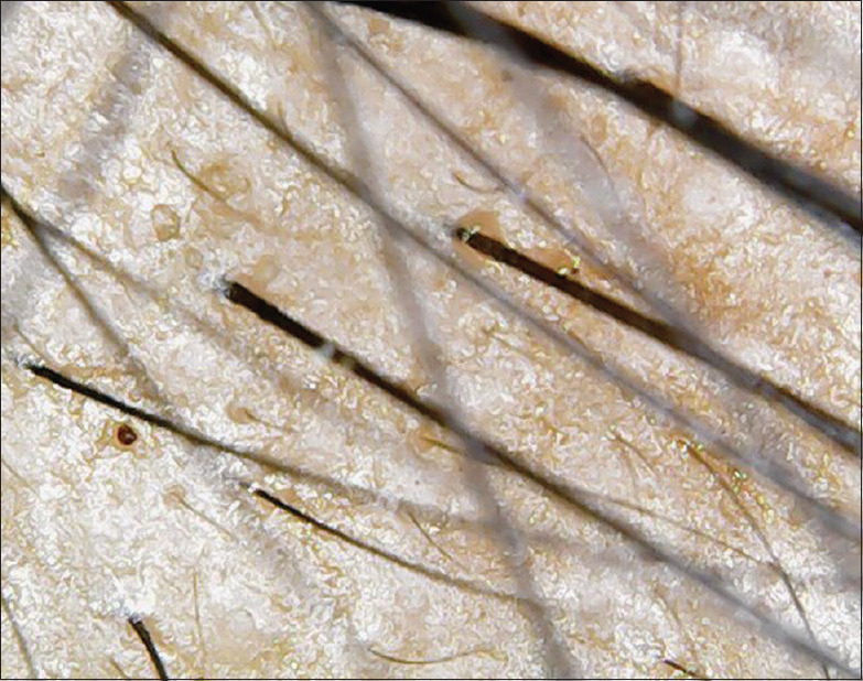
Dermascopic preoperative frontal view
Figure 8.
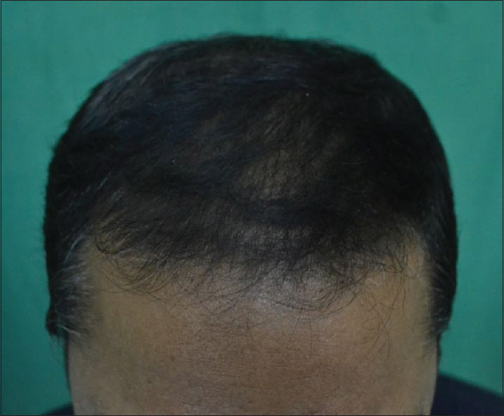
Postoperative frontal view
Figure 9.
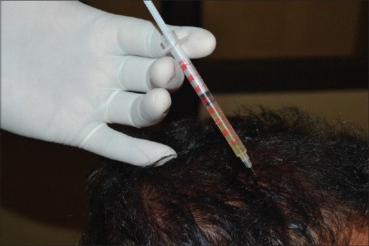
Injecting platelet-rich plasma using insulin syringe
Figure 11.
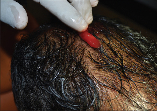
Applying concentrated growth factor gel after microneedling
Figure 6.

Dermascopic postoperative frontal view
Figure 7.
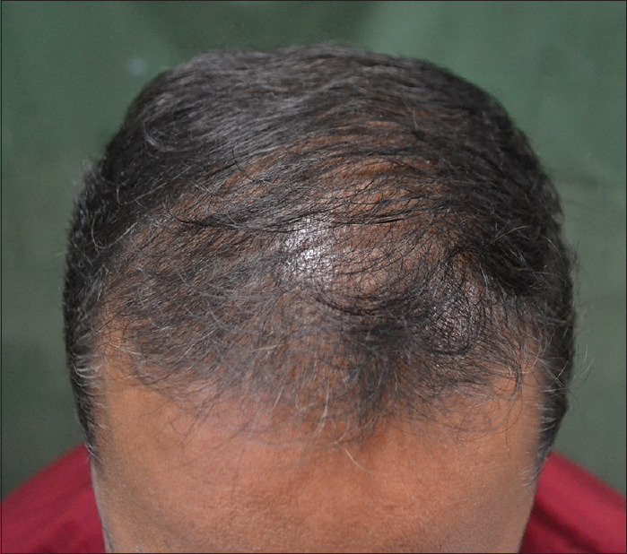
Preoperative frontal view
Figure 10.
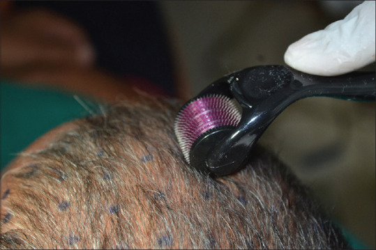
Micro-needling with 1.5 mm dermaroller having 540 needles
DISCUSSION
AGA is still the most common hair disorder without satisfactory treatment. It is characterized by the decrease in the anagen or growing phase of the hair cycle and increase in the miniaturization of the terminal hair to vellus hair.[14]
Lately, it had been proposed that growth factors released from PRP which is a 1st generation platelet concentrate, may act on the stem cells in the bulge area of the follicles, stimulating the development of new follicles, and promoting neovascularization.[7] Platelet-derived growth factor (PDGF) and vascular endothelial growth factor (VEGF) are important for hair formation and follicle size. PDGF plays a role in hair canal formation,[15] while VEGF-mediated angiogenesis involvement is demonstrated in the control of hair growth and follicle size.[16] Primitive stem cells of ectodermal origin are found giving origin to epidermal cells and sebaceous glands in the bulge area. In matrix, the germinative cells of mesenchymal origin are found at the dermal papilla. Interactions between these two kinds of cells and with binding growth factors activate the proliferative phase of hair giving rise to future follicular units.[2] Activated PRP stimulates the proliferation of dermal papillary cells which thus increases the survival of the follicle by its anti-apoptotic effect of dermal papillary cells and stimulates hair growth by prolonging anagen phase. In vitro findings correlated with the in vivo findings showed faster growth of mice's hair treated with PRP every 3 days for 3 weeks than that in untreated controls.[17] Intradermal injections of PRP have been proven to markedly increase the hair diameter while histologic results reveal thickened epithelium, proliferation of collagen fibers and fibroblast and increased blood vessels around the hair follicle.[10,18]
MN produces proliferation as well as migration of fibroblasts and produces collagen and elastin along with creating micro-trauma in the scalp increasing neo-vascularization.[19] MN stimulates follicular stem cells and activates growth factors in the dermal papilla. In our study, MN can be done using dermaroller or a dermapen which would assist the delivery of cosmeceutical products to benefit the scalp by increasing its penetration in addition to its healing effects.
Dhurat et al.[20] concluded that 12 sessions of MN treatment in combination with topical 5% minoxidil application increased hair count by four times in comparison with minoxidil application alone. The mean change in hair count at week 12 was significantly greater (where n = 50) which is 91.40 ± 49.27 for the microneedling group compared to the Minoxidil group (where n = 44) 22.20 ± 19.34 in 1 cm2 region while in our study, the mean change in hair count after 6 months was 28.4 ± 6.68, 35.95 ± 7.59, 35.3 ± 10.12, 37.45 ± 9.46 in frontal, left parietal, right parietal, and occipital region in 0.4 cm2 area using MN, PRP-CGF therapy.
CGF is a 2nd generation platelet concentrate which is applied topically on scalp fibrin rich growth matrix (Sacco in 2006) which releases PDGF, transforming growth factor-β1 (TGF-β1) and β2 (TGF-β2), fibroblast growth factor (FGF), VEGF, brain-derived growth factor (BDGF) and insulin-like growth factor (IGF) stimulating cell proliferation, matrix remodeling and angiogenesis.[21] In vitro study has proved that growth factors such as TNF-α and BDGF showed fast kinetic release from the concentrate and reached its maximum accumulation in the 1st and 3rd day, respectively. Similarly, PDGF-AB, TGF-β1, and IGF-I had constant kinetic release and reached its maximum in the 3rd and 6th day, respectively. VEGF and BMP-2 had slow kinetic release and reached its maximum in the 8th day. These growth factors predominantly play a role in osteoblast proliferation and differentiation.[22] CGF acts by degranulation of the alpha granules in platelets that contain growth factors which play a vital role in early wound healing.[23] The biphasic platelets in CGF are accelerated by thrombin, induce the release of growth factors and other substances which enhance the wound-healing process by increasing cellular proliferation, matrix formation, osteoid production, connective tissue healing, angiogenesis, and collagen synthesis.
This is the first case series to describe the combined use of MN, PRP, and CGF for treating AGA without using any other medicines or serum or laser therapy. PRP is an autologous preparation rich in growth factors-rich platelets and has about 20 different growth factors including PDGF, VEGF, FGF, and TGF.[10] Similarly, CGF has a wide range of healing properties in patients undergoing cosmetic procedures such as rhytidectomy, neck lifts, breast augmentation, and various maxillofacial hard and soft-tissue procedures.
The hair pull test results got much improved as compared to the preoperative result of the same which is comparable to the study performed by Jha et al.[24] According to Jha et al., more than 75% satisfaction rate was seen in 18 patients subjective hair growth assessment scale. In post-PRP-treated patients of AGA, increase in the number of vellus and total hairs and increased hair shaft diameter were appreciated after three sessions. Hair pull test was negative after treatment in 14 patients (70%).
According to Borhan et al.,[25] the mean hair density improved in 14 patients in the area of 0.64 cm2 from 128.8 ± 47.9 at W0-131.9 ± 48 at W16 or the end of 4 months using PRP injections. In our study, the mean hair density increased from 107.3 ± 29.4 at W0-137.1 ± 27.1 at the end of 6 months in 0.4 cm2 area stating the combined effect of MN, PRP-CGF therapy had a better effect. Decrease in the hair density was seen in only one patient (5%) in our study due to greater genetic predisposition and inception of hair loss before 25 years of age while according to Borhan et al.[25] The decrease in the hair density was present in three patients (21.42%).
Khatu et al.[11] observed a significant difference in hair count using PRP therapy in a study having 11 patients from 71 follicular units to 93 thus the gain being 22 units/cm2 while in our study, the mean difference 20 follicular units per 0.4 cm2 with improvement from 107 follicular units to 137 units after four sessions of PRP CGF therapy post 6 months.
Alves et al.[26] conducted a randomized, placebo-controlled, double-blind study having 25 patients where in half head was injected using PRP while the other half head was injected with placebo (saline). Total three treatments of PRP were given 1 month apart. After 6 months, the treatment area with PRP had a mean increase of 12.8 ± 32.6 hairs/cm2 and the control area a decrease of 2.1 ± 31.3 hairs/cm2 while in our study, the mean increase was 30.2 ± 2.3 hairs per 0.4 cm2.
There was significantly moderate improvement in hair growth and coverage as seen through the global pictures and the dermascopic pictures but our study also had some limitations. The sample size is small. Trichoscopic evaluation of hair could have much more accurately improved the data. Follow-up of patients is for a short time to conclude of the efficacy of the treatment for a longer period of time but as far as this particular population of patients is concerned, six patients out of the 20 needed a booster session after 6–9 months. Finally, half head design could have been opted for to know the comparative efficacy of PRP and CGF. This limited case series does not provide a conclusion for the use of CGF but further opens up the area of research for conducting more trials for the same.
CONCLUSION
In our study, it was demonstrated that the administration of PRP and CGF has a positive effect on male pattern alopecia without much significant side effects. The following can be concluded from the study.
Administration of PRP and CGF attended a statistically significant improvement in mean hair density, terminal hair density, mean anagen hairs, and telogen hairs when compared to the baseline at 6 months
An increase in the anagen/telogen ratio
A statistically significant correlation between the demographic data and dermascopic analysis was found with two parameters: Mean total hair density and mean anagen/telogen hairs
Male sex and beginning of AGA after 25 years seem to respond better to the treatment in both parameters
Hair fall also significantly reduced at the end of 6 months as compared to the baseline.
In conclusion, the use of PRP and CGF is effective, safe, and worthwhile as a complementary treatment for AGA, but additional control studies need to be carried out to identify the role of CGF, and longer follow-ups are required.
Financial support and sponsorship
Nil.
Conflicts of interest
There are no conflicts of interest.
REFERENCES
- 1.Writers AM. Treat androgenetic alopecia with antiandrogens, as well as other pharmacological and non-pharmacological interventions. Drugs Ther Perspect. 2017;33:326–30. [Google Scholar]
- 2.Gentile P, Garcovich S, Bielli A, Scioli MG, Orlandi A, Cervelli V. The Effect of Platelet-Rich Plasma in Hair Regrowth: A Randomized Placebo-Controlled Trial. Stem Cells Transl Med. 2015;4:1317–23. doi: 10.5966/sctm.2015-0107. [DOI] [PMC free article] [PubMed] [Google Scholar]
- 3.Pantagosta P. The Complete Book of Hair Loss Answers: Your Comprehensive Guide to the Latest and Best Techniques. 2nd ed. Santa Rosa, CA: Elite Books; 2005. pp. 18–19. [Google Scholar]
- 4.Rathnayake D, Sinclair R. Male androgenetic alopecia. Expert Opin Pharmacother. 2010;11:1295–304. doi: 10.1517/14656561003752730. [DOI] [PubMed] [Google Scholar]
- 5.Choi N, Shin S, Song SU, Sung JH. Minoxidil promotes hair growth through stimulation of growth factor release from adipose-derived stem cells? Int J Mol Sci. 2018;19:691. doi: 10.3390/ijms19030691. doi:10.3390/ijms19030691. [DOI] [PMC free article] [PubMed] [Google Scholar]
- 6.Han JH, Kwon OS, Chung JH, Cho KH, Eun HC, Kim KH. Effect of minoxidil on proliferation and apoptosis in dermal papilla cells of human hair follicle. J Dermatol Sci. 2004;34:91–8. doi: 10.1016/j.jdermsci.2004.01.002. [DOI] [PubMed] [Google Scholar]
- 7.Tosti A, Piraccini BM. Finasteride and the hair cycle. J Am Acad Dermatol. 2004;42:848–9. doi: 10.1067/mjd.2000.103272. [DOI] [PubMed] [Google Scholar]
- 8.Fertig RM, Gamret AC, Darwin E, Gaudi S. Sexual side-effects of 5-α-reductase inhibitors finasteride and dutasteride: A comprehensive review. Dermatol Online J. 2017;23:13030/qt24k8q743. [PubMed] [Google Scholar]
- 9.Kuffler DP. Platelet-rich plasma promotes axon regeneration, wound healing, and pain reduction: Fact or fiction. Mol Neurobiol. 2015;52:990–1014. doi: 10.1007/s12035-015-9251-x. [DOI] [PubMed] [Google Scholar]
- 10.Li ZJ, Choi HI, Choi DK, Sohn KC, Im M, Seo YJ, et al. Autologous platelet-rich plasma: A potential therapeutic tool for promoting hair growth. Dermatol Surg. 2012;38:1040–6. doi: 10.1111/j.1524-4725.2012.02394.x. [DOI] [PubMed] [Google Scholar]
- 11.Khatu SS, More YE, Gokhale NR, Chavhan DC, Bendsure N. Platelet-rich plasma in androgenic alopecia: Myth or an effective tool. J Cutan Aesthet Surg. 2014;7:107–10. doi: 10.4103/0974-2077.138352. [DOI] [PMC free article] [PubMed] [Google Scholar]
- 12.Trink A, Sorbellini E, Bezzola P, Rodella L, Rezzani R, Ramot Y, et al. A randomized, double-blind, placebo-and active-controlled, half-head study to evaluate the effects of platelet-rich plasma on alopecia areata. Br J Dermatol. 2013;169:690–4. doi: 10.1111/bjd.12397. [DOI] [PubMed] [Google Scholar]
- 13.De Boer HC, Van Oeveren-Reitdijk AM, Rotmans JI, Dekkers OM, Rabelink TJ, Van Zonneveld AJ. Activated platelets correlate with mobilization of naïve CD34+ cells and generation of CD34+/KDR+ cells in the circulation.A meta-regression analysis. J Thromb Haemost. 2013;11:1583–92. doi: 10.1111/jth.12315. [DOI] [PubMed] [Google Scholar]
- 14.Blumeyer A, Tosti A, Messenger A, Reygagne P, Del Marmol V, Spuls PI, et al. Evidence-based (S3) guideline for the treatment of androgenetic alopecia in women and in men. J Dtsch Dermatol Ges. 2011;9(Suppl 6):S1–57. doi: 10.1111/j.1610-0379.2011.07802.x. [DOI] [PubMed] [Google Scholar]
- 15.Takakura N, Yoshida H, Kunisada T, Nishikawa S, Nishikawa SI. Involvement of platelet-derived growth factor receptor-alpha in hair canal formation. J Invest Dermatol. 1996;107:770–7. doi: 10.1111/1523-1747.ep12371802. [DOI] [PubMed] [Google Scholar]
- 16.Hou C, Miao Y, Wang J, Wang X, Chen CY, Hu ZQ. Collagenase IV plays an important role in regulating hair cycle by inducing VEGF, IGF-1, and TGF-β expression. Drug Des Dev Ther. 2015;9:5373–83. doi: 10.2147/DDDT.S8912. [DOI] [PMC free article] [PubMed] [Google Scholar]
- 17.Takikawa M, Nakamura S, Nakamura S, Ishirara M, Kishimoto S, Sasaki K, et al. Enhanced effect of platelet-rich plasma containing a new carrier on hair growth. Dermatol Surg. 2011;37:1721–9. doi: 10.1111/j.1524-4725.2011.02123.x. [DOI] [PubMed] [Google Scholar]
- 18.Ferneini EM, Beauvais D, Castiglione C, Ferneini MV. Platelet-rich plasma in androgenic alopecia: indications, technique, and potential benefits. J Oral Maxillofac Surg. 2017;75:788–95. doi: 10.1016/j.joms.2016.10.040. [DOI] [PubMed] [Google Scholar]
- 19.Schmitt L, Marquardt Y, Amann P, Heise R, Huth L, Wagner-Schiffler S, et al. Comprehensive molecular characterization of microneedling therapy in a human three-dimensional skin model. PLoS One. 2018;13:e0204318. doi: 10.1371/journal.pone.0204318. [DOI] [PMC free article] [PubMed] [Google Scholar]
- 20.Dhurat R, Sukesh M, Avhad G, Dandale A, Pal A, Pund P. A randomized evaluator blinded study of effect of microneedling in androgenetic alopecia: A pilot study. Int J Trichology. 2013;5:6–11. doi: 10.4103/0974-7753.114700. [DOI] [PMC free article] [PubMed] [Google Scholar]
- 21.Yu B, Wang Z. Effect of concentrated growth factors on beagle periodontal ligament stem cells in vitro. Mol Med Rep. 2014;9:235–42. doi: 10.3892/mmr.2013.1756. [DOI] [PubMed] [Google Scholar]
- 22.Schär MO, Diaz-Romero J, Kohl S, Zumstein MA, Nesic D. Platelet-rich concentrates differentially release growth factors and induce cell migration in vitro. Clin Orthop Relat Res. 2015;473:1635–43. doi: 10.1007/s11999-015-4192-2. [DOI] [PMC free article] [PubMed] [Google Scholar]
- 23.Kshirsagar JT, Rubine S. Innovation in regeneration-concentrated growth factor. Int J Appl Dent Sci. 2017;3:206–8. [Google Scholar]
- 24.Jha AK, Udayan UK, Roy PK, Amar AKJ, Chaudhary RKP. Original article: Platelet-rich plasma with microneedling in androgenetic alopecia along with dermoscopic pre-and post-treatment evaluation. J Cosmet Dermatol. 2018;17:313–8. doi: 10.1111/jocd.12394. [DOI] [PubMed] [Google Scholar]
- 25.Borhan R, Gasnier C, Reygagne P. Autologous platelet rich plasma as a treatment of male androgenetic alopecia: Study of 14 cases. J Clin Exp Dermatol Res. 2015;6:4. [Google Scholar]
- 26.Alves R, Grimalt R. Randomized placebo-controlled, double-blind, half-head study to assess the efficacy of platelet-rich plasma on the treatment of androgenetic alopecia. Dermatol Surg. 2016;42:491–7. doi: 10.1097/DSS.0000000000000665. [DOI] [PubMed] [Google Scholar]