Abstract
Remdesivir, formerly GS-5734, has recently become the first antiviral drug approved by the U.S. Food and Drug Administration (FDA) to treat COVID-19, the disease caused by SARS-CoV-2. Therapeutic dosing and pharmacokinetic studies require a simple, sensitive, and selective validated assay to quantify drug concentrations in clinical samples. Therefore, we developed a rapid and sensitive LC-MS/MS assay for the quantification of remdesivir in human plasma with its deuterium-labeled analog, remdesivir-2H5, as the internal standard. Chromatographic separation was achieved on a Phenomenex® Synergi™ HPLC Fusion-RP (100 × 2 mm, 4 μm) column by gradient elution. Excellent accuracy and precision (<5.2% within-run variations and.
<9.8% between-run variations) were obtained over the range of 0.5–5000 ng/mL. The assay met the FDA Bioanalytical Guidelines for selectivity and specificity, and low inter-matrix lot variability (<2.7%) was observed for extraction efficiency (77%) and matrix effect (123%) studies. Further, stability tests showed that the analyte does not degrade under working conditions, nor during freezing and thawing processes.
Keywords: COVID-19, SARS-CoV-2, Remdesivir, Mass spectrometry, Bioanalysis
Abbreviations: HPLC, high performance liquid chromatography; MS, mass spectrometry; MS/MS, tandem mass spectrometry; QC, quality control; LLOQ, lower limit of quantification; ULOQ, upper limit of quantification
1. Introduction
Remdesivir (GS-5734) is a monophosphate prodrug of an adenosine nucleoside analog (GS-441524) [1]. Classified as a broad-spectrum antiviral drug, remdesivir exhibits in vitro therapeutic efficacy against multiple pathogenic RNA virus families including filoviruses, pneumoviruses, paramyxoviruses, and coronaviruses [2]. Remdesivir has exhibited antiviral activity against a wide variety of RNA viruses, including Ebola virus (EBOV) [3], MERS-CoV [4], and SARS-CoV [5]; however, until recently, it had never been approved for treatment of any one indication. The current COVID-19 pandemic highlighted the potential therapeutic role of remdesivir, and as a result, several clinical trials were swiftly developed to test the antiviral drug against SARS-CoV-2, the virus responsible for COVID-19 [3]. In May 2020, remdesivir received authorization by the U.S. Food and Drug Administration (FDA) for emergency treatment of hospitalized patients with severe cases of the novel coronavirus disease [4].
Remdesivir, however, has since become the first and only antiviral drug approved by the FDA to treat SARS-CoV-2: based on the recent findings from three completed, randomized clinical trials (NCT04292730, NCT04292899, NCT04280705), the FDA announced on October 22, 2020, that remdesivir is indicated in adults and pediatric patients (12 years old and above, or weighing more than 40 kg) who have been hospitalized due to COVID-19 [5].
The pharmacologically active metabolite (GS-441524) of remdesivir [6] is a nucleoside triphosphate that inhibits viral replication by inducing early chain termination through selective binding to the viral RNA-dependent RNA polymerase (RdRp), thereby inhibiting genome replication and further dissemination [7].
While many of these in vitro studies advanced onto in vivo animal studies, including those for MERS-CoV [8] and SARS-CoV-1 [5], clinical trials have only been developed to test the therapeutic efficacy of remdesivir against EBOV [9] and now SARS-CoV-2 (NCT04292730, NCT04292899, NCT04280705). The former compared the effectiveness of four different drugs to prevent mortality from EBOV; however, remdesivir was eventually dropped from this study as patients who received the drug exhibited higher death rates [10]. Meanwhile, the three aforementioned SARS-CoV-2 clinical trials used various clinical markers to gauge remdesivir efficacy [11], [12], [13]. For instance, in their recent publication of the results from the Adaptive COVID-19 Treatment Trial (ACTT; NCT04280705), the National Institute of Allergies and Infectious Diseases (NIAID, NIH) determined the safety and efficacy of remdesivir by using an eight-point ordinal scale of patient status, ranging from death to not hospitalized with no remaining activity-limiting symptoms [14]. Using data from preclinical studies in animal models, patients who received the drug were given an initial dose of 200 mg intravenous remdesivir, then subsequent daily doses of 100 mg for up to 10 days [11].
Directly adapting human dosing regimens from animal models can result in adverse drug reactions as well as failure to achieve clinical utility [15]. In humans, hepatic and renal toxicity and anaphylactic reactions are known adverse events associated with remdesivir treatment in COVID-19 patients, as outlined in the FDA label [16]. However, an understanding of hepatocellular toxicity with respect to remdesivir dosing in this patient population is lacking [17]. Clinical pharmacokinetics offers the ability to better determine therapeutic dose ranges by predicting the therapeutic efficacy and safety of remdesivir in humans as well as assessing potential drug-drug interactions.
Only two validated remdesivir assays in human plasma using LC-MS/MS exist in the literature; however, both procedures implement more straightforward methods that introduce higher variability and sacrifice sensitivity as indicated by their lowest calibrators (1 ng/mL and 3.91 ng/mL, respectively) [18], [19]. Given the indication for remdesivir in such a high-risk demographic of patients (hospitalized adults and young children), a robust and sensitive method for quantifying remdesivir in human plasma at clinically relevant concentrations, complete with validation and stability data, is essential for ongoing treatment as well as future studies. The methodology described herein also provides a simple tool for measuring the exposure of remdesivir in patients with hepatic impairment, a subgroup whose pharmacokinetics have yet to be characterized. This assay, which represents a sensitivity improvement over previously published assays as well as crucial stability data, offers a calibration range of 0.5–5000 ng/mL, demonstrates strong specificity and selectivity for remdesivir by using reliable techniques, and is validated for accuracy, precision, and improved long-term stability under a variety of conditions.
2. Experimental
2.1. Materials
Remdesivir (>99% pure) was purchased from Selleck Chemicals (Houston, TX), and the internal standard, remdesivir-2H5 (>99% pure), was purchased from MedChemExpress (Monmouth, NJ). Analyte and internal standard chemical structures are provided in Fig. 1 .
Fig. 1.
Structures of the analyte remdesivir (1) and the internal standard remdesivir-2H5 (2) utilized in the reported assay.
Drug-free human plasma (heparinized, pooled mixed gender) was purchased from BioIVT (Westbury, NY). Optima® HPLC-grade acetonitrile was purchased from Fisher Scientific (Pittsburgh, PA). Dimethyl sulfoxide (DMSO; molecular biology grade) and formic acid (reagent grade) were purchased from Sigma-Aldrich (St. Louis, MO), and water used for the preparation of mobile phases was deionized and ultra-filtered (18.2 MΩ·cm) on a MilliPore system (EMD MilliPore, Billerica, MA).
2.2. Preparation of stock solutions
Master stock solutions (1 mg/mL) of remdesivir and remdesivir-2H5 were prepared in DMSO. To prepare working stock solutions, a remdesivir master stock solution was serially diluted with DMSO to the following concentrations: 125000, 50000, 25000, 6250, 1250, 625, 250, 125, 50, 25, and 12.5 ng/mL. All master and working stock solutions were stored at −80 °C when not in use. For preparation of the internal standard solution, a remdesivir-2H5 master stock solution was diluted into 1% formic acid (v/v) in acetonitrile to a final concentration of 0.5 ng/mL and stored at −80 °C when not in use.
2.3. Sample preparation
Drug-free human plasma (heparinized, pooled mixed gender) was used as matrix for the preparation of calibration and quality control (QC) standards. All calibration standards were prepared daily in duplicate by diluting the remdesivir working stock solutions 25-fold into matrix to the following concentrations: 5000, 2000, 1000, 250, 50, 25, 10, 5, 2, 1, and 0.5 ng/mL. In all calibration standards, DMSO content was held constant at 4% (v/v). Concentrations for low (LQC), mid (MQC) and high (HQC) quality control standards, prepared daily in quintuplet, were 1.5, 1000, and 4000 ng/mL, respectively. A lower limit of quantitation quality control (LLOQ) of 0.5 ng/mL was also prepared daily in quintuplet. Per FDA Bioanalytical Guidelines, QC and calibration standards were prepared from the same stock solutions once accuracy and precision were established using separate stock solutions. In all QC standards, final DMSO content was held constant at 4% (v/v).
Calibrators and QC plasma standards were transferred to a Waters Ostro™ 96-well plate in 100 μL aliquots to capture and remove phospholipids and filter precipitated proteins. Protein precipitation was initiated by the addition of 300 μL of internal standard solution (0.5 ng/mL of remdesivir-2H5 in 1% formic acid (v/v) in acetonitrile) to all samples except for the double blank plasma controls, which received blank 1% formic acid (v/v) in acetonitrile. Prior to sample analysis, the contents of each well were: mixed using a multichannel pipette, pushed through the Ostro™ plate phospholipid-removal sorbent via a Waters Positive Pressure-96 Manifold using nitrogen gas at a flow of 10 psi for approximately 5 min., evaporated to dryness under a stream of nitrogen at 40 °C, reconstituted in 300 μL 1% formic acid (v/v) in acetonitrile, and finally centrifuged for 5 min, 1800 RPM, at 20 °C.
2.4. Instrument conditions
A Waters ACQUITY UPLC H-Class® with a quaternary solvent pump, temperature- controlled column compartment (35 °C) and refrigerated autosampler (6 °C) was fitted with a Phenomenex® Synergi™ HPLC Fusion-RP, 100 × 2 mm, 4 μm column. The injection volume for each calibration and QC sample was 5 μL. Mobile phases were 1% formic acid in water (v/v, aqueous) and 1% formic acid in acetonitrile (v/v, organic). The following gradient was employed over the course of a 4 min run: 1% organic at a 0.25 mL/min flow rate (initial), 80% organic at a
0.5 mL/min flow rate (2.0 min), 1% organic at a 0.25 mL/min flow rate (3.5 min to 4.0 min). A Waters Xevo® TQ-S micro tandem quadrupole mass spectrometer was used to monitor primary (m/z 603.2 → 200.1, collision energy [CE] = 38 V) and secondary (qualifier) ion transitions (m/z 603.2 → 402.2, CE = 12 V) of remdesivir and remdesivir-2H5 (primary: m/z 607.9 → 204.9, CE = 40; secondary: 607.9 → 406.9, CE = 14) in the ESI positive ion mode using multiple reaction monitoring (MRM). All mass transitions were independently determined via direct infusion of the analytes into the mass spectrometer. An optimal cone voltage of 36 V was identified for remdesivir, and 12 V for remdesivir-2H5. General ionization settings included a capillary voltage of 2 kV, source temperature of 150 °C and desolvation temperature of 450 °C. For remdesivir, cone voltage and collision energy values were optimized using the IntelliStart™ tuning software. The remaining instrument parameters were optimized manually. All MRM peak integration and subsequent data analyses were performed with the TargetLynx XS™ program.
2.5. Validation
2.5.1. Linearity
Assay calibration for remdesivir was achieved on an 11-point standard curve (0.5–5000 ng/mL) using least-squares quadratic regression by plotting the peak area ratio (analyte:internal standard) versus the concentration ratio (analyte:internal standard) in ng/mL. A weighting factor of 1/x2, which is recommended for bioanalytical LC-MS/MS assays [20], was implemented for each calibration curve where x is the ratio of nominal analyte:internal standard concentration. Calibrator response functions, as well as choice of regression analysis, were investigated via percent deviation (% DEV), defined as the relative error of back calculated concentrations for all calibrators, and correlation coefficient (r2).
2.5.2. Accuracy and precision
Accuracy and precision were evaluated on 4 separate days with fresh sets of 4 different QC standards: a LLOQ, LQC, MQC, and HQC. Each run consisted of a double blank plasma control (no analyte or internal standard), an internal standard plasma control (no analyte), and calibration standards prepared fresh daily in duplicate (n = 8 for each calibration standard); all QC and LLOQ standards were also prepared fresh in quintuplet on each day (n = 20 for each QC level). Accuracy (% DEV) was calculated as the percent difference between the mean observed analyte concentration and the nominal concentration. Precision was investigated using within-run precision (WRP) and between-run precision (BRP), which were determined using one-way analysis of variance (ANOVA) with run day as the classification variable. The WRP and BRP were calculated according to the equations below:
Here, grand mean (the mean of all observed concentrations for each concentration level) is represented by GM, within-group mean squared by MSWIT, between-group mean squared by MSBET, and the number of repetitions by n. The parameters GM, MSWIT, and MSBET were obtained using the software Prism 8 (v.8.3.0). As per FDA Bioanalytical Guidelines [21], ±15% variability in accuracy and precision was deemed acceptable with the exception of LLOQ standards, for which ±20% variability was permissible.
2.5.3. Stability
The stability of remdesivir in plasma at room temperature was assessed over a 4 hr period. Samples at two concentrations (10 ng/mL and 2000 ng/mL) were either extracted immediately once spiked in plasma (n = 5) or after sitting at room temperature for 4 hr (n = 4). To demonstrate analyte working stock stability over this 4 hr time interval, additional samples at each concentration were treated with working stocks kept at room temperature for 4 hr (n = 4 for each concentration). Samples processed after 4 hr were compared to the freshly prepared samples in the same analytical run for comparison of analyte and internal standard concentrations.
The stability of remdesivir in frozen DMSO as a stock solution was also assessed over a short-term period of 15 days and a long-term period of 5 months. Low concentration (10 ng/mL; n = 6) and high concentration (2000 ng/mL; n = 6) samples were spiked in plasma with working stocks prepared either 15 days prior or 5 months prior and stored at −80 °C. These samples were then compared in the same analytical run to plasma samples spiked with fresh working stocks prepared from a fresh analyte master stock (1 mg/mL remdesivir in DMSO).
Additionally, the stability of remdesivir in the 6 °C autosampler was tested. QC samples in plasma (0.5, 1.5, 1000, and 4000 ng/mL; n = 5 for each concentration) were re-injected 24 hr after the initial analysis and then compared to the previous values obtained for those same samples.
To evaluate the potential for remdesivir degradation throughout several freeze/thaw cycles, plasma samples were assayed in quintuplet at two concentrations (1.5 ng/mL and 4000 ng/mL). The samples were subjected to four freeze/thaw cycles from −80 °C to room temperature, with each freeze cycle lasting at minimum 12 hr. The analyte concentration after each cycle was compared to the analyte concentration of fresh samples in the same analytical run.
As per FDA Bioanalytical Guidelines [21], ±15% variability in accuracy at each concentration level was deemed acceptable for auto-sampler, freeze/thaw, stock solution, and benchtop plasma stability experimental data.
2.5.4. Extraction efficiency and matrix effects
The efficiency of the protein precipitation extraction was evaluated in 6 different lots of blank matrix by comparing the analyte peak areas of pre-extracted spiked samples to post- extraction spiked samples from both a low (10 ng/mL; n = 5) and a high (2000 ng/mL; n = 5) analyte concentration.
Matrix effects from plasma on the remdesivir mass spectrometric signal were investigated by comparing the peak areas of samples spiked post-extraction (n = 5) into 6 different lots of blank matrix to samples of the same concentration spiked in clean mobile phase (n = 5).
The concentrations evaluated matched those above, allowing for assessment throughout the calibration range.
Processing efficiency was evaluated at a low (10 ng/mL) and a high (2000 ng/mL) concentration by comparing the peak areas of samples spiked in mobile phase to pre-extracted spiked samples.
2.5.5. Specificity, Selectivity, and carryover
Specificity was evaluated at the lowest end of the calibration range (0.5 ng/mL) in 6 different lots of blank matrix to ensure that any endogenous interferences were resolvable from the analyte and internal standard peaks. To test selectivity, chromatograms from spiked matrix and blank matrix were compared. Carryover was evaluated in blank samples injected directly after the ULOQ and was deemed acceptable if detected signal did not exceed 20% of the LLOQ.
3. Results and discussion
3.1. Specificity, selectivity, and carryover
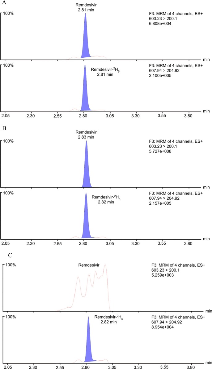
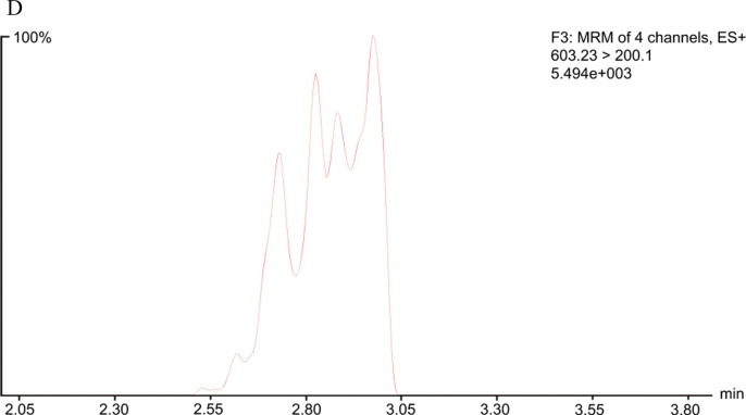
Remdesivir was selectively detected and quantified based on baseline separation from any endogenous interfering peaks, as well as from any potential crosstalk from the internal standard, in each of the six different lots of blank matrix. The LC-MS/MS chromatograms of remdesivir at 0.5 ng/mL (LLOQ) and 5000 ng/mL (ULOQ) are shown in Fig. 2 A and 2B, with their accompanying chromatograms of remdesivir-2H5 at 0.5 ng/mL. Both remdesivir and remdesivir-2H5 demonstrate sharp peaks that are adequately resolved and separated from interfering matrix peaks and background noise. Fig. 2 C and 2D display the chromatograms of plasma extract containing only internal standard (no analyte) and blank plasma extract (no analyte or internal standard) to confirm our identification of the remdesivir signal. The retention time of remdesivir and remdesivir-2H5 is 2.83 min. In blank samples analyzed immediately after the ULOQ, a signal less than 5% of the LLOQ was observed at the retention time of remdesivir and remdesivir-2H5, indicating no significant carryover.
Fig. 2.
LC-MS/MS chromatograms of A) the lower limit of quantification (LLOQ) with the accompanying internal standard, B) the upper limit of quantification (ULOQ) with the accompanying internal standard, C) an internal standard only plasma extract, and D) a blank plasma extract. All chromatograms shown correspond to primary/quantifier ion transitions.
3.2. Validation
An 11-point calibration curve ranging from 0.5 to 5000 ng/mL was run in duplicate on each of four days (n = 8). The calibration standards exhibited strong accuracy (DEV < 4.7%) and precision (CV < 11.5%) (Table 1A ). Only one outlier calibration curve point at the LLOQ level was excluded due to its failure to meet the ±20% variability acceptance criterion, which is well within FDA Bioanalytical Guidance that allows up to 25% of calibrators to be excluded in each validation run. The calibration standards formed a signal response best captured using quadratic regression and 1/x2 weighting, with a model correlation (r2) higher than 0.9935 for all curves (n = 4). Quality control was assessed at the LLOQ, LQC, MQC, and HQC concentration levels. QC standards were prepared alongside calibration standards across four days with five replicates each (n = 20 per concentration level). Within-run precision was below 5.2% CV, while the between-run precision was below 9.8% CV (Table 1B ).
Table 1A.
Accuracy and precision of calibration standards.
| Nominal (ng/mL) | GM (ng/mL) | SD (ng/mL) | CV (%) | DEV (%) | n |
|---|---|---|---|---|---|
| 0.5 | 0.500 | 0.0577 | 11.5 | – | 7 |
| 1 | 0.988 | 0.0641 | 6.49 | −1.25 | 8 |
| 2 | 1.99 | 0.164 | 8.26 | −0.625 | 8 |
| 5 | 5.14 | 0.200 | 3.88 | 2.75 | 8 |
| 10 | 9.98 | 0.656 | 6.58 | −0.250 | 8 |
| 25 | 25.1 | 1.20 | 4.80 | 0.300 | 8 |
| 50 | 47.7 | 1.76 | 3.70 | −4.70 | 8 |
| 250 | 251 | 7.44 | 2.96 | 0.370 | 8 |
| 1000 | 1020 | 33.5 | 3.28 | 2.12 | 8 |
| 2000 | 2050 | 73.9 | 3.60 | 2.56 | 8 |
| 5000 | 4930 | 159 | 3.22 | −1.34 | 8 |
Abbreviations: GM, grand mean; SD, standard deviation; CV (%), coefficient of variation; DEV (%) relative deviation from nominal value; n, total number of samples. Each calibration standard was prepared fresh daily in duplicate on each of four days. A dash (-) indicates no observed DEV.
Table 1B.
Accuracy and precision of quality controls.
| Nominal (ng/mL) | GM (ng/mL) | SD (ng/mL) | CV (%) | DEV (%) | WRP | BRP | n |
|---|---|---|---|---|---|---|---|
| 0.5 | 0.510 | 0.0553 | 10.8 | 2.00% | 5.19 | 9.80 | 20 |
| 1.5 | 1.55 | 0.100 | 6.45 | 3.33% | 1.99 | 6.20 | 20 |
| 1000 | 1060 | 44.3 | 4.18 | 5.90% | 3.41 | 3.69 | 20 |
| 4000 | 4180 | 201 | 4.81 | 4.48% | – | 4.98 | 20 |
Abbreviations: GM, grand mean; SD, standard deviation; CV (%), coefficient of variation; DEV (%) relative deviation from nominal value; WRP, within-run precision; BRP, between-run precision; n, total number of samples. Each QC level was prepared fresh daily in quintuplet on each of four days. A dash (–) indicates no observed CV as a result of performing the assay in different runs.
3.3. Stability
Benchtop stability was demonstrated for remdesivir in human plasma and DMSO over 4 hr to establish the non-degradation of assay materials over the duration of sample preparation (Table 2 ). In the 4 hr study, remdesivir dissolved in DMSO demonstrated nonsignificant deviation (<8%) from a nominal concentration of 10 ng/mL. Additionally, remdesivir was found to be stable in DMSO for at least 5 months at −80 °C, indicating that working stocks can be reused over multiple days and freeze/thaw cycles. Extracted plasma samples incubated at 6 °C for 24 hr in the autosampler of the LC-MS/MS instrument deviated <3% over freshly-prepared samples (Table 3 ). These stability tests allow for the re-analysis of prepared samples in case of instrument hardware failure.
Table 2.
Benchtop, short-term, long-term, and freeze/thaw stability of remdesivir.
| Matrix, Conditions | Nominal (ng/mL) | GM (ng/mL) | SD (ng/mL) | CV (%) | DEV from Fresh (%) | n |
|---|---|---|---|---|---|---|
| Plasma, RT, 4 h | 10 | 9.73 | 0.364 | 3.74 | −4.04 | 4 |
| 2000 | 1770 | 40.1 | 2.27 | −5.47 | 4 | |
| DMSO, RT, 4 h | 10 | 9.38 | 0.190 | 2.02 | −7.44 | 4 |
| 2000 | 1860 | 56.0 | 3.01 | −0.53 | 4 | |
| DMSO (−80 °C, 15 days) | 10 | 9.79 | 0.571 | 5.83 | 11.6 | 6 |
| 2000 | 1.90 × 10³ | 115 | 6.02 | 1.63 | 6 | |
| DMSO (−80 °C, 5 months) | 10 | 10.9 | 0.400 | 3.67 | 9.00 | 4 |
| 2000 | 1930 | 54.4 | 2.82 | −3.90 | 4 | |
| Freeze/thaw (4 cycles) | 1.5 | 1.60 | – | – | 3.90 | 5 |
| 4000 | 3.80 × 10³ | 85.0 | 2.24 | −9.61 | 5 |
Abbreviations: RT, room temperature; GM, grand mean; SD, standard deviation; CV (%), coefficient of variation; DEV from Fresh (%) relative deviation from GM of freshly prepared samples; n, total number of samples. A dash (-) indicates no observed SD/CV.
Table 3.
24-hour autosampler (6 °C) stability of remdesivir.
| Nominal (ng/mL) | GM (ng/mL) | SD (ng/mL) | CV (%) | DEV from Fresh (%) | n |
|---|---|---|---|---|---|
| 0.5 | 0.500 | – | – | – | 5 |
| 1.5 | 1.50 | – | – | −2.60 | 5 |
| 1000 | 1.00 × 10³ | 10.8 | 1.08 | −0.873 | 5 |
| 4000 | 4.20 × 10³ | 43.1 | 1.03 | −0.102 | 5 |
Abbreviations: GM, grand mean; SD, standard deviation; CV (%), coefficient of variation; DEV from Fresh (%) relative deviation from GM of freshly prepared samples; n, total number of samples. A dash (-) indicates no observed SD/CV/DEV.
Although not considered significant (i.e. >15%), slight remdesivir degradation (4% at 10 ng/mL and 5.5% at 2000 ng/mL [Table 2)]) was observed in unextracted plasma samples left at room temperature for 4 hr, possibly due to the presence of plasma esterases. Interestingly, only 10% remdesivir degradation was observed after 4 freeze/thaw cycles in unextracted plasma, which may be attributed to heat-induced esterase structure/activity loss [19]. While we find that a 4 hr stability study is more than sufficient to establish the integrity of benchtop working conditions for an assay that typically takes less than 1 hr to complete, these results emphasize the importance of using proper sample transportation conditions (e.g. dry ice).
3.4. Extraction efficiency and matrix effects
The protein precipitation with phospholipid removal procedure demonstrated consistent extraction efficiency throughout the calibration range (77% at 10 ng/mL and 75% at 2000 ng/mL) and exhibited low inter-matrix lot variability (<2.6%) (Table 4 ). While matrix effects were observed, with ion enhancement increasing remdesivir signals (when in plasma) by 23% at 10 ng/mL and 24% at 2000 ng/mL, it is widely established in the literature that inter-matrix lot variability rather than mean percentage is a more important parameter when assessing analytical accuracy and precision [22], [23]. Importantly, a CV < 4% has been used as a benchmark to indicate high method ruggedness [23]. Overall, the process efficiency of analyzing remdesivir in human heparinized plasma varied between 93 and 95% over a range of 10–2000 ng/mL.
Table 4.
Extraction efficiency and matrix effects.
| Concentration Level (ng/mL) | GM (%) | SD (%) | CV (%) | n | |
|---|---|---|---|---|---|
| Extraction efficiency (%) | 10 | 77.3 | 0.773 | 1.00 | 6 |
| 2000 | 75.0 | 1.98 | 2.63 | 6 | |
| Matrix effects (%) | 10 | 123 | 1.24 | 1.01 | 6 |
| 2000 | 124 | 3.30 | 2.65 | 6 |
Abbreviations: GM, grand mean; SD, standard deviation; CV (%), coefficient of variation; n, total number of samples.
4. Conclusions
Here, we present a fully-validated LC-MS/MS assay for remdesivir, the only antiviral drug approved by the FDA for the treatment of SARS-CoV-2. This remdesivir assay, which to our knowledge is the most sensitive to date, exhibits a dynamic range from 0.5 to 5000 ng/mL and is suitable for therapeutic dosing studies. In addition, the precision, accuracy, and selectivity all meet the FDA Bioanalytical Guidelines. Further, remdesivir is stable under the working conditions, as well as throughout four freezing and thawing cycles. Thus, this assay would be complementary to any ongoing or future remdesivir clinical trials.
5. Financial support
This project has been funded in whole or in part with federal funds from the National Cancer Institute, National Institutes of Health, grant ZIA BC 011974.
6. Disclaimer
The content of this publication does not necessarily reflect the views or policies of the Department of Health and Human Services, nor does mention of trade names, commercial products, or organizations imply endorsement by the U.S. Government. The views in this manuscript are those of the authors and may not necessarily reflect NIH policy. No official endorsement is intended nor should be inferred.
CRediT authorship contribution statement
Ryan Nguyen: Methodology, Validation, Formal analysis, Writing - original draft, Writing - review & editing. Jennifer C. Goodell: Methodology, Validation, Formal analysis, Writing - original draft, Writing - review & editing. Priya S. Shankarappa: Methodology, Validation, Formal analysis, Writing - original draft, Writing - review & editing. Sara Zimmerman: Methodology, Validation, Formal analysis, Writing - original draft, Writing - review & editing. Tyler Yin: Supervision, Project administration, Writing - original draft, Writing - review & editing. Cody J. Peer: Supervision, Project administration, Writing - original draft, Writing - review & editing. William D. Figg: Funding acquisition, Resources.
Declaration of Competing Interest
The authors declare that they have no known competing financial interests or personal relationships that could have appeared to influence the work reported in this paper.
Acknowledgements
This work was supported in part by the Intramural Research Program of the NIH and the National Cancer Institute. This is a US Government study and there are no restrictions on its use. Any opinions expressed herein do not necessarily reflect those of the US Government. The authors would like to acknowledge the frontline workers at the NIH Clinical Center for their ongoing efforts amidst the COVID-19 pandemic, as well as the patients who enroll in clinical trials to advance the knowledge of this disease.
Footnotes
Supplementary data to this article can be found online at https://doi.org/10.1016/j.jchromb.2021.122641.
Appendix A. Supplementary material
The following are the Supplementary data to this article:
References
- 1.Wang Y., Zhang D., Du G., et al. Remdesivir in adults with severe COVID-19: a randomised, double-blind, placebo-controlled, multicentre trial. The Lancet. 2020 doi: 10.1016/S0140-6736(20)31022-9. [DOI] [PMC free article] [PubMed] [Google Scholar]
- 2.Lo M.K., Jordan R., Arvey A., et al. GS-5734 and its parent nucleoside analog inhibit Filo-, Pneumo-, and Paramyxoviruses. Sci. Rep. 2017;7:43395. doi: 10.1038/srep43395. [DOI] [PMC free article] [PubMed] [Google Scholar]
- 3.Warren T.K., Jordan R., Lo M.K., et al. Therapeutic efficacy of the small molecule GS-5734 against Ebola virus in rhesus monkeys. Nature. 2016;531:381–385. doi: 10.1038/nature17180. [DOI] [PMC free article] [PubMed] [Google Scholar]
- 4.Hinton DM. Emergency Use Authorization (EUA) Of Remdesivir (GS-5734™). Food and Drug Administration, 2020.
- 5.Hinton DM. Veklury (remdesivir) EUA Letter Of Approval, Reissued 10/22/2020. Food and Drug Administration, 2020.
- 6.Eastman R.T., Roth J.S., Brimacombe K.R., et al. Remdesivir: A Review of Its Discovery and Development Leading to Emergency Use Authorization for Treatment of COVID-19. ACS Cent. Sci. 2020 doi: 10.1021/acscentsci.0c00489. [DOI] [PMC free article] [PubMed] [Google Scholar]
- 7.M.L. Agostini, E.L. Andres, A.C. Sims et al., Coronavirus Susceptibility to the Antiviral Remdesivir (GS-5734) Is Mediated by the Viral Polymerase and the Proofreading Exoribonuclease, mBio (2018) 9. 10.1128/mBio.00221-18. [DOI] [PMC free article] [PubMed]
- 8.de Wit E., Feldmann F., Cronin J., et al. Prophylactic and therapeutic remdesivir (GS-5734) treatment in the rhesus macaque model of MERS-CoV infection. Proc. Natl. Acad. Sci. USA. 2020;117:6771–6776. doi: 10.1073/pnas.1922083117. [DOI] [PMC free article] [PubMed] [Google Scholar]
- 9.Investigational Therapeutics for the Treatment of People With Ebola Virus Disease (NCT03719586).
- 10.Mulangu S., Dodd L.E., Davey R.T., Jr, et al. A Randomized, Controlled Trial of Ebola Virus Disease Therapeutics. N Engl. J. Med. 2019;381:2293–2303. doi: 10.1056/NEJMoa1910993. [DOI] [PMC free article] [PubMed] [Google Scholar]
- 11.Adaptive COVID-19 Treatment Trial (ACTT) (NCT04280705), 2020.
- 12.Study to Evaluate the Safety and Antiviral Activity of Remdesivir (GS-5734™) in Participants With Moderate Coronavirus Disease (COVID-19) Compared to Standard of Care Treatment, 2020.
- 13.Study to Evaluate the Safety and Antiviral Activity of Remdesivir (GS-5734™) in Participants With Severe Coronavirus Disease (COVID-19), 2020.
- 14.Beigel J.H., Tomashek K.M., Dodd L.E., et al. Remdesivir for the Treatment of Covid-19 — Final Report. N. Engl. J. Med. 2020 doi: 10.1056/NEJMoa2007764. [DOI] [PMC free article] [PubMed] [Google Scholar]
- 15.Amirian E.S., Levy J.K. Current knowledge about the antivirals remdesivir (GS-5734) and GS-441524 as therapeutic options for coronaviruses. One Health. 2020;9 doi: 10.1016/j.onehlt.2020.100128. [DOI] [PMC free article] [PubMed] [Google Scholar]
- 16.VEKLURY (remdesivir) [package insert]. U.S. Food and Drug Administration. Foster City, CA: Gilead Sciences, 2020.
- 17.Zampino R., Mele F., Florio L.L., et al. Liver injury in remdesivir-treated COVID-19 patients. Hepat Int. 2020:1–3. doi: 10.1007/s12072-020-10077-3. [DOI] [PMC free article] [PubMed] [Google Scholar]
- 18.Alvarez J.C., Moine P., Etting I., Annane D., Larabi I.A. Quantification of plasma remdesivir and its metabolite GS-441524 using liquid chromatography coupled to tandem mass spectrometry. Application to a Covid-19 treated patient. Clin. Chem. Lab Med. 2020;58:1461–1468. doi: 10.1515/cclm-2020-0612. [DOI] [PubMed] [Google Scholar]
- 19.Avataneo V., de Nicolò A., Cusato J., et al. Development and validation of a UHPLC-MS/MS method for quantification of the prodrug remdesivir and its metabolite GS-441524: a tool for clinical pharmacokinetics of SARS-CoV-2/COVID-19 and Ebola virus disease. J. Antimicrob. Chemother. 2020;75:1772–1777. doi: 10.1093/jac/dkaa152. [DOI] [PMC free article] [PubMed] [Google Scholar]
- 20.Gu H., Liu G., Wang J., Aubry A., Arnold M.E. Selecting the correct weighting factors for linear and quadratic calibration curves with least-squares regression algorithm in bioanalytical LC-MS/MS assays and impacts of using incorrect weighting factors on curve stability, data quality, and assay performance. Anal. Chem. 2014;86:8959–8966. doi: 10.1021/ac5018265. [DOI] [PubMed] [Google Scholar]
- 21.Guidance for Industry. Bioanalytical Method Validation. U.S. Department of Health and Human Services, Food and Drug Administration, Center for Drug Evaluation and Research (CDER), Center for Veterinary Medicine (CVM), May 2018.
- 22.De Nicolò A., Cantù M., D’Avolio A. Matrix effect management in liquid chromatography mass spectrometry: the internal standard normalized matrix effect. Bioanalysis. 2017;9:1093–1105. doi: 10.4155/bio-2017-0059. 10.415/bio-2017-0059. [DOI] [PubMed] [Google Scholar]
- 23.Matuszewski B.K. Standard line slopes as a measure of a relative matrix effect in quantitative HPLC-MS bioanalysis. J. Chromatogr. B Analyt. Technol. Biomed. Life Sci. 2006;830:293–300. doi: 10.1016/j.chromb.2005.11.009. [DOI] [PubMed] [Google Scholar]
Associated Data
This section collects any data citations, data availability statements, or supplementary materials included in this article.


