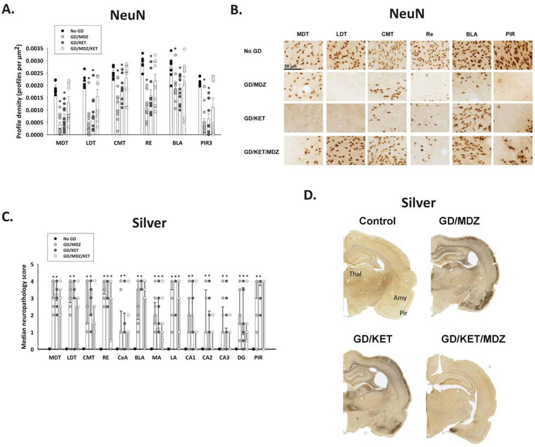
Figure 10. Delayed ketamine and midazolam combination therapy reduced neuronal cell death and neuronal fiber degeneration following soman (GD)-induced seizure.

Ketamine (KET; 30 mg/kg) with or without midazolam (MDZ; 3 mg/kg) was administered at 40 min after GD-induced (132 µg/kg, SC) seizure onset and compared with no agent control rats (n = 10–11/group). Brain samples were processed for immunohistochemistry with NeuN antibody to visualize mature neurons (A, B) and a silver stain to visualize neuronal fiber degeneration (C, D). (A) Rats treated with MDZ monotherapy (GD/MDZ) or KET monotherapy (GD/KET) had significantly reduced NeuN-positive (NeuN+) cells compared to No GD control in the dorsomedial thalamus (MDT), dorsolateral thalamus (LDT), central medial thalamus (CMT), reuniens (RE) nucleus of the thalamus, basolateral amygdala (BLA), and layer 3 of the piriform cortex (PIR3); Data shown are mean ± SEM. (B) Representative images are shown for brain tissue stained with NeuN. (C) Rats in GD/MDZ and GD/KET groups had significantly higher neuropathology scores shown via silver stain compared to No GD control in the MDT, LDT, CMT, RE, central amygdala (CeA), BLA, medial amygdala (MA), lateral amygdala (LA), CA1, CA2, CA3, and dentate gyrus (DG) regions of the hippocampus, and the piriform cortex (PIR). (D) Representative images of silver-stained brain tissue are shown. Data shown are median ± SD. *p < 0.05, compared to No GD group.
