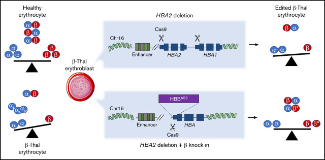
Key Points
HBA2 gene deletion in human HSPCs reduces α-globin precipitates in erythroblasts and ameliorates β+-thalassemia phenotype.
Synergistic HBA2 gene deletion and HBB gene replacement in HSPCs ameliorate β0-thalassemia phenotype
Abstract
β-thalassemias (β-thal) are a group of blood disorders caused by mutations in the β-globin gene (HBB) cluster. β-globin associates with α-globin to form adult hemoglobin (HbA, α2β2), the main oxygen-carrier in erythrocytes. When β-globin chains are absent or limiting, free α-globins precipitate and damage cell membranes, causing hemolysis and ineffective erythropoiesis. Clinical data show that severity of β-thal correlates with the number of inherited α-globin genes (HBA1 and HBA2), with α-globin gene deletions having a beneficial effect for patients. Here, we describe a novel strategy to treat β-thal based on genome editing of the α-globin locus in human hematopoietic stem/progenitor cells (HSPCs). Using CRISPR/Cas9, we combined 2 therapeutic approaches: (1) α-globin downregulation, by deleting the HBA2 gene to recreate an α-thalassemia trait, and (2) β-globin expression, by targeted integration of a β-globin transgene downstream the HBA2 promoter. First, we optimized the CRISPR/Cas9 strategy and corrected the pathological phenotype in a cellular model of β-thalassemia (human erythroid progenitor cell [HUDEP-2] β0). Then, we edited healthy donor HSPCs and demonstrated that they maintained long-term repopulation capacity and multipotency in xenotransplanted mice. To assess the clinical potential of this approach, we next edited β-thal HSPCs and achieved correction of α/β globin imbalance in HSPC-derived erythroblasts. As a safer option for clinical translation, we performed editing in HSPCs using Cas9 nickase showing precise editing with no InDels. Overall, we described an innovative CRISPR/Cas9 approach to improve α/β globin imbalance in thalassemic HSPCs, paving the way for novel therapeutic strategies for β-thal.
Visual Abstract
Introduction
Adult hemoglobin consists of 2 pairs of globin subunits (α2β2), whose production is strictly regulated to ensure their balanced expression in erythroid cells. Disorders in hemoglobin synthesis cause thalassemia, a severe anemia requiring lifelong supportive treatments.1 β-thalassemia is the most common and severe form of thalassemia, with >70 000 new patients per year worldwide, caused by mutations in the β-globin gene (HBB) cluster, which result in reduced or absent synthesis of β-globin chain.2 As a consequence, the free α-globin in excess forms toxic precipitates that cause intramedullary apoptosis of erythroid precursors (ineffective erythropoiesis) and death of mature red blood cells (hemolysis). Healthy individuals carry 4 α-globin genes per cell (αα/αα), 2 genes in cis on each chromosome 16 (HBA1 and HBA2), which often undergo genomic rearrangements and HBA2 gene loss because of their sequence homology (96.67%, GRCh38). Clinical data have shown that the severity of β-thalassemia directly correlates with the number of α-globin genes, with HBA deletions having a beneficial effect for patients.3 This positive effect is especially pronounced in patients with hemoglobin variant E (HbE) β-thalassemia, where the HbE allele still allows residual expression of β-globin chain (∼50% of all β-thalassemia patients).4-7
Current clinical management of β-thalassemia patients relies on regular blood transfusion and iron chelation therapy, required to reduce organ damage and iron overload, respectively.8 Allogeneic hematopoietic stem cell (HSC) transplantation is a definitive cure, but it is limited by the availability of compatible donors.9-12
Two alternative strategies have been proposed to counteract the α/β globin imbalance in β-thal. The first consists in the expression/induction of β-globins or β-like globins, such as γ-globin, to complex with the α-globins in excess and produce adult or fetal hemoglobin.13-18 In particular, ex vivo HSC gene therapy with lentiviral vectors (LV) encoding for HBB transgene significantly ameliorates or resolves β+-thalassemia in patients, where the residual endogenous HBB expression contributes to the clinical benefit; however, its effects are less pronounced for β0-thalassemia (no residual HBB expression).13,14 The second approach to treat β-thalassemia aims at reducing α-globin expression to avoid the formation of toxic precipitates in erythroid cells.3,19,20
Here, we propose to use the CRISPR/Cas9 system to combine these 2 strategies: (1) α-globin chain reduction by recreating the natural α-thalassemia trait (-α3.7 deletion, -α/αα or -α/-α) and (2) targeted integration and expression of a HBB transgene under the control of the endogenous HBA promoter. Concomitant downregulation of α-globin and upregulation of β-globin allows successful amelioration of α/β globin imbalance in both β+ and β0 thalassemia cells.
Methods
Plasmids
We designed 2 different cassettes for the expression of HBB (gene ID: 3043): (1) the first cassette contains a β-globin complementary DNA (cDNA), with 3 antisickling point mutations (βAS3),21 followed by a woodchuck posttranscriptional regulatory element and a SV40polyA; and (2) the second cassette contains the whole βAS3 gene with introns and its original 3′ untranslated region (UTR) and polyA (pA) site, with 3 antisickling point mutations.
Each cassette, followed by a green fluorescent protein (GFP) reporter gene under the control of the constitutive human phosphoglycerate kinase 1 promoter with an SV40 pA, was flanked by arms of homology (250 bp each) and cloned in a standard AAV vector backbone (AAV2) in sense orientation with respect to its inverted terminal repeat.
To generate the double reporter AAV, we substituted the βAS3 gene with the human low-affinity nerve growth factor receptor cDNA. Each cassette was synthetized by Genscript (Piscataway, NJ).
The LV encoding the βAS3 gene under the control of the erythroid specific β-globin enhancer/promoter was already described.22
All reagents and detailed sequence information are available upon request.
Vector productions
All recombinant single-stranded AAV2/6 used in this study were produced using a triple transfection protocol and purified by 2 sequential cesium chloride density gradients or chromatography, as described earlier.23 The vector titer of each preparation was determined by real-time quantitative polymerase chain reaction (qPCR)–based titration method using primers and probe corresponding to the inverted terminal repeat region of the AAV genome.24
LV were produced by transient transfection of 293T using third-generation packaging plasmid pMDLg/p.RRE and pK.REV and pseudotyped with the vesicular stomatitis virus glycoprotein G envelope. LV were titrated in HCT116 cells and HIV-1 Gag p24 content was measured by enzyme-linked immunosorbent assay (PerkinElmer, Waltham, MA) according to the manufacturer’s instructions.
Cell culture and reagents
K562 cells (ATCC CCL243) were maintained in RPMI 1640 medium containing 2 mM glutamine and supplemented with 10% fetal bovine serum (Lonza, Basel, Switzerland), 10 mM N-2-hydroxyethylpiperazine-N′-2-ethanesulfonic acid, 1 mM sodium pyruvate, and penicillin and streptomycin (100 U/mL each; Gibco, Waltham, MA).25
HUDEP-2 β0 cells were derived from a HUDEP-226 clone where both β-globin alleles were knocked out using CRISPR/Cas9 and a guide RNA (gRNA) targeting HBB exon 1 (HBB gRNA), as previously described.17 Both HUDEP-2 and HUDEP-2 β0 cells erythroid differentiation was performed in presence of doxycycline in 2 phases, with and without stem cell factor to promote erythroid maturation.27
HUDEP-2 β0 single cell clones with different HBA2 copy number were obtained by limiting dilution.
Streptococcus pyogenes Cas9 protein (with 2 SV40 nuclear localization signals) was provided by J.P. Concordet and was expressed and purified as previously described.28 Streptococcus pyogenes Cas9 D10A protein (Alt-R S.p. Cas9 D10A Nickase V3; with 3 SV40 nuclear localization signals) was purchased from Integrated DNA Technologies (Coralville, IA).
Chemically modified single guide RNA were purchased from Synthego (with protospacer adjacent motif):
HBB gRNA: TCTGCCGTTACTGCCCTGT(GGG)
5′ UTR (HBA15) gRNA: GGGUUCUCUCUGAGUCUGUG(GGG)
HBA knockout (KO) gRNA: GUCGGCAGGAGACAGCACCA(TGG)
HBA20 gRNA: CATAAACCCTGGCGCGCTCG(CGG)
Primer and probes for PCRs were purchased from Sigma-Aldrich (St Louis, MO) and Integrated DNA Technologies (Coralville, IA).
CD34+ cell culture, transfection, and transduction
Human umbilical cord blood (UCB) samples were provided by Centre Hospitalier Sud-Francilien (CHSF; Evry, France) and processed according to France bioethics laws (Declaration DC-2012-1655 to the French Ministry of Higher Education and Research). CD34+ cells were selected from patients affected by β-thalassemia as described29 upon signed informed consent approved by the Ethical Committee of the San Raffaele Hospital (Milan, Italy).
CD34+ cells were purified by immunomagnetic selection with AUTOMACS PRO (Miltenyi Biotec, Paris, France) after immunostaining with CD34 MicroBead Kit (Miltenyi Biotec). Mobilized peripheral blood (MPB) and UCB CD34+ were also purchased from Cliniscience (Nanterre, France).
MPB- or UCB-derived HSPCs were thawed and cultured in prestimulation media for 48 hours (StemSpan, Stemregenin-1 0.75 μM, UM171 0.35 μM; StemCell Technologies, Vancouver, BC, Canada; rhSCF 300 ng/mL, rhFlt3-L 300 ng/mL, rhTPO 100 ng/mL, and rhIL-3 20 ng/mL; CellGenix, Freiburg, Germany).
Single guide RNA (sgRNA) was diluted following the manufacturer’s instruction and ribonucleoprotein complexes were formed with 30 pmol of spCas9 (ratio 1:1.5). A total of 2.5 × 10⁵ hematopoietic stem/progenitor cells (HSPCs) per condition were transfected with ribonucleoprotein (RNP) using a P3 Primary Cell 4D-Nucleofector Kit (CA137 program).25 In knock-in experiments, 10 minutes after transfection, HSPCs were transduced with AAV6 for 6 hours (multiplicity of infection, 10 000-30 000), washed, and left in prestimulation media for additional 24 to 48 hours.
Lentiviral transduction was performed as previously described.25
After manipulation, HSPCs were cultured in erythroid differentiation medium (StemSpan, StemCell Technologies; rhSCF 20 ng/mL, rhEpo 1 U/mL, IL3 5 ng/mL, dexamethasone 2 µM and beta-estradiol 1 µM; Sigma-Aldrich) or in semisolid MethoCult medium (colony-forming cells [CFC] assay, H4435; StemCell Technologies) for 14 days. Colonies were counted and identified according to morphological criteria (burst-forming units-erythroid [BFU-E], colony-forming unit-granulocyte/granulocyte macrophage, and colony-forming unit-granulocyte, erythrocyte, monocyte, megakaryocyte). In some experiments, BFU-E were picked and cultured in erythroid progenitor expansion medium, as previously described.30
Flow cytometry
Cells were fixed and/or permeabilized using Cytofix/Cytoperm (BD Bioscience, San Jose, CA) according to the manufacturer's instructions. For live cell analysis, viability was assessed using Zombie Yellow dye (BioLegend, San Diego, CA) per manufacturers’ instructions to exclude dead cells from the analysis. Negative controls were obtained by staining cells with isotype control antibodies. For engraftment studies, an Fc Receptor Binding Inhibitor antibody was used to block unspecific binding of mouse antibody to human cells, as per manufacturers’ instructions. Cells were analyzed using CytoFLEX S (Beckman Coulter, Pasadena, CA) or SP6800 Spectral Analyzer (Sony, Tokyo, Japan); data were elaborated with CytExpert (Beckman Coulter) or FlowJo software (Tree Star, Woodburn, OR).
We used MoFlocell sorter (Beckman Coulter) to select live GFP+ cells.
See supplemental Table 1 for the antibodies list and supplemental Figure 3A for gating strategy of human engrafted cells.
DNA analysis
Genomic DNA was extracted with QIAamp DNA Micro Kit, AllPrep DNA/RNA 96 Kit (Qiagen, Hilden, Germany) or QuickExtract DNA Extraction Solution (Lucigen, Middleton, WI).
Quantification of editing efficiency (InDels).
Fifty nanograms of genomic DNA were used to amplify the region that spans the cutting site of each gRNA using KAPA2G Fast ReadyMix (Kapa Biosystem, Wilmington, MA). After Sanger sequencing (Genewiz, Takeley, United Kingdom), the percentage of insertions and deletions (InDels) was calculated using TIDE31 and ICE (Synthego) softwares. See supplemental Table 2 for primer sequences.
ddPCR.
Digital droplet PCR (ddPCR) was performed according to manufacturer’s instruction using ddPCR Supermix for Probes No dUTP (BioRad, Hercules, CA) and 1 to 50 ng of genomic DNA digested with HindIII (New England Biolabs, Ipswich, MA). Droplets were generated using AutoDG Droplet Generator and analyzed with a QX200 Droplet Reader; data analysis was performed with QuantaSoft (BioRad).
To quantify HBA2 copy number, primers and probes were designed on the 3′UTR of HBA2 gene because it differs significantly from HBA1.
To quantify on-target transgene integration events, primers and probes were designed spanning the donor DNA-genome 3′ junction. Human albumin (ALB) or ZNF843 genes were used as reference for copy number evaluation (assay ID: dHsaCP2506312, BioRad). See supplemental Table 2 for primer and probe sequences.
RNA extraction and reverse transcription-qPCR
Total RNA was purified using RNeasy Micro Kit or, in case of individual BFU-E colonies, AllPrep DNA/RNA 96 Kit (Qiagen). RNA was reverse-transcribed using Transcriptor First Strand cDNA Synthesis Kit (Roche, Basel, Switzerland). qPCR was performed using Maxima Syber Green/Rox (Life Scientific, Thermo Fisher Scientific, Waltham, MA). Primers and probe were optimized using the standard curve method to reach 100% ± 5% efficiency. The expression of each target gene was normalized using human GAPDH as a reference gene (NM_002046.6) and represented as 2ΔCt for each sample or as fold changes (2ΔΔCt) relative to the control. See supplemental Table 2 for primer sequences.
Off-target analyses
GUIDE-seq.
GUIDE-Seq experiments were designed as previously described.32 Human embryonic kidney 293T/17 cells (2.5 × 10⁵) were transfected using Lipofectamine 3000 (Life Scientific, Thermo Fisher Scientific) with 750 ng of SpCas9-coding and 250 ng of sgRNA-coding plasmid or an empty pUC19 vector (background control), 10 pmol of the bait double-stranded oligonucleotide containing phosphorothioate bonds at both ends and 50 ng of a pEGFP-IRES-Puro plasmid, expressing both enhanced GFP and the puromycin resistance genes. One day after transfection, cells were replated and selected with puromycin (1 μg/mL) for 48 hours to enrich for transfected cells.
Genomic DNA extraction was performed using DNeasy Blood and Tissue Kit (Qiagen) following manufacturer’s instructions. Genomic DNA was sheared using a Covaris S200 sonicator to an average length of 500 bp and subsequently end-repaired. Library preparation was performed using adapters and primers previously described32 and quantified with Qubit double-stranded DNA High Sensitivity Assay Kit (Life Scientific, Thermo Fisher Scientific). The sequencing reaction was performed with MiSeq sequencing system (Illumina) using an Illumina Miseq Reagent Kit V2 - 300 cycles (2 × 150 bp paired-end) and raw sequencing data (FASTQ files) were analyzed using the GUIDE-Seq computational pipeline.33 Briefly, after demultiplexing, putative PCR duplicates were consolidated into single reads and mapped to the human reference genome GrCh37. Reads with a mapping quality lower than 50 were filtered out. Upon identification of the genomic regions integrating double-stranded oligonucleotides in aligned data, off-target sites were identified if, at most, 7 mismatches against the target were present and if absent in the background controls. GUIDE-Seq datasets are available in the BioProject online repository (ID PRJNA676022; http://www.ncbi.nlm.nih.gov/bioproject/676022).
Amplicon-sequencing.
A total of 200 ng of genomic DNA of RNP-edited HSPCs were used to amplify top 3 off-targets identified with Guide-Seq using Phusion High-Fidelity polymerase (New England Biolabs). After checking the correct amplification by Sanger sequencing (Genewiz), amplicons were purified using NucleoSpin Gel and PCR Clean-up Kit (Macherey-Nagel, Hoerdt, France), and 500 ng of DNA were used for library preparation. Illumina-compatible barcoded DNA amplicon libraries were prepared using TruSeq DNA PCR-Free Kit (Illumina). PCR amplification was then performed using 1 ng of double-stranded ligation product and Kapa Taq polymerase reagents (KAPA HiFi HotStart ReadyMix PCR Kit; Sigma-Aldrich). After purification (Ampure XP beads; Beckman Coulter), libraries were pooled and sequenced with Illumina NovaSeq 6000 (paired-end sequencing 100 × 100 bases). Data were analyzed using CRISPResso2.34
See supplemental Table 2 for primer sequences.
HPLC analysis of globin chains and tetramers
HPLC analysis was performed using a NexeraX2 SIL-30AC chromatograph (Shimadzu, Kyoto, Japan) and analyzed with LC Solution software. HSPC-derived erythroblasts were lysed in water and globin chains were separated using a 250 × 4.6-mm, 3.6-µm Aeris Widepore column (Phenomenex, Torrance, CA). Samples were eluted with a gradient mixture of solution A (water/acetonitrile/trifluoroacetic acid, 95:5:0.1) and solution B (water/acetonitrile/trifluoroacetic acid, 5:95:0.1), monitoring absorbance at 220 nm. Hemoglobin tetramers were separated by HPLC using a cation-exchange column (PolyCAT A; PolyLC, Columbia, MD). Samples were eluted with a gradient mixture of solution A (20 mM bis Tris, 2 mM KCN; pH 6.5) and solution B (20 mM bis Tris, 2 mM KCN, 250 mM NaCl; pH 6.8). The absorbance was measured at 415 nm.
In vivo experiments
NOD.Cg-PrkdcscidIl2rgtm1Wjl/SzJ (NSG) mice were purchased from The Jackson Laboratory, Bar Harbor, ME (strain 005557) and maintained in specific-pathogen-free conditions. This study was approved by Ethical Committee CEEA-51, and conducted according to French and European legislation on animal experimentation (APAFiS#16499-2018071809263257_v4).
Forty-eight hours after editing, 5 to 7 × 10⁵ CD34+ cells were injected intravenously into 8-week-old female NSG mice after sublethal irradiation (150 cGy). Human cell engraftment and knockin (KI) levels were monitored at different time points in peripheral blood by flow cytometry using anti-human CD45 and HLA-ABC antibodies (supplemental Table 1). Sixteen weeks after transplantation, blood, bone marrow, and spleen were harvested and analyzed. Peripheral blood was stained and red blood cells lysed during sample fixation (VersaLyse Lysing Solution and IOTest3 Fixative Solution; Beckman Coulter).
Cell purification and enrichment
Human CD34+ or CD45+ cells were purified from mouse peripheral blood or bone marrow by immunomagnetic selection with CD34 or CD45 MicroBead Kit UltraPure in combination with AUTOMACS PRO (PosselD2 Separation Program; Miltenyi Biotec).
Statistical analyses
Statistical analyses were performed using GraphPad Prism, version 6.00, for Windows (GraphPad Software, La Jolla, CA). One- or 2-way analysis of variance (ANOVA) with Tukey’s multiple comparison posttest for 3 or more groups was performed as indicated. Values are expressed as mean ± standard deviation (SD) as otherwise indicated, with "n" indicating the number of independent biological replicates used in each group. Differences were considered significant at *P < .05 **P < .01, and ***P < .001.
Results
Deletion of HBA2 gene reduces α-globin precipitates in a β0 adult erythroid cell line
Clinical data suggest that a reduction of α-globin to 75% to 25% of its physiological levels is safe and beneficial to patients with β-thalassemia.3,35 The most common natural mutations affecting α-globin synthesis are gene deletions that remove a single HBA gene from 1 or both chromosomes generating -α/αα or -α/-α genotypes (-α3.7 and -α4.2deletions36) (Figure 1A). To mimic these beneficial deletions with Streptococcus pyogenes (Sp)Cas9 nuclease, we designed a single gRNA that cuts both HBA1 and HBA2 alleles removing 1 HBA2 gene per chromosome. To minimize the possibility of generating a α-globin KO, the sgRNA was designed to target the 5′UTR (HBA15) of HBA1 and HBA2 (Figure 1A), where the presence of InDels resulting from double-strand breaks (DSBs) does not affect α-globin production.37 As a β0-thalassemia cell model, we used immortalized HUDEP-2, which can differentiate and express adult hemoglobin (supplemental Figure 1A-B), and we knocked out β-globin genes (HUDEP-2 β0) (supplemental Figure 1C).
Figure 1.
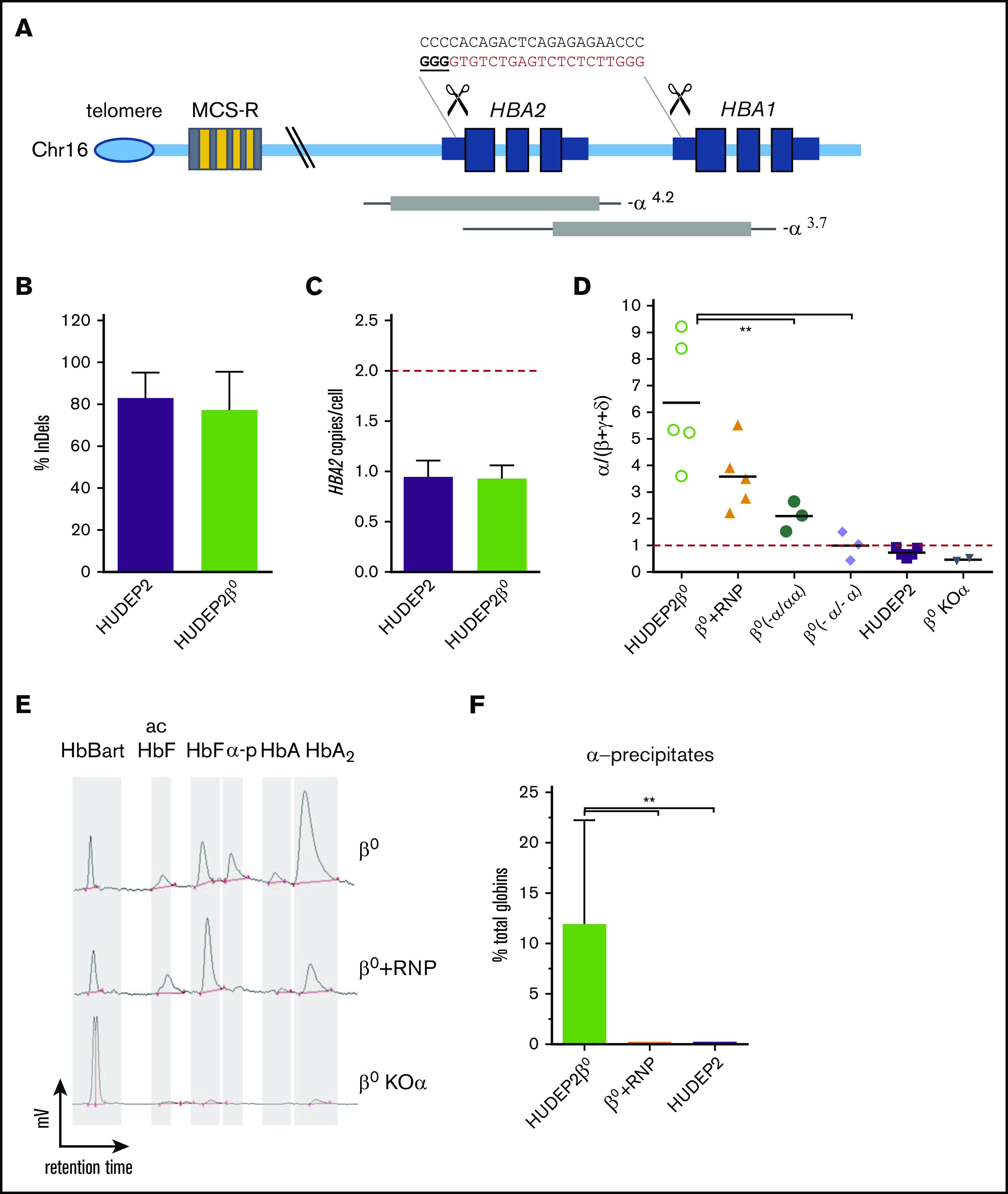
Deletion of HBA2 reduces α-globin precipitates in HUDEP-2 β0cells. (A) Schematic representation of the α-globin locus. HBA1 and HBA2 genes are located in the subtelomeric region of chromosome (Chr) 16. Expression is regulated by 4 erythroid-specific enhancers (multispecies conserved elements [MCS-R]) located 10 to 50 kb upstream. Common occurring deletions are shown as gray bars and thin lines indicate regions of uncertainty of the breakpoints (adapted from Harteveld and Higgs98). gRNA cutting sites are indicated as scissors. gRNA nucleotide sequence is indicated on top (in bold/underlined the protospacer adjacent motif sequence). (B) Editing efficiency in HUDEP-2 cells is expressed as percentage of modified HBA alleles. Bars represent mean ± SD (n = 3). (C) HBA2 copies in edited cells in panel B. Red dashed line indicates the number of expected HBA2 alleles in normal cells. (D) Reverse transcription-qPCR quantification of α/β-like globin mRNA ratios in HUDEP-2 β0 (n = 3-5) and in single-cell clones with monoallelic (α-/αα; n = 3) or biallelic (α-/α-; n = 3) HBA2 deletions. β0 + RNP = HUDEP2 β0 bulk population edited with gRNA HBA15/Cas9. β0 KOα = HUDEP2 β0 bulk population edited with gRNA HBB/Cas9 to induce α-globin KO. Red dashed line indicates the physiological α/β-like ratio of 1. Bars represent mean ± SD (**P < .01, ANOVA, Tukey test). (E) Representative HPLC chromatograms of globin tetramer analysis in edited HUDEP-2 β0. γ4, Hb Bart; acHbF, acetylated fetal hemoglobin; HbF, fetal hemoglobin (α2γ2); α-p, α-precipitates; HbA, adult hemoglobin (α2β2). (F) Quantification of α-precipitates. Every tetramer is reported as percent of total hemoglobins; bars show mean ± SD (n = 3; **P < .01; ANOVA, Tukey test).
We transfected both wild-type HUDEP-2 and HUDEP-2 β0 with RNP targeting HBA and we achieved efficient editing (83.1% ± 12.1 and 77.3% ± 18.2, respectively, n = 3; Figure 1B) and genomic deletion of HBA2 gene (0.9 ± 0.2 and 0.9 ± 0.1 HBA2 copy per cell, n = 3; Figure 1C), which resulted in a decrease of α-globin messenger RNA (mRNA) expression upon erythroid differentiation (Figure 1D). To establish a correlation between α-globin expression and number of HBA genes, we generated multiple cell clones with mono- or biallelic HBA2 deletions (-α/αα and -α/-α, respectively, n = 3 per genotype) and we showed a significant amelioration of the α/β-like globin imbalance upon deletion of HBA2, with the -α/-α clones being indistinguishable from wild-type HUDEP-2 cells (Figure 1D; supplemental Figure 1D). Importantly, our HBA2 deletion strategy reduced but did not abolish α-globin production, as observed in α-globin KO control cells obtained with a gRNA targeting the coding sequence within the first exon of HBA1 and HBA2 genes (Figure 1D).
We also measured the relative abundance of the different hemoglobin forms by HPLC analysis. In the absence of β-globin, a portion of the α-globin pool complexes with β-like globins, such as γ- and δ-globins (to form HbF and HbA2, respectively), whereas the excess precipitates in insoluble aggregates (α-precipitates). Remarkably, RNP deletion of HBA2 genes significantly reduced α-precipitates without affecting hemoglobin synthesis (Figure 1E-F). This is in sharp contrast with α-globin KO cells, where the predominant hemoglobin observed was the toxic HbBart (γ-globin tetramers), typical of severe forms of α-thalassemia (Figure 1E). This result clearly indicates that only a controlled reduction of α-globin results in a beneficial effect.
Targeted integration of βAS3 transgene restores HbA expression in a β0 adult erythroid cell line
To treat β0-thalassemia patients, it is essential to express β-like globin chains; however, reaching expression levels to balance the endogenous α-globin has proven very challenging.14 Therefore, we aim to lower the therapeutic threshold of β-globin expression by reducing α-globin abundance. For this purpose, we combined HBA2 deletion with the KI of a β-globin transgene under the control of the endogenous HBA promoter. The same gRNA that deletes the HBA2 gene will facilitate the integration of the β-globin transgene at this locus, whereas the endogenous HBA promoter will provide strong erythroid β expression, as previously suggested.38-40 To discriminate exogenous vs endogenous β-globin expression, we used an HBB transgene containing 3 antisickling point mutations (βAS3).21
To optimize βAS3 expression, we designed 2 DNA donor cassettes: (1) a βAS3 cDNA followed by a posttranscriptionally regulatory element41 and SV40 polyadenylation signal (βAS3 cDNA); and (2) a βAS3 transgene that includes full-length HBB introns and the endogenous 3′UTR and polyadenylation signal (βAS3 full). Both cassettes were cloned in an AAV6 vector with a GFP reporter gene under the control of a constitutive promoter and flanked by homology arms to favor homologous DNA recombination (HDR)42 (Figure 2A).
Figure 2.
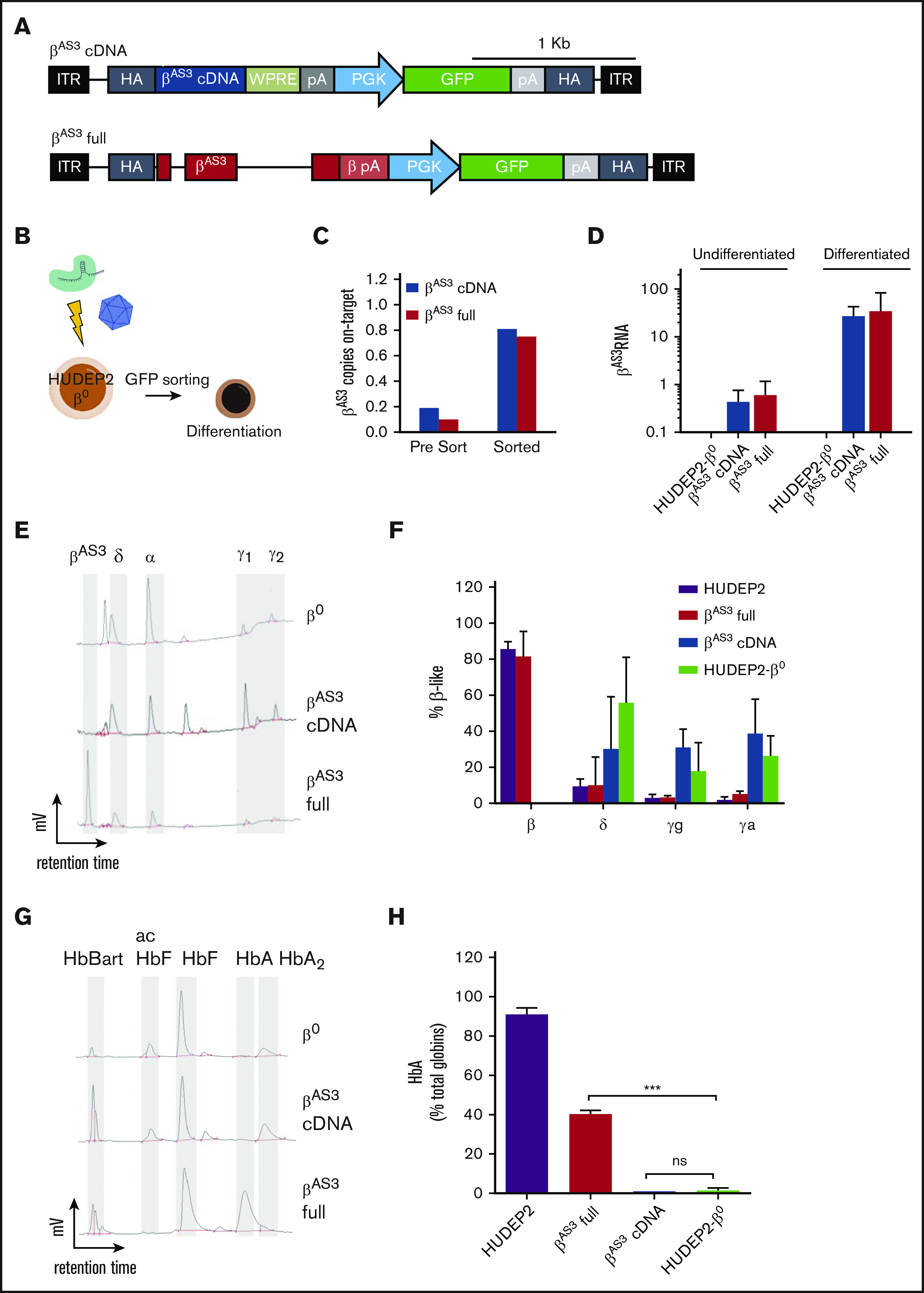
Targeted integration of a βAS3transgene in the α-globin locus corrects thalassemic phenotype in HUDEP-2 β0cells. (A) AAV6 donors used for KI experiments. Both vectors contain a promoterless βAS3 transgene, followed by a phosphoglycerate kinase (PGK) promoter with a GFP reporter and simian virus pA. This cassette is flanked by 250-bp homology arms (homology) to gRNA genomic target. ITR, inverted terminal repeats. Top: βAS3 cDNA followed by the woodchuck posttranscriptionally regulatory element (WPRE) and SV40 pA (βAS3 cDNA); bottom: βAS3 transgene that includes endogenous introns, 3′UTR and pA (βAS3 full). A 1-kb scale bar is indicated at top. (B) Schematic representation of HUDEP-2 β0 targeting experiments. (C) KI efficiency of βAS3 cDNA (blue) or βAS3 full (red) in HUDEP-2 β0 cells measured by on-target ddPCR before and after sorting. (D) βAS3 transcript upregulation in targeted HUDEP-2 β0 upon erythroid differentiation (qPCR, n = 2, mean ± SD). (E-F) HPLC analysis of globin monomers in differentiated HUDEP-2 β0. Representative chromatograms (E) and relative quantification (F) of β-like subunits are shown; (mean ± SD; n = 3). (G-H) HPLC analysis of globin tetramers in differentiated HUDEP-2 β0. Representative chromatograms (G) and HbA (α2β2) quantification (H) are shown (mean ± SD, n = 3; **P < .01; ANOVA, Tukey test). ns, not significant.
To perform KI HUDEP-2 β0 cells were transfected with RNP, transduced with AAV6, and then GFP sorted to enrich for βAS3 integration (Figure 2B). A specific ddPCR confirmed on-target integration at molecular level (∼0.8 βAS3 on-target copies per cell), in good agreement with GFP expression (∼95% GFP positive cells after sorting) (Figure 2C).
Analysis of βAS3 mRNA expression in sorted cells showed an upregulation of about 100-fold for both cassettes upon erythroid differentiation, as expected from the endogenous α-globin promoter (Figure 2D). Although RNA levels were similar, we could detect βAS3 globin protein only with the βAS3 full, but not with the βAS3 cDNA cassette (Figure 2E-F). This observation was further confirmed by hemoglobin tetramer analysis, in which only βAS3 full cassette successfully restored ∼40% of HbA (α2β2) (Figure 2G-H). Overall, these results demonstrate that KI of βAS3 full cassette under the endogenous HBA promoter can restore HbA expression in β0 thalassemic cells.
HSPCs can be efficiently edited and retain long-term and multilineage engraftment potential
To evaluate this strategy in clinically relevant cells, we transfected human umbilical cord blood HSPCs with RNP and then transduced with AAV, as described for HUDEP-2 cells (Figure 3A). Without any selection, we achieved robust genome cutting (Figure 3B), with an InDel pattern consisting mostly of 1 T nucleotide insertion (supplemental Figure 2A-C), together with efficient HBA2 deletion and donor DNA KI (Figure 3C-D).
Figure 3.
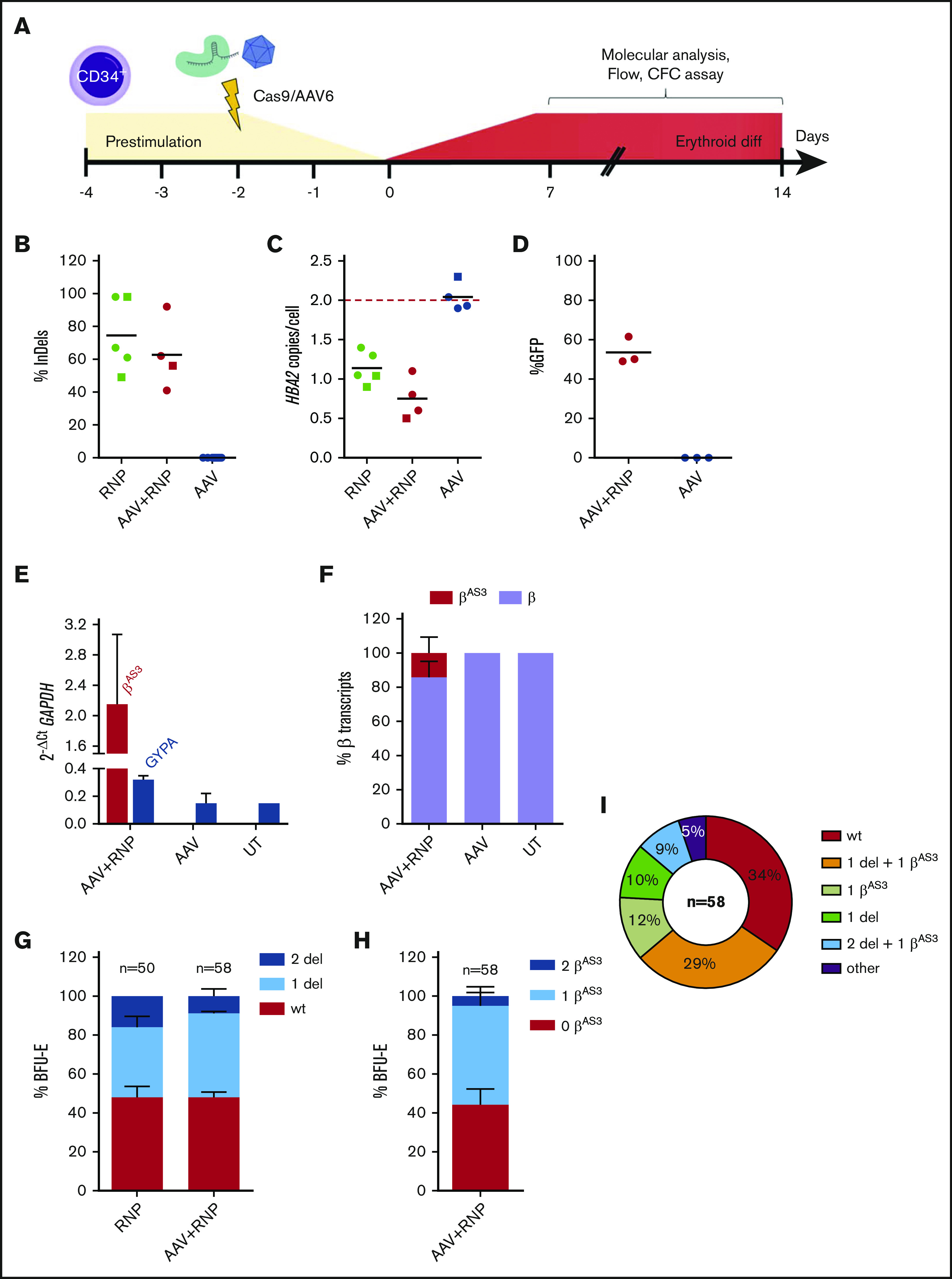
HBA2 deletion and βAS3integration efficiency in HSPCs. (A) Schematic representation of HSPC targeting experiments. (B) Editing efficiencies in HSPCs at day 12 of erythroid differentiation. Lines represent mean. HBA2 copies (C) and KI efficiency (D) in edited HSPCs in erythroid liquid culture (●) or in BFU-E (■). Black lines represent mean; red line indicates the number of expected HBA2 alleles in untreated HSPCs. (E) βAS3 and glycophorin A (GYPA) transcripts in HSPC-derived erythroblasts (qPCR, n = 2, mean ± SD). (F) Relative abundance of endogenous β and KI-βAS3 mRNA at day 12 of erythroid liquid culture (n = 6, mean ± SD). HBA2 deletion (G) and βAS3 integration (H) pattern in single BFU-E. Bars represent mean ± SD (colonies derived from 2 independent experiments); no integration (0), monoallelic (1), and biallelic (2) KI or deletion (del). (I) Genotypes distribution of single KI-βAS3 BFU-E. Percentages are indicated (n = 58).
Upon differentiation, HSPC-derived erythroblasts expressed βAS3-mRNA, which accounted for ∼15% (14.2 ± 9.4, n = 6) of total β globin RNA (Figure 3E-F).
We then plated HSPCs in methylcellulose containing cytokines supporting erythroid and myeloid differentiation (CFC assay), and we confirm that modified progenitors retained their multilineage potential, although some toxicity was observed (supplemental Figure 2D-E).
Single BFU-E genotyping showed that the majority of colonies had HBA2 deletion (Figure 3G) and βAS3 KI (Figure 3H) and 41% of them harbored both modifications (Figure 3I; supplemental Figure 2F).
Both HBA2 deleted and βAS3 KI HSPCs were then transplanted in immunodeficient NOD/SCID/γ (NSG) mice43 to evaluate their in vivo homing, engraftment, and multilineage potential (Figure 4A). Both edited HSPCs showed successful engraftment in bone marrow, spleen and blood, and multilineage differentiation (Figure 4B; supplemental Figure 3B). Because NSG mice do not support human erythroid differentiation,44 we confirmed differentiation by CFC assay of isolated human CD34+ cells from bone marrow of engrafted mice (supplemental Figure 3C).
Figure 4.
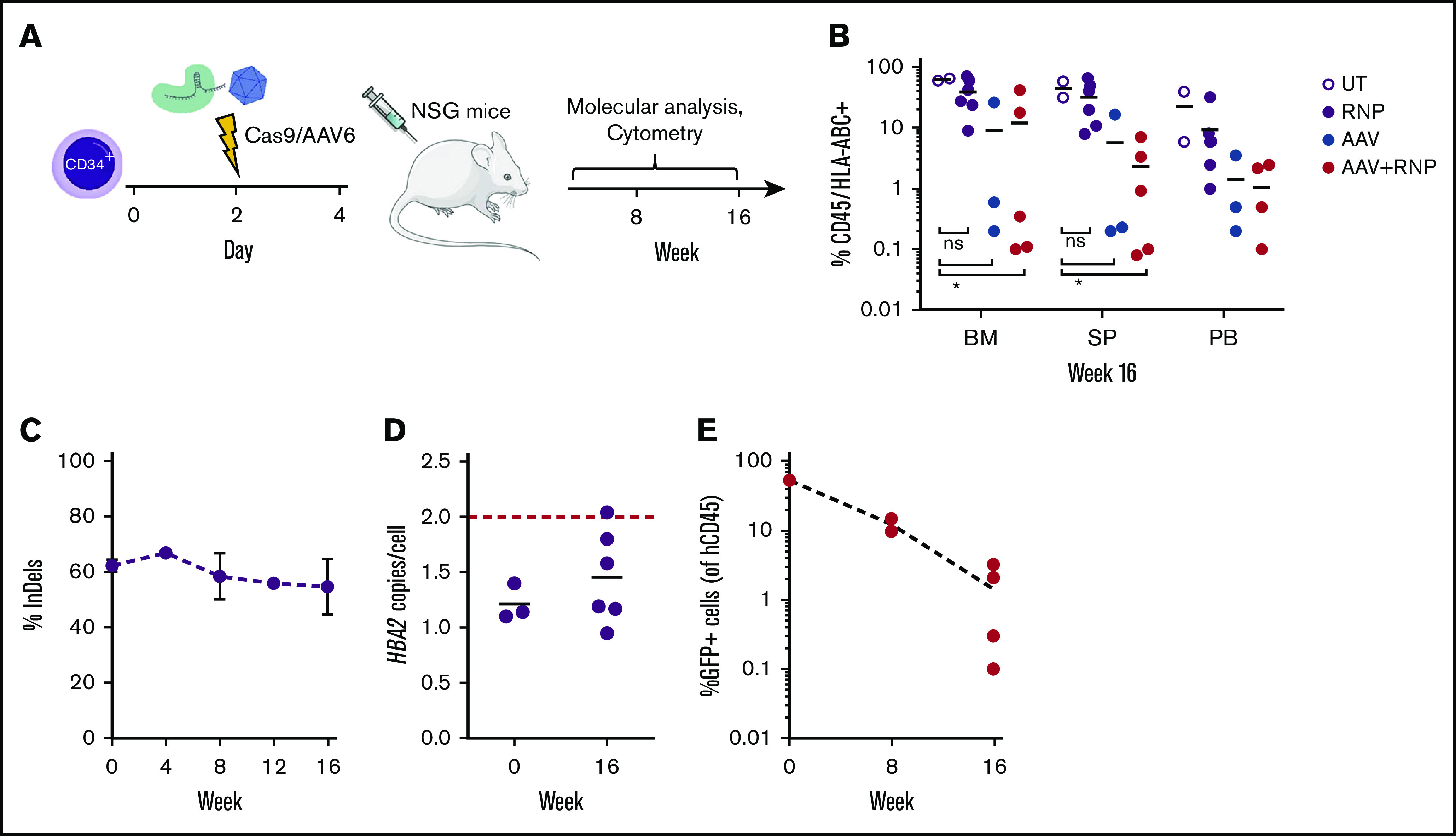
HBA2-deleted and βAS3KI HSPCs engraft NSG mice and maintain their multilineage potential. (A) Schematic representation of engraftment experiments. (B) Percentage of human CD45+/HLA-ABC+ cells in hematopoietic organs of mice. BM, bone marrow; PB, peripheral blood; SP, spleen. Black lines indicate mean. (C) InDel efficiency in PB of RNP-engrafted mice at different timepoints (mean ± SD; n = 2-4). (D) HBA2 copies in RNP-treated HSPCs at day 0 (injection) and in BM of engrafted mice at week 16. Black lines indicate mean; red line indicates the number of expected HBA2 alleles in untreated HSPCs. (E) GFP+ cells in PB of transplanted mice over time. GFP is expressed as percentage of CD45+ cells; line indicates mean.
In addition, the percentage of InDels, as well as the extent of HBA2 deletion in RNP-treated HSPCs, remained similar ex vivo and in vivo, confirming a similar efficiency in both stem and progenitor cells (Figure 4C-D).
βAS3KI HSPCs (GFP+) were present at different time points (Figure 4E) in different lineages (supplemental Figure 3D); however, their engraftment was lower compared with unedited or HBA2-deleted HSPCs (Figure 4B,E), in accordance with previous reports describing AAV toxicity in HSPCs45,46 and less efficient KI in noncycling HSCs.47 Overall, these data show that we can achieve efficient HBA2 deletion and βAS3 KI in HSPCs while preserving their in vivo long-term engraftment.
Editing patients’ HSPCs ameliorates β+- and β0-thalassemia phenotype
To test our strategy in therapeutic conditions, we assessed correction of globin imbalance in patients’ HSPCs. In particular, we tested the HBA2 deletion approach in β+ cells and the combination of the α-deletion and βAS3 KI strategy in β+- and β0-thalassemic HSPCs (supplemental Figure 4A-B).
HSPCs were transfected with RNP and transduced with AAV as described previously (Figure 3A). As positive control, HSPCs were transduced with a LV encoding for a βAS3 transgene under the control of the β-globin gene promoter and its mini-locus control region, currently in clinical trial for thalassemia (LV βAS3).13,48
RNP transfection was very efficient both in erythroid liquid culture and CFC in generating InDels (90.5% ± 7.1 for RNP and 90.4% ± 9.0 for RNP+AAV, mean ± SD; Figure 5A), HBA2 deletion (0.95 ± 0.06 HBA2 copies/cell for RNP and 0.77 ± 0.15 for RNP+AAV, mean ± SD; Figure 5B) and βAS3 KI (0.80 ± 0.21 copies/cell for RNP+AAV, mean ± SD; Figure 5C).
Figure 5.
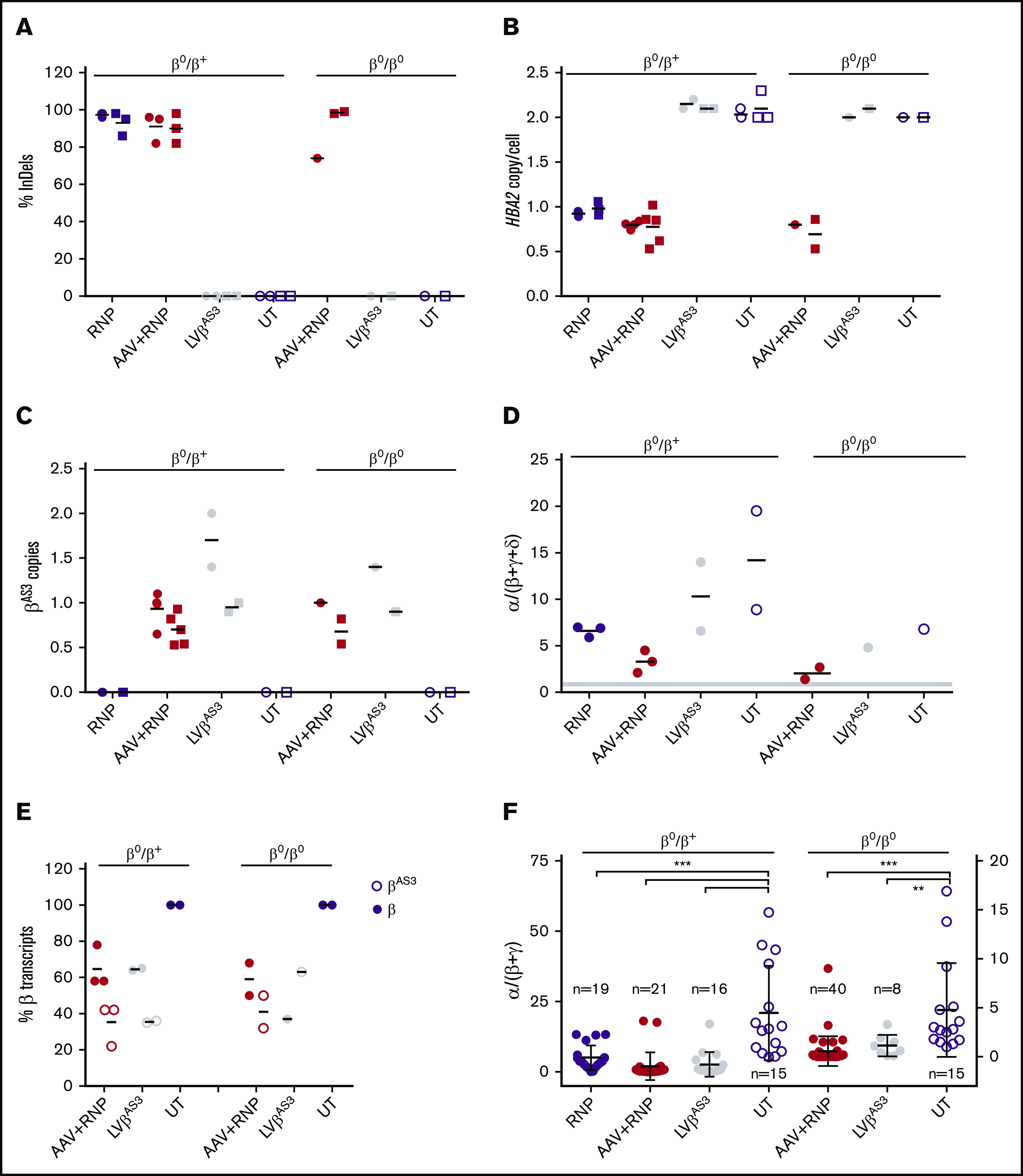
Genome editing of the α-globin locus ameliorates globin balance in thalassemic HSPCs. InDels (A), HBA2 copies (B), and βAS3 integrated copies (C) quantification in edited thalassemic HSPCs in erythroid liquid culture (●) or in BFU-E (■). Black lines represent mean. HSPCs from 1 patient of each genotype were used. (D) α/β-like globin mRNA ratios in edited thalassemic erythroblasts at day 12. Black lines indicate mean; gray line shows the range of α/β-like globin ratio in healthy donor MPB and UCB HSPCs (n = 5). (E) Relative abundance of endogenous β and integrated βAS3 mRNA at day 12 of erythroid liquid culture. Black lines represent mean. (F) α/β-like globin mRNA ratios in edited thalassemic BFU-E. β0/β+colonies (n = 71) are plotted on the left axis, β0/β0 (n = 63) on the right axis. Each dot represents a single colony. Black lines indicate mean ± SD (***P < .001; **P < .01; ANOVA, Tukey test). HSPCs derived from 1 patient of each genotype were used for this figure.
Globin expression was monitored during erythroid differentiation. mRNA globin imbalance (measured as α/β-like globins ratio) was ameliorated in all conditions (Figure 5D), with βAS3 KI cells performing better than HBA2-deleted erythroblasts because of βAS3 expression (Figure 5E; supplemental Figure 4C). Of note, modified HSPCs retained proper erythroid differentiation (supplemental Figure 4D) and multilineage potential (CFC assay; supplemental Figure 4F), although we observed some toxicity associated with the editing procedure (supplemental Figure 4E). Globin mRNA analysis of single BFU-E colonies derived from β0- and β+-edited HSPCs showed that α/β imbalance was improved in all conditions, in accordance with the reduced number of HBA2 genes (supplemental Figure 4G) and with the stronger effect resulting from the concomitant α downregulation and βAS3 expression (Figure 5F).
Overall, these data show that we can modify β-thalassemia HSPCs and reduce their α/β globin balance, without affecting HSPCs potential.
Cas9 nickase represents a safe tool for HSPC editing
To reduce HSPC toxicity associated with nuclease-induced DSBs, we evaluated the use of Cas9 D10A nickase (Cas9 D10A). Because nicked genomic DNA is corrected by the endogenous base-excision repair pathway,49 Cas9 D10A is expected to have minimal genotoxicty.50,51
To compare the efficiency of HBA2 deletion and transgene targeted integration with single vs trans paired nicking of genomic DNA,52 we selected another gRNA targeting α-globin 5′UTR (HBA20; supplemental Figure 5A-C) to combine with gRNA HBA15. K562 erythroleukemia cells were transfected with 1 or both gRNA complexed with Cas9 or Cas9D10A (RNP D10A) and transduced with a dual-reporter AAV vector to detect HDR (via a promoterless low-affinity nerve growth factor receptor) and non-HDR (via a phosphoglycerate kinase 1-GFP) integrations (supplemental Figure 5D). We first confirmed that gRNA HBA20 was comparable to HBA15 in generating InDels and HBA2 deletions when complexed with wild-type Cas9 (RNP) (supplemental Figure 5E-F). When using RNP D10A, we observed that, without inducing any InDels (supplemental Figure 5E), both single and dual gRNA induced efficient HBA2 deletion and HDR integration, although to a lower extent compared with RNP (supplemental Figure 5F-H). Finally, we confirmed that RNP and RNP D10A gave the same ratio of HDR and non-HDR-mediated DNA integration (supplemental Figure 5I). Single gRNA generating 2 in cis nicks could induce genomic deletion by strand displacement, possibly facilitated by the homology of HBA1 and 2 genes, whereas nick induced AAV integration can proceed via genome-AAV alignment, followed by Holiday junction resolution and completion of homologous recombination.53 Because we did not observe any difference between HBA15 and dual gRNA RNP D10A, we decided to continue on HSPCs using only HBA15.
HSPCs were transfected with RNP or RNP D10A and then transduced with the AAV6 βAS3 donor DNA. In both conditions, we observed HBA2 deletion, βAS3 KI and, upon erythroid differentiation, βAS3 mRNA upregulation (Figure 6A-D), although on-target InDels were observed only with Cas9 as a result of DSB repair (Figure 6E). The lower number of DSBs could also explain the reduced toxicity observed in CFC edited with Cas9 D10A (Figure 6F-G).
Figure 6.
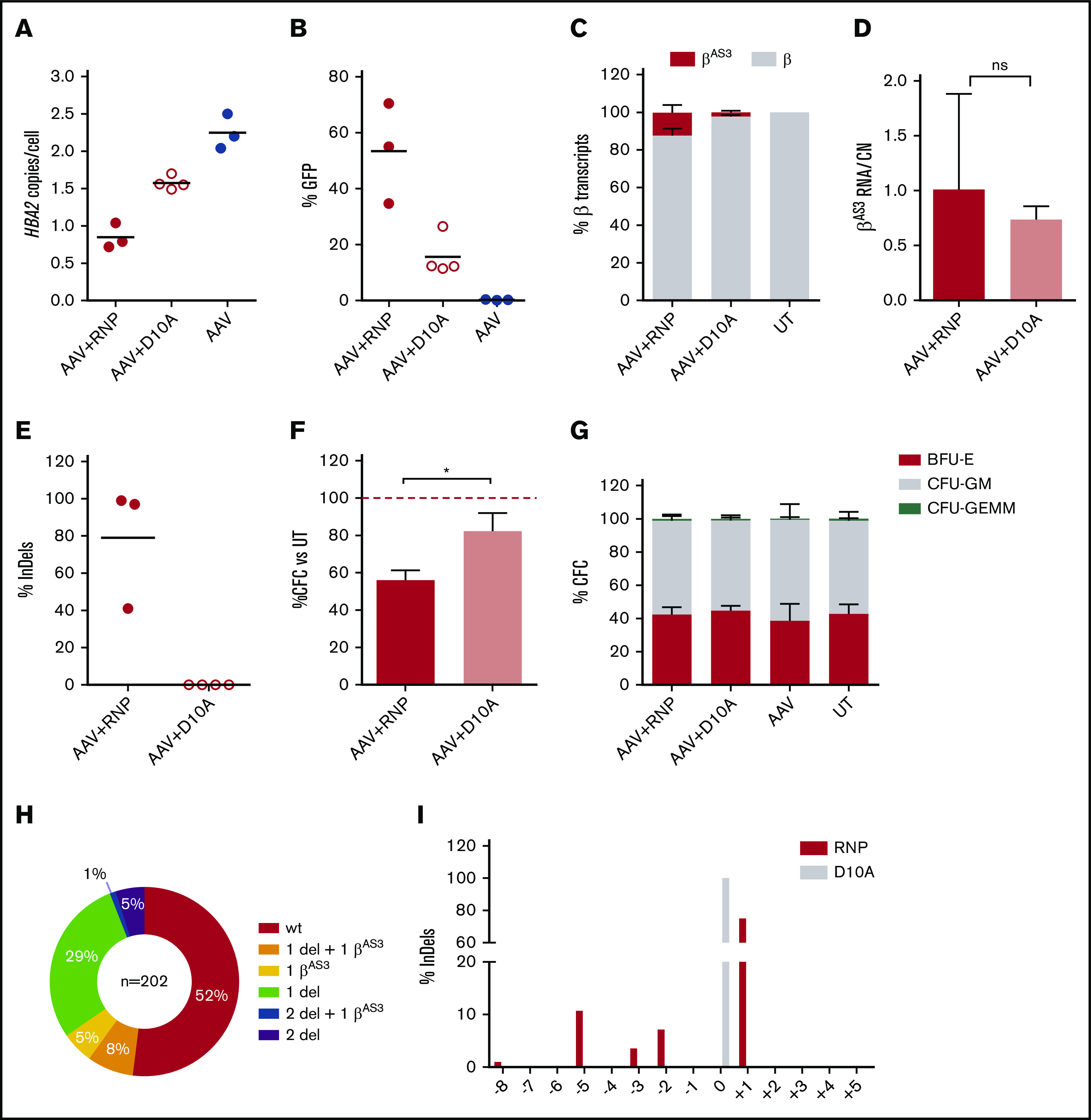
Cas9 nickase editing results in seamless HBA2 deletion and reduced HSPCs toxicity. (A-B) HBA2 copies (A) and KI efficiency (B) in Cas9 (RNP) or Cas9 nickase (RNP D10A) edited HSPCs in erythroid liquid culture. Black lines represent mean. (C) Relative abundance of endogenous β and KI-βAS3 mRNA at day 12 of erythroid liquid culture (mean ± SD, n = 3-4). (D) expression of βAS3 transcripts normalized by copy number (CN) in Cas9- or Cas9 nickase-treated erythroblasts (mean ± SD, n = 4-5). (E) InDel quantification at gRNA target site in edited HSPCs. Black lines represent mean. (F) CFC number expressed as percentage of untreated control (UT). Bars represent mean ± SD (n = 3-4); red dashed line indicates 100%. (G) CFU frequency in edited HSPCs. BFU-E, burst-forming unit-erythroid; CFU-GM, CFU-granulocyte, macrophage. Colony-forming unit-granulocyte, erythroid, macrophage, megakaryocyte; Bars represent mean ± SD (n = 2-4). (H) Genotypes of BFU-E (n = 202) derived from RNP D10A + AAV HSPCs. Percentages are indicated. (I) Frequency of different InDel patterns in HBA2 deleted BFU-E treated with Cas9 (RNP, red; n = 28) or Cas9 nickase (RNP D10A, gray; n = 96). Editing was measured across HBA2 deletion junctions.
By genotyping single BFU-E colonies, we observed that most of RNP D10A-edited BFU-E had 1 HBA2 deletion, followed by 1 HBA2 deletion and 1 βAS3 KI (Figure 6H; supplemental Figure 6A-B). In addition, we performed Sanger sequencing of PCR products spanning deletion junctions and observed that, whereas Cas9 left a composite pattern of InDels, Cas9 D10A deletions were seamless, suggesting absence of exonuclease processing (Figure 6I).
Last, in bulk populations of RNP and RNP D10A edited HSPC, deep sequencing of 3 top-scoring off-targets identified by GUIDE-Seq32 in 293T cells (supplemental Figure 6C) showed low to undetectable off-target activity for both Cas9 and Cas9 D10A (supplemental Figure 6D). This analysis, together with our previous off-target analyses,37 confirms the target specificity of the selected gRNA HBA15 sequence.
Overall, these data indicate that Cas9 D10A performs HBA2 deletion and βAS3 KI, although less efficiently than Cas9, with no InDels and lower cellular toxicity, representing a promising alternative strategy for HSPC editing.
Discussion
In this study, we demonstrated the possibility of correcting α/β globin imbalance in β-thalassemia cells using the CRISPR/Cas9 system. In particular, we showed an amelioration in both β+- and β0-thalassemia patients’ HSPC-derived erythroblasts by combining 2 approaches: α-globin reduction by deleting HBA2 gene to mimic α-thalassemia (-α/αα or -α/-α) and concomitant targeted integration and expression of a HBB transgene under the control of the endogenous HBA promoter. Because edited HSPCs retained engraftment and multilineage differentiation potential in vivo, the proposed strategy has clear potential for future clinical testing.
Inspired by clinical data showing that α-thalassemia ameliorates β-thalassemia,3 several groups have proposed artificial downregulation of α-globin expression as potential treatment of β+-thalassemia, where residual β-globin expression guarantees sufficient hemoglobin formation (such as HbE, about one-half of all β-thalassemias).3
Using a single gRNA targeting the 5′ untranslated region of both α-globin genes, we achieved efficient α-globin reduction by deleting 1 α-globin gene (HBA2), which corrected α/β imbalance and reduced α-precipitates in HUDEP-2 β0. Because HBA1 and HBA2 are similarly expressed,54 removal of 1 or 2 HBA2 genes should reduce α-globin expression to ∼75% to 50% of its normal level, thus falling in the therapeutic window to benefit β-thal.3 This α-globin reduction strategy could be used to treat moderate β+-thalassemia or to complement pharmacological treatment aimed at increasing expression of β-like globin in severe β+ or β0 patients.55-57 To treat β0-thalassemia, where there is no residual β-globin expression, we combined α-globin reduction with β-globin replacement. We optimized, delivered, and integrated an AAV6 βAS3 expression cassette under the control of the endogenous HBA promoter by HDR using the same gRNA deleting HBA2 gene. Remarkably, the expression of 1 βAS3 transgene was sufficient to reestablish almost one-half of normal hemoglobin level in HUDEP-2 β0.
Hijacking the endogenous HBA promoter for the expression of βAS3 transgene has 2 main advantages: (1) it does not require the genomic insertion of an exogenous promoter, which could result in transactivation of neighboring genes; and (2) compared with HBB gene correction strategies, it is a viable approach also for point mutations and deletions that inactivate regulatory elements in the HBB promoter.58
We confirmed that this “double” editing is feasible and efficient in primary human HSPCs from healthy donors and β+- or β0-thalassemia patients and does not affect erythroblast differentiation.
Both HBA2 deletion and βAS3 KI improved α/β globin imbalance to levels that were comparable or better than current HBB gene addition strategy based on LV. Additional experiments will be performed to systematically compare these 2 gene therapy strategies; however, our encouraging results support the development of a CRISPR/Cas9-based gene therapy approach. In addition, by transplanting HSPCs in humanized NSG, we demonstrated that both HBA2 deletion and βAS3 KI occur in long-term HSCs and that edited HSCs can give rise to multiple progenitors and differentiated hematopoietic lineages.
The idea of downregulating the expression of α-globin alone or in combination with increased expression of β-globin has already been proposed by several groups in mice and human cells with mixed outcomes.19,59,60 The only test on thalassemic patient’s HSPCs was performed using a foamy viral vector encoding an HBB transgene and a short hairpin against α-globin mRNA61; however, this approach is hindered by limitations of this RNA interference technology62-64 and the mutagenic potential of semirandom integrating viral vector.65,66
Alternatively, α-globin mRNA downregulation was obtained in HSPCs with epigenetic drugs, such as broad-spectrum histone demethylase or deacetylase inhibitor; nevertheless, the effect on α-globin protein was not established and drug specificity, toxicity, and mechanism of action still need to be assessed.67-69
Using genome editing tools, Mettananda et al elegantly demonstrated the possibility of reducing α-globin expression in HbE patients’ cells by deleting a powerful α-globin enhancer.20 Although promising, this modification is beneficial only for β+ patients and requires plasmid transfection combined with GFP-sorting enrichment, a strategy that is not suited for clinical translation.25 Finally, the need for 2 gRNAs to perform enhancer deletion increases the risk of on- and off-target side effects and can be technically challenging.
Our approach has the advantage of combining α reduction with β expression to restore α/β-globin balance and increase hemoglobin level to treat both β0- and β+-thalassemia patients’ HSPCs. Technically, we deliver Cas9/gRNA as RNP, which allows efficient HSPC modification without any selection and is already used in clinical trials (eg, NCT03164135, NCT03655678). In addition, our strategy requires only 1 gRNA, thus minimizing the possibility of on- and off-target side effects.
To further reduce this risk and general cellular toxicity associated with Cas9-induced DSB, we investigated the use of Cas9 D10A nickase.51,70-72 Inspired by Metzger et al, who demonstrated that site-specific nicking enzymes can induce HDR with an AAV-delivered template in 293 cells,53 we showed for the first time that a single Cas9 D10A/gRNA RNP can be used in HSPCs to achieve seamless genomic deletion and AAV targeted integration, without generating any InDels. Because the efficiency of targeted integration obtained with Cas9 D10A is lower than with Cas9, we will further optimize this approach by improving RNP transfection with nanoparticles,73 reducing AAV donor size,74 and using Cas9 nickase variants75 and drugs affecting cellular DNA repair.76,77 Further in vivo studies will be performed with this strategy to demonstrate effective editing of repopulating HSPCs.
By performing GUIDE-Seq analysis,32 we verified the target specificity of the selected gRNA HBA15, confirming our previous off-target analyses.37 However, additional safety aspects need to be carefully evaluated before clinical translation of this HSPC gene therapy platform. It will be particularly challenging to qualitatively and quantitatively assess all the possible on-target editing outcomes, especially low-frequency ones. We quantified HBA2 deletions, but we could not detect any HBA2 inversion by PCR analysis of edited HSPCs, although inversions are reported to be a frequent side product when generating CRISPR genomic deletions.17,78 This observation could be a true negative result, as previously reported for HBG editing in HSPCs,79,80 or because of technical difficulties associated with amplification of repetitive inverted sequences with high GC content present in the α-globin locus.81 In addition, on-target cleavage can generate undesired chromosomal alterations,70,82-84 whereas integration of AAV donor DNA can occur via HDR as well as via partial HDR or nonhomologous end joining with or without the HBA2 deletion.85-87 A combination of whole genome sequencing,88 long-range PCR,89 long-read sequencing,90 next-generation mapping,91 and/or directional sequencing92 will help to address these concerns by unraveling the full spectrum of editing outcomes.
In summary, we describe a novel CRISPR strategy to correct α/β globin imbalance in thalassemic HSPCs by α-globin downregulation with or without HBB transgene expression. Future experiments will elucidate the therapeutic potential of this strategy for treating β+- and β0-thalassemia and, eventually, sickle cell disease, where lower α-globin levels reduce HbS polymerization and sickle hemoglobin concentration and βAS3 compete with β sickling for binding α-globin.21,93-97
Supplementary Material
The full-text version of this article contains a data supplement.
Acknowledgments
The authors thank Chiara Antoniani for the generation of HUDEP-2 β0 cells, Anna De Cian for Cas9 protein production, Samantha Scaramuzza for help with patients HSPCs, Fanny Collaud, Genethon “Vector Core Facility” for AAV production, Genethon “Imaging and Cytometry Core Facility” for image analysis and fluorescence-activated cell sorting, Genethon “Functional Evaluation Facility” for help with mice experimentation, and the Imagine “Genomic Platform” for amplicon sequencing. The authors also thank the entire M.A. laboratory, Marina Cavazzana, Anne Galy, and Ronzitti for fruitful discussion. In addition, the authors thank Ryo Kurita and Yukio Nakamura for proving HUDEP-2 cells under a material transfer agreement with Genethon. The authors gratefully acknowledge the Conseil Général de l’Essonne (ASTRES) and Genopole Research in Evry for financial help for the purchase of equipment and are grateful to consenting mothers and to L. Rigonnot and staff of the Maternity at the Centre Hospitalier Sud-Francilien (CHSF; Evry, France) for providing umbilical cord blood samples.
This work was supported by grants from Bayer Hemophilia Awards Program (G.P.) and AFM-Telethon, INSERM, Genopole Chaire Fondagen and the Agence Nationale de la Recherche (ANR-16-CE18 STaHR) (M.A.). G.P. was supported by European Union’s Horizon 2020 (SCIDNET No 666908).
Footnotes
GUIDE-seq datasets are available in the BioProject online repository (ID PRJNA676022; http://www.ncbi.nlm.nih.gov/bioproject/676022).
Requests for original data should be sent to Giulia Pavani (gpavani@genethon.fr).
Authorship
Contribution: G.P. conceived the study, designed and performed experiments, analyzed data, and wrote the manuscript; A.F. performed experiments and analyzed data on HSPCs; M.L. performed experiments and analyzed data on HUDEP-2 β0 cells; F.A. performed experiments and analyzed data on Cas9 (D10A); E.C. performed experiments and analyzed data on HSPCs; A.C. performed HPLC analyses; G. M. and A.C. performed and analyzed GUIDE-Seq experiments; A.T. performed molecular analyses; J.-P.C. provided purified SpCas9 protein; F.M. provided scientific advice and financial support; G.F. provided β-thalassemia HSPCs; A.M. provided HUDEP-2 β0 cells, performed and analyzed HPLC, and edited the manuscript; and M.A. conceived the study, designed experiments, analyzed data, and wrote the manuscript.
Conflict-of-interest disclosure: G.P. and M.A. are the inventors of a patent describing this HSC-based gene therapy strategy for treating β-thalassemia (correction of β-thalassemia phenotype by genetically engineering hematopoietic stem cell; EP19305484.8). The remaining authors declare no competing financial interests.
Correspondence: Mario Amendola, INTEGRARE, Genethon, UMR S951 INSERM, University Evry, University Paris-Saclay, 1 bis, Rue de I’Internationale, 91000 Evry, France; e-mail: mamendola@genethon.fr.
References
- 1.Cao A, Galanello R. Beta-thalassemia. Genet Med. 2010;12(2):61-76. [DOI] [PubMed] [Google Scholar]
- 2.Weatherall DJ. The challenge of haemoglobinopathies in resource-poor countries. Br J Haematol. 2011;154(6):736-744. [DOI] [PubMed] [Google Scholar]
- 3.Mettananda S, Gibbons RJ, Higgs DR. α-Globin as a molecular target in the treatment of β-thalassemia. Blood. 2015;125(24):3694-3701. [DOI] [PMC free article] [PubMed] [Google Scholar]
- 4.Sharma V, Saxena R. Effect of alpha-gene numbers on phenotype of HbE/beta thalassemia patients. Ann Hematol. 2009;88(10):1035-1036. [DOI] [PubMed] [Google Scholar]
- 5.Fucharoen S, Weatherall DJ. The hemoglobin E thalassemias. Cold Spring Harb Perspect Med. 2012;2(8):a011734. [DOI] [PMC free article] [PubMed] [Google Scholar]
- 6.Sripichai O, Munkongdee T, Kumkhaek C, Svasti S, Winichagoon P, Fucharoen S. Coinheritance of the different copy numbers of alpha-globin gene modifies severity of beta-thalassemia/Hb E disease. Ann Hematol. 2008;87(5):375-379. [DOI] [PubMed] [Google Scholar]
- 7.Charoenkwan P, Teerachaimahit P, Sanguansermsri T. The correlation of α-globin gene mutations and the XmnI polymorphism with clinical severity of Hb E/β-thalassemia. Hemoglobin. 2014;38(5):335-338. [DOI] [PubMed] [Google Scholar]
- 8.Allali S, de Montalembert M, Brousse V, Chalumeau M, Karim Z. Management of iron overload in hemoglobinopathies. Transfus Clin Biol. 2017;24(3):223-226. [DOI] [PubMed] [Google Scholar]
- 9.Besse K, Maiers M, Confer D, Albrecht M. On modeling human leukocyte antigen-identical sibling match probability for allogeneic hematopoietic cell transplantation: estimating the need for an unrelated donor source. Biol Blood Marrow Transplant. 2016;22(3):410-417. [DOI] [PubMed] [Google Scholar]
- 10.Chandrakasan S, Malik P. Gene therapy for hemoglobinopathies: the state of the field and the future. Hematol Oncol Clin North Am. 2014;28(2):199-216. [DOI] [PMC free article] [PubMed] [Google Scholar]
- 11.Saber W, Opie S, Rizzo JD, Zhang MJ, Horowitz MM, Schriber J. Outcomes after matched unrelated donor versus identical sibling hematopoietic cell transplantation in adults with acute myelogenous leukemia. Blood. 2012;119(17):3908-3916. [DOI] [PMC free article] [PubMed] [Google Scholar]
- 12.Sadelain M, Boulad F, Galanello R, et al. Therapeutic options for patients with severe beta-thalassemia: the need for globin gene therapy. Hum Gene Ther. 2007;18(1):1-9. [DOI] [PubMed] [Google Scholar]
- 13.Marktel S, Scaramuzza S, Cicalese MP, et al. Intrabone hematopoietic stem cell gene therapy for adult and pediatric patients affected by transfusion-dependent ß-thalassemia. Nat Med. 2019;25(2):234-241. [DOI] [PubMed] [Google Scholar]
- 14.Thompson AA, Walters MC, Kwiatkowski J, et al. Gene therapy in patients with transfusion-dependent β-thalassemia. N Engl J Med. 2018;378(16):1479-1493. [DOI] [PubMed] [Google Scholar]
- 15.Wu Y, Zeng J, Roscoe BP, et al. Highly efficient therapeutic gene editing of human hematopoietic stem cells. Nat Med. 2019;25(5):776-783. [DOI] [PMC free article] [PubMed] [Google Scholar]
- 16.Martyn GE, Wienert B, Yang L, et al. Natural regulatory mutations elevate the fetal globin gene via disruption of BCL11A or ZBTB7A binding. Nat Genet. 2018;50(4):498-503. [DOI] [PubMed] [Google Scholar]
- 17.Antoniani C, Meneghini V, Lattanzi A, et al. Induction of fetal hemoglobin synthesis by CRISPR/Cas9-mediated editing of the human β-globin locus. Blood. 2018;131(17):1960-1973. [DOI] [PubMed] [Google Scholar]
- 18.Traxler EA, Yao Y, Wang YD, et al. A genome-editing strategy to treat β-hemoglobinopathies that recapitulates a mutation associated with a benign genetic condition. Nat Med. 2016;22(9):987-990. [DOI] [PMC free article] [PubMed] [Google Scholar]
- 19.Xie SY, Ren ZR, Zhang JZ, et al. Restoration of the balanced alpha/beta-globin gene expression in beta654-thalassemia mice using combined RNAi and antisense RNA approach. Hum Mol Genet. 2007;16(21):2616-2625. [DOI] [PubMed] [Google Scholar]
- 20.Mettananda S, Fisher CA, Hay D, et al. Editing an α-globin enhancer in primary human hematopoietic stem cells as a treatment for β-thalassemia. Nat Commun. 2017;8(1):424. [DOI] [PMC free article] [PubMed] [Google Scholar]
- 21.Levasseur DN, Ryan TM, Reilly MP, McCune SL, Asakura T, Townes TM. A recombinant human hemoglobin with anti-sickling properties greater than fetal hemoglobin. J Biol Chem. 2004;279(26):27518-27524. [DOI] [PubMed] [Google Scholar]
- 22.Weber L, Poletti V, Magrin E, et al. An optimized lentiviral vector efficiently corrects the human sickle cell disease phenotype. Mol Ther Methods Clin Dev. 2018;10:268-280. [DOI] [PMC free article] [PubMed] [Google Scholar]
- 23.Ayuso E, Mingozzi F, Bosch F. Production, purification and characterization of adeno-associated vectors. Curr Gene Ther. 2010;10(6):423-436. [DOI] [PubMed] [Google Scholar]
- 24.Rohr UP, Wulf MA, Stahn S, Steidl U, Haas R, Kronenwett R. Fast and reliable titration of recombinant adeno-associated virus type-2 using quantitative real-time PCR. J Virol Methods. 2002;106(1):81-88. [DOI] [PubMed] [Google Scholar]
- 25.Lattanzi A, Meneghini V, Pavani G, et al. Optimization of CRISPR/Cas9 delivery to human hematopoietic stem and progenitor cells for therapeutic genomic rearrangements. Mol Ther. 2019;27(1):137-150. [DOI] [PMC free article] [PubMed] [Google Scholar]
- 26.Kurita R, Suda N, Sudo K, et al. Establishment of immortalized human erythroid progenitor cell lines able to produce enucleated red blood cells. PLoS One. 2013;8(3):e59890. [DOI] [PMC free article] [PubMed] [Google Scholar]
- 27.Vinjamur DS, Bauer DE. Growing and genetically manipulating human umbilical cord blood-derived erythroid progenitor (HUDEP) cell lines. Methods Mol Biol. 2018;1698:275-284. [DOI] [PubMed] [Google Scholar]
- 28.Ménoret S, De Cian A, Tesson L, et al. Homology-directed repair in rodent zygotes using Cas9 and TALEN engineered proteins. Sci Rep. 2015;5(1):14410. [DOI] [PMC free article] [PubMed] [Google Scholar]
- 29.Lidonnici MR, Aprile A, Frittoli MC, et al. Plerixafor and G-CSF combination mobilizes hematopoietic stem and progenitors cells with a distinct transcriptional profile and a reduced in vivo homing capacity compared to plerixafor alone. Haematologica. 2017;102(4):e120-e124. [DOI] [PMC free article] [PubMed] [Google Scholar]
- 30.Wen J, Tao W, Hao S, Zu Y. Cellular function reinstitution of offspring red blood cells cloned from the sickle cell disease patient blood post CRISPR genome editing. J Hematol Oncol. 2017;10(1):119. [DOI] [PMC free article] [PubMed] [Google Scholar]
- 31.Brinkman EK, Chen T, Amendola M, van Steensel B. Easy quantitative assessment of genome editing by sequence trace decomposition. Nucleic Acids Res. 2014;42(22):e168. [DOI] [PMC free article] [PubMed] [Google Scholar]
- 32.Tsai SQ, Zheng Z, Nguyen NT, et al. GUIDE-seq enables genome-wide profiling of off-target cleavage by CRISPR-Cas nucleases. Nat Biotechnol. 2015;33(2):187-197. [DOI] [PMC free article] [PubMed] [Google Scholar]
- 33.Tsai SQ, Topkar VV, Joung JK, Aryee MJ. Open-source guideseq software for analysis of GUIDE-seq data. Nat Biotechnol. 2016;34(5):483. [DOI] [PubMed] [Google Scholar]
- 34.Clement K, Rees H, Canver MC, et al. CRISPResso2 provides accurate and rapid genome editing sequence analysis. Nat Biotechnol. 2019;37(3):224-226. [DOI] [PMC free article] [PubMed] [Google Scholar]
- 35.Badens C, Joly P, Agouti I, et al. Variants in genetic modifiers of β-thalassemia can help to predict the major or intermedia type of the disease. Haematologica. 2011;96(11):1712-1714. [DOI] [PMC free article] [PubMed] [Google Scholar]
- 36.Mettananda S, Higgs DR. Molecular basis and genetic modifiers of thalassemia. Hematol Oncol Clin North Am. 2018;32(2):177-191. [DOI] [PubMed] [Google Scholar]
- 37.Pavani G, Laurent M, Fabiano A, et al. Ex vivo editing of human hematopoietic stem cells for erythroid expression of therapeutic proteins [published correction appears in Nat Commun. 2020;11(11):4146]. Nat Commun. 2020;11(1):3778. [DOI] [PMC free article] [PubMed] [Google Scholar]
- 38.Morianos I, Siapati EK, Pongas G, Vassilopoulos G. Comparative analysis of FV vectors with human α- or β-globin gene regulatory elements for the correction of β-thalassemia. Gene Ther. 2012;19(3):303-311. [DOI] [PubMed] [Google Scholar]
- 39.Moreau-Gaudry F, Xia P, Jiang G, et al. High-level erythroid-specific gene expression in primary human and murine hematopoietic cells with self-inactivating lentiviral vectors. Blood. 2001;98(9):2664-2672. [DOI] [PubMed] [Google Scholar]
- 40.Papanikolaou E, Georgomanoli M, Stamateris E, et al. The new self-inactivating lentiviral vector for thalassemia gene therapy combining two HPFH activating elements corrects human thalassemic hematopoietic stem cells. Hum Gene Ther. 2012;23(1):15-31. [DOI] [PMC free article] [PubMed] [Google Scholar]
- 41.Huang ZM, Yen TS. Role of the hepatitis B virus posttranscriptional regulatory element in export of intronless transcripts. Mol Cell Biol. 1995;15(7):3864-3869. [DOI] [PMC free article] [PubMed] [Google Scholar]
- 42.Wang J, Exline CM, DeClercq JJ, et al. Homology-driven genome editing in hematopoietic stem and progenitor cells using ZFN mRNA and AAV6 donors. Nat Biotechnol. 2015;33(12):1256-1263. [DOI] [PMC free article] [PubMed] [Google Scholar]
- 43.Ishikawa F, Yasukawa M, Lyons B, et al. Development of functional human blood and immune systems in NOD/SCID/IL2 receptor gamma chain(null) mice. Blood. 2005;106(5):1565-1573. [DOI] [PMC free article] [PubMed] [Google Scholar]
- 44.Hu Z, Van Rooijen N, Yang YG. Macrophages prevent human red blood cell reconstitution in immunodeficient mice. Blood. 2011;118(22):5938-5946. [DOI] [PMC free article] [PubMed] [Google Scholar]
- 45.Romero Z, Lomova A, Said S, et al. Editing the sickle cell disease mutation in human hematopoietic stem cells: comparison of endonucleases and homologous donor templates. Mol Ther. 2019;27(8):1389-1406. [DOI] [PMC free article] [PubMed] [Google Scholar]
- 46.Schiroli G, Conti A, Ferrari S, et al. Precise gene editing preserves hematopoietic stem cell function following transient p53-mediated DNA damage response. Cell Stem Cell. 2019;24(4):551-565.e8. [DOI] [PMC free article] [PubMed] [Google Scholar]
- 47.Genovese P, Schiroli G, Escobar G, et al. Targeted genome editing in human repopulating haematopoietic stem cells. Nature. 2014;510(7504):235-240. [DOI] [PMC free article] [PubMed] [Google Scholar]
- 48.Miccio A, Cesari R, Lotti F, et al. In vivo selection of genetically modified erythroblastic progenitors leads to long-term correction of beta-thalassemia. Proc Natl Acad Sci USA. 2008;105(30):10547-10552. [DOI] [PMC free article] [PubMed] [Google Scholar]
- 49.Caldecott KW. Single-strand break repair and genetic disease. Nat Rev Genet. 2008;9(8):619-631. [DOI] [PubMed] [Google Scholar]
- 50.Cong L, Ran FA, Cox D, et al. Multiplex genome engineering using CRISPR/Cas systems. Science. 2013;339(6121):819-823. [DOI] [PMC free article] [PubMed] [Google Scholar]
- 51.Ran FA, Hsu PD, Lin CY, et al. Double nicking by RNA-guided CRISPR Cas9 for enhanced genome editing specificity [published correction appears in Cell. 2013;155(2):479-480]. Cell. 2013;154(6):1380-1389. [DOI] [PMC free article] [PubMed] [Google Scholar]
- 52.Chen X, Janssen JM, Liu J, et al. In trans paired nicking triggers seamless genome editing without double-stranded DNA cutting. Nat Commun. 2017;8(1):657. [DOI] [PMC free article] [PubMed] [Google Scholar]
- 53.Metzger MJ, McConnell-Smith A, Stoddard BL, Miller AD. Single-strand nicks induce homologous recombination with less toxicity than double-strand breaks using an AAV vector template. Nucleic Acids Res. 2011;39(3):926-935. [DOI] [PMC free article] [PubMed] [Google Scholar]
- 54.Molchanova TP, Pobedimskaya DD, Huisman TH. The differences in quantities of alpha 2- and alpha 1-globin gene variants in heterozygotes. Br J Haematol. 1994;88(2):300-306. [DOI] [PubMed] [Google Scholar]
- 55.Bradai M, Abad MT, Pissard S, Lamraoui F, Skopinski L, de Montalembert M. Hydroxyurea can eliminate transfusion requirements in children with severe beta-thalassemia. Blood. 2003;102(4):1529-1530. [DOI] [PubMed] [Google Scholar]
- 56.Biswas S, Ray R, Roy K, Bandyopadhyay A, Ghosh K, Bhattacharyya M. Alpha globin gene mutation: a major determinant of hydroxyurea response in transfusion-dependent HbE-β-thalassaemia. Acta Haematol. 2019;142(3):132-141. [DOI] [PubMed] [Google Scholar]
- 57.Thein SL. The emerging role of fetal hemoglobin induction in non-transfusion-dependent thalassemia. Blood Rev. 2012;26(Suppl 1):S35-S39. [DOI] [PubMed] [Google Scholar]
- 58.Thein SL. The molecular basis of β-thalassemia. Cold Spring Harb Perspect Med. 2013;3(5):a011700. [DOI] [PMC free article] [PubMed] [Google Scholar]
- 59.Xie SY, Li W, Ren ZR, Huang SZ, Zeng F, Zeng YT. Correction of β654-thalassaemia mice using direct intravenous injection of siRNA and antisense RNA vectors. Int J Hematol. 2011;93(3):301-310. [DOI] [PubMed] [Google Scholar]
- 60.Voon HP, Wardan H, Vadolas J. siRNA-mediated reduction of alpha-globin results in phenotypic improvements in beta-thalassemic cells. Haematologica. 2008;93(8):1238-1242. [DOI] [PubMed] [Google Scholar]
- 61.Amendola M, Giustacchini A, Gentner B, Naldini L. A double-switch vector system positively regulates transgene expression by endogenous microRNA expression (miR-ON vector). Mol Ther. 2013;21(5):934-946. [DOI] [PMC free article] [PubMed] [Google Scholar]
- 62.Martin JN, Wolken N, Brown T, Dauer WT, Ehrlich ME, Gonzalez-Alegre P. Lethal toxicity caused by expression of shRNA in the mouse striatum: implications for therapeutic design. Gene Ther. 2011;18(7):666-673. [DOI] [PMC free article] [PubMed] [Google Scholar]
- 63.Grimm D, Streetz KL, Jopling CL, et al. Fatality in mice due to oversaturation of cellular microRNA/short hairpin RNA pathways. Nature. 2006;441(7092):537-541. [DOI] [PubMed] [Google Scholar]
- 64.Manjunath N, Wu H, Subramanya S, Shankar P. Lentiviral delivery of short hairpin RNAs. Adv Drug Deliv Rev. 2009;61(9):732-745. [DOI] [PMC free article] [PubMed] [Google Scholar]
- 65.Rothe M, Modlich U, Schambach A. Biosafety challenges for use of lentiviral vectors in gene therapy. Curr Gene Ther. 2013;13(6):453-468. [DOI] [PubMed] [Google Scholar]
- 66.Cavazzana-Calvo M, Payen E, Negre O, et al. Transfusion independence and HMGA2 activation after gene therapy of human β-thalassaemia. Nature. 2010;467(7313):318-322. [DOI] [PMC free article] [PubMed] [Google Scholar]
- 67.Mai A, Jelicic K, Rotili D, et al. Identification of two new synthetic histone deacetylase inhibitors that modulate globin gene expression in erythroid cells from healthy donors and patients with thalassemia. Mol Pharmacol. 2007;72(5):1111-1123. [DOI] [PubMed] [Google Scholar]
- 68.Mettananda S, Fisher CA, Sloane-Stanley JA, et al. Selective silencing of α-globin by the histone demethylase inhibitor IOX1: a potentially new pathway for treatment of β-thalassemia. Haematologica. 2017;102(3):e80-e84. [DOI] [PMC free article] [PubMed] [Google Scholar]
- 69.Mettananda S, Yasara N, Fisher CA, Taylor S, Gibbons R, Higgs D. Synergistic silencing of α-globin and induction of γ-globin by histone deacetylase inhibitor, vorinostat as a potential therapy for β-thalassaemia. Sci Rep. 2019;9(1):11649. [DOI] [PMC free article] [PubMed] [Google Scholar]
- 70.Cullot G, Boutin J, Toutain J, et al. CRISPR-Cas9 genome editing induces megabase-scale chromosomal truncations. Nat Commun. 2019;10(1):1136. [DOI] [PMC free article] [PubMed] [Google Scholar]
- 71.Chen X, Tasca F, Wang Q, et al. Expanding the editable genome and CRISPR-Cas9 versatility using DNA cutting-free gene targeting based on in trans paired nicking. Nucleic Acids Res. 2020;48(2):974-995. [DOI] [PMC free article] [PubMed] [Google Scholar]
- 72.Hyodo T, Rahman ML, Karnan S, et al. Tandem paired nicking promotes precise genome editing with scarce interference by p53. Cell Rep. 2020;30(4):1195-1207.e7. [DOI] [PubMed] [Google Scholar]
- 73.Nguyen DN, Roth TL, Li PJ, et al. Polymer-stabilized Cas9 nanoparticles and modified repair templates increase genome editing efficiency. Nat Biotechnol. 2020;38(1):44-49. [DOI] [PMC free article] [PubMed] [Google Scholar]
- 74.Pavel-Dinu M, Wiebking V, Dejene BT, et al. Gene correction for SCID-X1 in long-term hematopoietic stem cells [published corrections appear in Nat Commun. 2019;10(1):2021 and 2019;10(1):5264]. Nat Commun. 2019;10(1):1634. [DOI] [PMC free article] [PubMed] [Google Scholar]
- 75.Rees HA, Yeh WH, Liu DR. Development of hRad51-Cas9 nickase fusions that mediate HDR without double-stranded breaks. Nat Commun. 2019;10(1):2212. [DOI] [PMC free article] [PubMed] [Google Scholar]
- 76.Metzger MJ, Stoddard BL, Monnat RJ Jr.. PARP-mediated repair, homologous recombination, and back-up non-homologous end joining-like repair of single-strand nicks. DNA Repair (Amst). 2013;12(7):529-534. [DOI] [PMC free article] [PubMed] [Google Scholar]
- 77.Vriend LE, Krawczyk PM. Nick-initiated homologous recombination: protecting the genome, one strand at a time. DNA Repair (Amst). 2017;50:1-13. [DOI] [PubMed] [Google Scholar]
- 78.Li J, Shou J, Guo Y, et al. Efficient inversions and duplications of mammalian regulatory DNA elements and gene clusters by CRISPR/Cas9. J Mol Cell Biol. 2015;7(4):284-298. [DOI] [PMC free article] [PubMed] [Google Scholar]
- 79.Métais JY, Doerfler PA, Mayuranathan T, et al. Genome editing of HBG1 and HBG2 to induce fetal hemoglobin. Blood Adv. 2019;3(21):3379-3392. [DOI] [PMC free article] [PubMed] [Google Scholar]
- 80.Li C, Psatha N, Sova P, et al. Reactivation of γ-globin in adult β-YAC mice after ex vivo and in vivo hematopoietic stem cell genome editing. Blood. 2018;131(26):2915-2928. [DOI] [PMC free article] [PubMed] [Google Scholar]
- 81.Flint J, Tufarelli C, Peden J, et al. Comparative genome analysis delimits a chromosomal domain and identifies key regulatory elements in the alpha globin cluster. Hum Mol Genet. 2001;10(4):371-382. [DOI] [PubMed] [Google Scholar]
- 82.Kosicki M, Tomberg K, Bradley A. Repair of double-strand breaks induced by CRISPR-Cas9 leads to large deletions and complex rearrangements [published correction appears in Nat Biotechnol. 2018;36:899]. Nat Biotechnol. 2018;36(8):765-771. [DOI] [PMC free article] [PubMed] [Google Scholar]
- 83.Zuccaro MV, Xu J, Mitchell C, et al. Allele-specific chromosome removal after Cas9 cleavage in human embryos. Cell. 2020;183(6):1650-1664. [DOI] [PubMed] [Google Scholar]
- 84.Adikusuma F, Piltz S, Corbett MA, et al. Large deletions induced by Cas9 cleavage. Nature. 2018;560(7717):E8-E9. [DOI] [PubMed] [Google Scholar]
- 85.Nelson CE, Wu Y, Gemberling MP, et al. Long-term evaluation of AAV-CRISPR genome editing for Duchenne muscular dystrophy. Nat Med. 2019;25(3):427-432. [DOI] [PMC free article] [PubMed] [Google Scholar]
- 86.Hanlon KS, Kleinstiver BP, Garcia SP, et al. High levels of AAV vector integration into CRISPR-induced DNA breaks. Nat Commun. 2019;10(1):4439. [DOI] [PMC free article] [PubMed] [Google Scholar]
- 87.Canaj H, Hussmann JA, Li H, et al. Deep profiling reveals substantial heterogeneity of integration outcomes in CRISPR knock-in experiments. bioRxiv. 2019:841098. </prpt> [Google Scholar]
- 88.Zuo E, Huo X, Yao X, et al. CRISPR/Cas9-mediated targeted chromosome elimination. Genome Biol. 2017;18(1):224. [DOI] [PMC free article] [PubMed] [Google Scholar]
- 89.Dolan AE, Hou Z, Xiao Y, et al. Introducing a spectrum of long-range genomic deletions in human embryonic stem cells using type I CRISPR-Cas. Mol Cell. 2019;74(5):936-950.e5. [DOI] [PMC free article] [PubMed] [Google Scholar]
- 90.Bi C, Wang L, Yuan B, et al. Long-read individual-molecule sequencing reveals CRISPR-induced genetic heterogeneity in human ESCs. Genome Biol. 2020;21(1):213. [DOI] [PMC free article] [PubMed] [Google Scholar]
- 91.Barseghyan H, Tang W, Wang RT, et al. Next-generation mapping: a novel approach for detection of pathogenic structural variants with a potential utility in clinical diagnosis. Genome Med. 2017;9(1):90. [DOI] [PMC free article] [PubMed] [Google Scholar]
- 92.Stangl C, de Blank S, Renkens I, et al. Partner independent fusion gene detection by multiplexed CRISPR-Cas9 enrichment and long read nanopore sequencing. Nat Commun. 2020;11(1):2861. [DOI] [PMC free article] [PubMed] [Google Scholar]
- 93.Rumaney MB, Ngo Bitoungui VJ, Vorster AA, et al. The co-inheritance of alpha-thalassemia and sickle cell anemia is associated with better hematological indices and lower consultations rate in Cameroonian patients and could improve their survival. PLoS 1. 2014;9(6):e100516. [DOI] [PMC free article] [PubMed] [Google Scholar]
- 94.Higgs DR, Aldridge BE, Lamb J, et al. The interaction of alpha-thalassemia and homozygous sickle-cell disease. N Engl J Med. 1982;306(24):1441-1446. [DOI] [PubMed] [Google Scholar]
- 95.Raffield LM, Ulirsch JC, Naik RP, et al. ; NHLBI Trans-Omics for Precision Medicine (TOPMed) Consortium, Hematology & Hemostasis, Diabetes, and Structural Variation TOPMed Working Groups . Common α-globin variants modify hematologic and other clinical phenotypes in sickle cell trait and disease. PLoS Genet. 2018;14(3):e1007293. [DOI] [PMC free article] [PubMed] [Google Scholar]
- 96.Serjeant GR, Vichinsky E. Variability of homozygous sickle cell disease: the role of alpha and beta globin chain variation and other factors. Blood Cells Mol Dis. 2018;70:66-77. [DOI] [PubMed] [Google Scholar]
- 97.Levasseur DN, Ryan TM, Pawlik KM, Townes TM. Correction of a mouse model of sickle cell disease: lentiviral/antisickling beta-globin gene transduction of unmobilized, purified hematopoietic stem cells. Blood. 2003;102(13):4312-4319. [DOI] [PubMed] [Google Scholar]
- 98.Harteveld CL, Higgs DR. Alpha-thalassaemia. Orphanet J Rare Dis. 2010;5(1):13. [DOI] [PMC free article] [PubMed] [Google Scholar]
Associated Data
This section collects any data citations, data availability statements, or supplementary materials included in this article.


