Abstract
Wound healing is a complex cellular and biochemical process and can be affected by several systemic and local factors. In this study, we aimed to discuss the aetiologic factors of non‐healing wounds and the management of this complicated process with current information. The medical data of the patients who were admitted to our clinic due to non‐healing or chronic wounds were analysed retrospectively. A total of 27 patients were evaluated retrospectively during the 14 months of the study. The data of 6 patients who were followed up for chronic wound that developed after abdominal incisional hernia repair and pilonidal sinus surgery were not included in the study as their data could not be reached. A total of 21 patients were included in the study. Malignancy was diagnosed in two patients and granulomatous disease was found in four patients. The aetiology of the other cases included foreign body reaction, infection, and mechanical causes. Non‐healing wounds are a serious social and economic problem for patients. Further studies on the pathophysiology of various aetiologies in non‐healing wounds in both clinical settings and experimental animal models would be a useful step in treatment.
Keywords: chronic, foreign body, infection, non‐healing, wound
1. INTRODUCTION
Our skin, which covers a large part of our body, is a structure that forms an effective barrier against the environment and plays a vital role in protecting against factors such as bacteria and dehydration. Following a cutaneous trauma, the body initiates a series of complex events, defined as wound healing, to restore this protection.1, 2
The earliest known information about wound healing dates back to 2000 bc. The discovery of antiseptics and thereby reduction of wound infections is the biggest step in this process as knowledge about the wound increases over time. Wound healing is a complex cellular and biochemical process that regenerates tissue integrity and function. Although specific tissues may have single healing characteristics, all tissues show healing by similar mechanisms including inflammation, cell migration, proliferation, matrix deposition, and remodelling. The optimal outcome of an acute wound depends on the evaluation of the patient and the wound as a whole and the use of best practice and technique.3
Normal healing may be affected by several systemic and local factors (Table 1. Factors affecting wound healing). For a healthy wound healing, the clinician should know and be able to manage these factors. Complications developing in the wound for any reason may cause a delay in healing.3 Such wounds have an ongoing inflammation and the healing fails. Such non‐healing wounds result in infections, loss of function, and financial loss and are often the cause of tissue defect or sepsis. These wounds often occur secondary to factors such as aging, obesity, and diabetes. Unfortunately, these health problems are increasing rapidly in many parts of the world and are recognised as a silent epidemic affecting the quality of life of more than 40 million people worldwide.1
Table 1.
Factors affecting wound healing
| Systemic | Local |
|---|---|
| Age | Mechanical injury |
| Nutrition | Infection |
| Trauma | Oedema |
| Metabolic diseases | Ischaemia/necrotic tissue |
| Immunosuppression | Topical agents |
| Connective tissue | Ionising radiation |
| Disorders | Low oxygen tension |
| Smoking | Foreign bodies |
In this study, we aimed to discuss the aetiologic factors and management of this complex process under the light of current knowledge.
2. METHODS
The medical data of patients aged 18 years or older who were treated for non‐healed wounds between December 2017 and January 2019 at the General Surgery Clinic of Alanya Alaaddin Keykubat University Training and Research Hospital were retrospectively analysed. Demographic characteristics, vital signs, state of consciousness, surgical history, diagnostic methods of any possible foreign bodies in the wound, recovery time, and follow‐up information of all cases were recorded.
2.1. Ethical considerations
Ethical approval was obtained from the Alanya Alaaddin Keykubat University School of Medicine Ethics Committee.
2.2. Statistics
Descriptive data of the patients were analysed using the SPSS package program.
3. RESULTS
A total of 27 patients were evaluated retrospectively during the 14 months of the study. The data of 6 patients who were followed up for chronic wound after abdominal incisional hernia repair and pilonidal sinus surgery were not included in the study since their data could not be reached. A total of 21 patients were included in the study. The average length of time to admission to the hospital after the onset of complaints was 8.5 months (2–25 months). The average age of the patients was 39 years (18–62 years). The number of female patients was 14 and the number of males was 7.
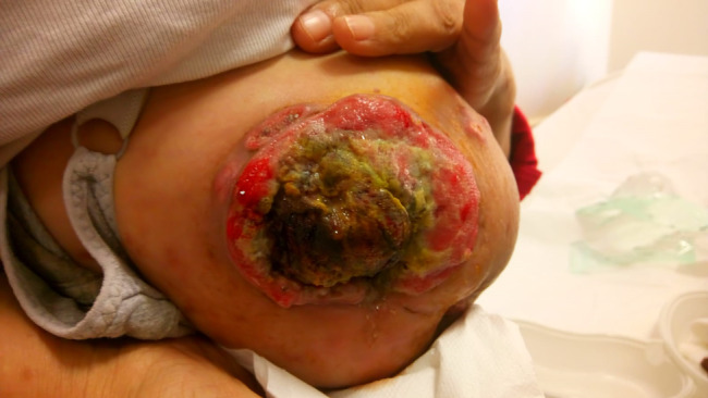
Among these, two patients had a draining wound in the breast. The wound biopsy results of these patients were consistent with malignancy. Interestingly, one of these patients had concomitant invasive breast cancer and differentiated squamous cell carcinoma as a rare diagnosis in the wound (Figure 1). Both patients are followed up in adjuvant chemoradiotherapy program after mastectomy.
Figure 1.

Concomitant invasive breast cancer and differentiated squamous cell carcinoma
The final pathologies of other four patients with a draining wound in breast were reported as idiopathic granulomatous mastitis.
One patient had a history of soft tissue tumour in the epigastric region. The patient had a draining wound and excisional biopsy was performed with debridement. The final pathology examination was reported as benign.
Four patients had foreign body reaction due to prolene suture and two patients had foreign body reaction due to silk suture. Treatment for these patients was achieved by suture removal following wound exploration.
Two patients had previously undergone incisional hernia repair. Draining wounds of these patients were explored and it was thought that foreign body reaction occurred due to prolene graft; therefore, these grafts were removed.
Four patients had undergone pilonidal sinus excision (two patients had primary repair and two patients had limberg transposition) in the postsacral area at different hospitals. On the 6th‐ to 12th postoperative days, wound dehiscence and drainage had developed. The average length of time to admission to the clinic after the onset of complaints was 3 months (2‐4 months). Culture was obtained and antibiotic therapy was started. The patients were then followed up with daily dressing. Approximately, all wounds were closed in the third months.
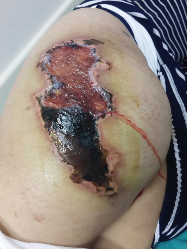
Deep tissue culture was taken after the debridement in a hemiplegic patient with decubitus ulcers of the right gluteal region and in the other two patients with necrosis and non‐healing wounds after intramuscular injection. Vacuum‐assisted closure was performed in these patients and antibiotic therapy was initiated (Figure 2). The characteristics of the patients are shown in Table 2.
Figure 2.

Necrosis and non‐healing wound after intramuscular injection
Table 2.
Characteristics of patients
| Patients | Age (years) | Gender | Time (month) | Before operation | Macroscopy | Pathology | Foreign body | Treatment |
|---|---|---|---|---|---|---|---|---|
| 1 | 48 | Male | 10 | Incisional hernia repair | Epigastric discharge | No | Prolene suture | Remove of suture |
| 2 | 24 | Female | 7 | Endometriosis | Suprapubic discharge | No | Silk suture | Remove of suture |
| 3 | 38 | Female | 10 | Ovarian cyst rupture | Suprapubic discharge | No | Silk suture | Remove of suture |
| 4 | 41 | Female | 7 | Thyroidectomy | Pretracheal discharge | No | Prolene suture | Remove of suture |
| 5 | 38 | Female | 12 | Inguinal hernia repair | Suprapubic discharge | No | Prolene suture | Remove of suture |
| 6 | 52 | Male | 25 | Left hemicolectomy | Umbilical discharge | No | Prolene suture | Remove of suture |
| 7 | 43 | Female | 10 | No | Left breast wound | Invasive ductal carcinoma | No | Mastectomy |
| 8 | 56 | Female | 12 | No | Right breast wound | Differentiated squamous cell and invasive breast carcinoma | No | Mastectomy |
| 9 | 49 | Female | 10 | No | Right breast discharge | Idiopathic granulomatous mastitis | No | Lumpectomy |
| 10 | 41 | Female | 8 | No | Left breast discharge | Idiopathic granulomatous mastitis | No | Tru‐cut biopsy |
| 11 | 37 | Female | 11 | No | Right breast discharge | Idiopathic granulomatous mastitis | No | Tru‐cut biopsy |
| 12 | 45 | Female | 7 | No | Right breast discharge | Idiopathic granulomatous mastitis | No | Tru‐cut biopsy |
| 13 | 18 | Female | 11 | No (hemiplegia) | Right gluteal wound | Benign | No | Vacuum‐assisted closure |
| 14 | 43 | Female | 4 | No (intramuscular Injection) | Right gluteal wound | Benign | No | Vacuum‐assisted closure |
| 15 | 38 | Male | 8 | Soft tissue tumour | Epigastric discharge | Benign | No | Excision |
| 16 | 58 | Male | 7 | Incisional hernia repair | Umbilical discharge | No | Prolene graft | Remove of graft |
| 17 | 62 | Female | 8 | Incisional hernia repair | Epigastric discharge | No | Prolene graft | Remove of graft |
| 18 | 19 | Female | 2 | Pilonidal sinus—Limberg transposition | Wound discharge | No | No | Wound dressing |
| 19 | 23 | Male | 4 | Pilonidal sinus—Primary repair | Wound discharge | No | No | Wound dressing |
| 20 | 27 | Male | 4 | Pilonidal sinus—Primary repair | Wound discharge | No | No | Wound dressing |
| 21 | 20 | Male | 2 | Pilonidal sinus—Limberg transposition | Wound discharge | No | No | Wound dressing |
4. DISCUSSION
Non‐healing wounds pose a serious social and financial cost to both patients and the health system. In addition to the factors that delay wound healing, other causative mechanisms may also play a role in the aetiology. Unresponsiveness to normal regulatory signals in the healing process has also been shown to be a predictive factor for non‐healing wounds. This process may occur as a failure of normal growth factor synthesis, overexpression of protease activity, or increased disintegration of growth factors in a significantly proteolytic wound environment due to lack of normal antiprotease inhibitor mechanisms. It has been found that the proliferation potential of fibroblasts in these wounds decreases as a result of ageing or decreased expression of growth factor receptors.3 Furthermore, foreign body reaction may also be responsible for the process in non‐healing wounds. This reaction of macrophages and foreign body giant cells is an inflammatory process following implantation of a medical device, prosthesis, or biomaterial representing the final stage of wound healing. Synthetic materials are used in hernia repair, which is one of the most common operations of general surgery. In surgical hernia repair, low‐weight, wide‐pore polypropylene materials are generally used.4 Inflammatory responses due to these materials may result in postoperative complications. The implantation of prostheses is generally associated with rapid and highly orchestrated processes, including inflammatory foreign body reaction or granuloma formation, humoral immune activation, coagulation, molecular pattern recognition, and release of the hazard signal from the damaged tissue.5, 6 If prolonged inflammation develops following implantation, it may lead to clinical complications such as chronic pain, defective wound healing, damage to the implant, and need for re‐surgery.7 The aetiology in our patients was due to non‐absorbable prolene suture in four cases, silk suture in two cases, and graft‐related reaction in two patients, resulting in non‐healing wounds. These patients were followed up for 7 to 25 months after the operation. Although suture‐related chronic reactions are mostly seen after silk sutures, they do not occur with absorbable and monofilament sutures. In addition, while there are not many publications in the literature about suture‐related non‐healing wounds, granulomas that usually develop due to suture and manifest cancer‐like findings are discussed.8, 9 Interestingly, our four patients developed excessive reaction due to prolene suture. A deeper understanding of the interaction between the material and the biological environment is essential to overcome side effects and to develop strategies and solutions in the use of these materials, which are still a major challenge in the biomedical field.
Any wound that does not heal for a long time is prone to malignant transformation and malignancy should be considered in the aetiology of non‐healing wounds. In a chronic wound, chronic ulcers may develop a malignant transformation3 (Marjolin's ulcer). This is a long‐term stable period but would transform to malignancy rapidly and progressively once ulceration formed.10 The underlying malignant transformation mechanism remains unclear. Sometimes, as in our patients, locally advanced tumours may appear as an ulcerated, non‐healing chronic wound. Malignant wounds differ clinically from non‐malignant wounds in the presence of overturned wound edges. In patients with suspected malignant transformation, biopsy of the wound margin should be performed to exclude malignancy. De novo cancers in chronic wounds include both squamous and basal cell carcinomas.3 Two patients included in the study were reported to have malignant biopsy results. However, since these diagnoses were the primary tumour of the relevant organ, we did not think that it was a malignant transformation of the chronic wound. Furthermore, one of these patients had two primary cancer diagnoses, a rare diagnosis of differentiated squamous cell along with invasive breast carcinoma (Figure 1).
In our study, vacuum‐assisted closure technique was applied to a hemiplegic patient with right gluteal region decubitus ulcers and to patients who developed necrosis and non‐healing wounds after intramuscular injection (Figure 2). This technique is a non‐invasive method that enables the use of controlled and localised negative pressure on the wound to facilitate healing in acute and chronic wounds. It is rather preferred in non‐healing and chronic wounds. It is a method of treatment based on the sterile closure of the wound site, applying continuous or intermittent negative pressure thereto. With the vacuum‐assisted closure system, partial oxygen pressure in tissues increases and bacterial growth is reduced. In addition, it provides a local interleukin‐8 and vascular endothelial growth factor increase that may cause the accumulation of neutrophils and angiogenesis.11
In non‐healing wounds, foreign bodies can be diagnosed in the postoperative period owing to pain, infection, palpable mass, or during radiological imaging. Computed tomography is generally used in patients with suspected foreign bodies.12 Since the foreign bodies were located superficially in our patient group, no radiological evaluation was needed.
5. CONCLUSIONS
Non‐healing wounds are a serious social and economic problem for patients. Both health care workers and patients will benefit from the early diagnosis and treatment of such non‐healing wounds. Understanding the pathophysiology of various aetiologies, especially in non‐healing wounds, in both clinical and experimental animal models will be a step in treatment. As a result, inflammation in particular plays a critical role. Therefore, dissolution of the inflammatory environment has been the target of both conventional wound care and experimental drug‐based therapies. Although non‐healing wounds are quite common, the number of studies on the aetiologic factors is quite low. In the clinical practice of many surgeons, such wounds are only followed by dressing. Surgeons or clinics especially dealing with chronic wounds apply various treatments based on the aetiology, and patients benefit from these treatments.
In addition to the retrospective nature of our study, although the number of cases was small and the study was single‐centred, the aetiology, diagnostics, and treatment methods in patients with non‐healing wounds were discussed in the light of the literature and attention was paid to the issues to be considered for early diagnosis and treatment.
CONFLICT OF INTEREST
The authors did not receive any financial assistance or have any personal relationships with other people organisations that could inappropriately influence (bias) their work.
Calis H, Sengul S, Guler Y, Karabulut Z. Non‐healing wounds: Can it take different diagnosis? Int Wound J. 2020;17:443–448. 10.1111/iwj.13292
REFERENCES
- 1. Zhao R, Liang H, Clarke E, Jackson C, Xue M. Inflammation in chronic wounds. Int J Mol Sci. 2016;17(12):E2085. [DOI] [PMC free article] [PubMed] [Google Scholar]
- 2. Reinke JM, Sorg H. Wound repair and regeneration. Eur Surg Res. 2012;49(1):35‐43. [DOI] [PubMed] [Google Scholar]
- 3. Barbul A, Efron DT, Kavaluka SL. Wound healing. In: Brunicardi FC, Andersen DK, Billiar TR, et al., eds. Schwartz's Principles of Surgery. 10th ed. New York, NY: Mc Graw Hill Education; 2015:241‐271. [Google Scholar]
- 4. Lake SP, Ray S, Zihni AM, Thompson DM Jr, Gluckstein J, Deeken CR. Pore size and pore shape—but not mesh density—alter the mechanical strength of tissue ingrowth and host tissue response to synthetic mesh materials in a porcine model of ventral hernia repair. J Mech Behav Biomed Mater. 2015;42:186‐197. [DOI] [PubMed] [Google Scholar]
- 5. Heymann F, von Trotha KT, Preisinger C, et al. Polypropylene mesh implantation for hernia repair causes myeloid cell‐driven persistent inflammation. JCI Insight. 2019;4(2):1‐20. 10.1172/jci.insight.123862. [DOI] [PMC free article] [PubMed] [Google Scholar]
- 6. Klopfleisch R, Jung F. The pathology of the foreign body reaction against biomaterials. J Biomed Mater Res A. 2017;105(3):927‐940. 10.1002/jbm.a.35958. [DOI] [PubMed] [Google Scholar]
- 7. Fitzgibbons RJ, Forse RA. Clinical practice. Groin hernias in adults. N Engl J Med. 2015;372(8):756‐763. 10.1056/NEJMcp1404068. [DOI] [PubMed] [Google Scholar]
- 8. Yoshioka Y, Nakatao H, Hamana T, et al. Suture granulomas developing after the treatment of oral squamous cell carcinoma. Int J Surg Case Rep. 2018;50:68‐71. 10.1016/j.ijscr.2018.07.021. [DOI] [PMC free article] [PubMed] [Google Scholar]
- 9. Molina‐Ruiz AM, Requena L. Foreign body granulomas. Dermatol Clin. 2015;33(3):497‐523. [DOI] [PubMed] [Google Scholar]
- 10. Xiao H, Deng K, Liu R, et al. A review of 31 cases of Marjolin's ulcer on scalp: is it necessary to preventively remove the scar? Int Wound J. 2019. Apr;16(2):479‐485. 10.1111/iwj.13058. [DOI] [PMC free article] [PubMed] [Google Scholar]
- 11. Ribeiro MA Jr, Barros EA, Carvalho SM, Nascimento VP, Neto CJ, Fonseca AZ. Comparative study of abdominal cavity temporary closure techniques for damage control. Rev Col Bras Cir. 2016;43(5):368‐373. 10.1590/0100-69912016005015. [DOI] [PubMed] [Google Scholar]
- 12. Chen CL, Cooper MA, Shapiro ML, Angood PB, Makar MA. Patient safety. In: Brunicardi FC, Andersen DK, Billiar TR, et al., eds. Schwartz's Principles of Surgery. 10th ed. New York, NY: Mc Graw Hill Education; 2015:377‐378. [Google Scholar]


