Dear Editors,
1.
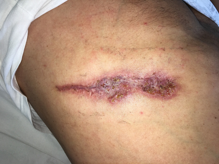
A 57 years old Caucasian male, attending the outpatient of University Hospital Federico II of Naples, section of Dermatology, presented to our attention in June 2018 with a painful ulcerated lesion characterised by a peripheral red halo with raised, red‐purple, edges and a purulent base on the right side of the trunk (Figure 1). The onset dates back to May 2018.
Figure 1.

A painful ulcerated lesion characterised by a peripheral red halo with raised, red‐purple, edges and a purulent base on the right side of the trunk
The patient had a history of idiopathic hypertension on therapy with hydrochlorothiazide. His erythrocyte sedimentation rate was 21 mm/h, C‐reactive protein was 17 mg/L, white blood cell count was 5.7 × 10⁸/L, and fibrinogen was 3.7 g/L. Other standard parameters were all normal. Pathogenic bacteria were not detected from the tissue or from the ulcer floor.
We performed a punch biopsy and histology showed hyperplasia of the epidermis and extensive neutrophilic infiltration in the dermis with focal microabscesses and no signs of vasculitis: these findings confirmed the pyoderma gangrenosum (PG) aspect.
All autoimmune markers such as rheumatoid factor, anti‐nuclear antibody, anti‐extractable nuclear antigens, and anti‐neutrophil cytoplasmic autoantibody were negative. A subclinical inflammatory bowel disease (IBD) was investigated with colonoscopy and faecal calprotectin both negative.
Following the diagnosis of PG, the patient was screened with blood tests for human immunodeficiency virus, hepatitis B and C, and tuberculosis in order to begin a therapy with the tumour necrosis factor inhibitor, adalimumab. Adalimumab therapy was started with a loading dose of 160 mg subcutaneous injection at week 0, 80 mg at week 2, and 40 mg weekly at week 4 and on. In addition to adalimumab, the patient started a topical therapy with topical calcineurin inhibitor, tacrolimus, twice daily.
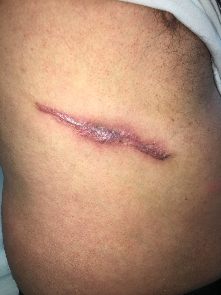
After 5 weeks of therapy with adalimumab and tacrolimus, a really rapid response was registered: more than 50% of the ulcer floor was reepithelialised and with a great pain improvement (Figure 2).
Figure 2.

After 5 weeks of therapy with adalimumab and tacrolimus, more than 50% of the ulcer floor was reepithelialised and with a great pain improvement
PG is a an ulcerative skin disease often destructive that usually presents with painful nodules or pustules that dramatically evolve into large ulcers with raised, red‐purple, edges.1
The incidence of PG is estimated at six persons/million of the population2 and it affects mainly individuals between 20‐50 years of age equally males and females.3, 4
The majority of cases (75%) are associated with underlying illnesses, such as IBD, haematological disorders, and inflammatory arthritis.5
PG is a diagnosis mainly clinical and by exclusion6: differential diagnoses, such as necrotising fasciitis, other infections (bacterial, fungal, and amoebic), vasculitis, and cutaneous malignancies need to be excluded.3, 4 The aetiology of PG remains uncertain, several reports suggest PG is related to a systemic immune dysfunction.
Several treatments have been investigated over the past few decades, but the combination of topical and systemic drugs (such as corticosteroids, immunosuppressants, and analgesia) and local wound care are the mainly used in clinical practice.
Nowadays, the management of many dermatological diseases, such as psoriasis, hidradenitis suppurativa, has been changed by targeted therapies. Also PG has been reported to improve with biologic therapy, especially with tumour necrosis factor alfa inhibitors.7
Regarding tacrolimus, it is a topical immunomodulatory: its mechanism of action is based on calcineurin inhibition, which results in decreased T‐cell activation and inflammatory cytokines' release. Tacrolimus has previously showed its efficacy in some case of hidradenitis suppurativa and PG.8, 9
In the present case, the combination of adalimumab and topical tacrolimus induced the formation of granulation tissue and wound's epithelialisation yet after 4 week, with normalisation of biochemical indices.
To the best of our knowledge, our case is the first PG treated with the combination of adalimumab plus tacrolimus, registering a really rapid clinical and biological response after one month of therapy.
Nevertheless, it has been described a paradoxical development of PG in psoriasis patient treated with Infliximab.10
Thus, we wondered if it was time to continue therapy and for how long in our patient.
CONFLICT OF INTEREST
The authors declare no potential conflict of interest.
REFERENCES
- 1. Blitz NM, Rudikoff D. Pyoderma gangrenosum. Mt Sinai J Med. 2001;68:287‐297. [PubMed] [Google Scholar]
- 2. Weizman A, Huang B, Berel D, et al. Clinical, serologic, and genetic factors associated with pyoderma gangrenosum and erythema nodosum in inflammatory bowel disease patients. Inflamm Bowel Dis. 2014;20:525‐533. [DOI] [PMC free article] [PubMed] [Google Scholar]
- 3. Ahronowitz I, Harp J, Shinkai K. Etiology and management of pyoderma gangrenosum: a comprehensive review. Am J Clin Dermatol. 2012;13(3):191‐211. [DOI] [PubMed] [Google Scholar]
- 4. Bhatti H, Khalid N, Rao B. Superficial pyoderma gangrenosum treated with infliximab: a case report. Cutis. 2012;90(6):297‐299. [PubMed] [Google Scholar]
- 5. Cozzani E, Gasparini G, Parodi A. Pyoderma gangrenosum: a systematic review. G Ital Dermatol Venereol. 2014;149(5):587‐600. [PubMed] [Google Scholar]
- 6. Ben Abdallah H, Fogh K, Bech R. Pyoderma gangrenosum and tumour necrosis factor alpha inhibitors: a semi‐systematic review. Int Wound J. 2019;16(2):511‐521. 10.1111/iwj.13067. [DOI] [PMC free article] [PubMed] [Google Scholar]
- 7. Streit M, Beleznay Z, Braathen LR. Topical application of the tumour necrosis factor‐alpha antibody infliximab improves healing of chronic wounds. Int Wound J. 2006;3:171‐179. [DOI] [PMC free article] [PubMed] [Google Scholar]
- 8. Marasca C, Annunziata MC, Masarà A, Fabbrocini G. A case of axillary hidradenitis suppurativa treated with topical tacrolimus. G Ital Dermatol Venereol. 2018. 10.23736/S0392-0488.18.05855-8. [DOI] [PubMed] [Google Scholar]
- 9. Napolitano M, Megna M, Patrì A, Monfrecola G, Balato N. Pyoderma gangrenosum successfully treated with topical tacrolimus. G Ital Dermatol Venereol. 2018. 10.23736/S0392-0488.18.05735-8. [DOI] [PubMed] [Google Scholar]
- 10. Vestita M, Guida S, Mazzoccoli S, Loconsole F, Foti C. Late paradoxical development of pyoderma gangrenosum in a psoriasis patient treated with infliximab. Eur J Dermatol. 2015;25:272‐273. [DOI] [PubMed] [Google Scholar]


