Abstract
Only a few papers have been published about unusual localisations of pressure ulcer. To date, no papers were published presenting pressure ulcer on external genitals in women. The paper presents the mechanism of origin of vulval pressure ulcer, surgical treatment (excision of lesion tissue of the pressure ulcer) and reconstruction of the vulva. The patient, aged 50, has been paraplegic for 20 years. During the last 3 years she has had a wound which was spreading in the region of the vulva. The pressure ulcer was surgically removed, external female genitals were reconstructed using advancement skin flap and the function and natural appearance of organs were re‐established. The presence of all three aetiological factors for the formation of pressure ulcer – presence of prolonged pressure, swelling and infection – were proven in the described patient. For this reason, we are able to claim that this was in fact a pressure ulcer of the vulva. Reconstruction was simple without any complications and donor‐site morbidity.
Keywords: Pressure, Region, Ulcer, Unusual, Vulva
INTRODUCTION
Wounds produced by pressure used to be called decubitus (Latin word decumbre, meaning ‘to lie down’) or bed sores. More suitable terms are ‘pressure sores' and ‘pressure ulcer’(1).
Pressure and tissue ischaemia are the basic mechanism for onset of pressure sores. This is usually the pressure of the body against the bed; however, any other prolonged pressure on the body can lead to pressure sores. Prolonged pressure of a nasogastric probe can lead to the nasal pressure sores wing, pressure of an oxygen mask on the nasal dorsum; pressure of the endotracheal cannula leads to sometimes fatal chondromalacia in patients with tracheostomy, disc pressure in the region of ileostomy leads to ulceration and so does pressure from an unadjusted section of a prosthesis (1). There is also data about very unusual localisation of wounds produced by pressure (regions of toes and the lower leg as a result of long‐term use of compression stockings) (2).
According to data from a large global study, encompassing developed countries (USA, Canada, and Saudi Arabia) between 5·6% and 15·5% of hospital patients develop pressure ulcus (3). Most cases of pressure ulcus are degrees I and II. Most frequent localisations in paraplegic patients are the sacrum (36%); heel (30%) and ischial, trochanteric or malleolar (34%).
Degrees I and II pressure sores (when the lesion affects only the skin) can be treated conservatively. Degrees III and IV are mostly treated surgically (pressure ulcus). In any case, a precondition for starting the treatment is to eliminate the causes that have led to the pressure ulceration. If surgical treatment is selected, adequate presurgical treatment must be performed.
CASE REPORT
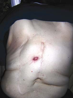
A 50‐year‐old female patient came for a check‐up to the plastic surgery OPD because of a large wound in the region of her external genitals. She had been paraplegic for 20 years. At the age of 25, she had first operated an echinococcus cyst in the region of thoracic vertebrae 10 to 12. Since that time, she moved only with the aid of device, a stick. Until the age of 31, she has had two more operations, and since that time, because of paraplegia, she mainly sits in a wheelchair. The mentioned two surgeries left behind fistulae which occasionally opened in the region of the surgical scar (Figure 1). Five years ago she was operated because of pelveoperitonitis resulting from an ingrown contraceptive device. At that time, hysterectomy was also performed. During this hospitalisation, she developed several pressure sores which were treated conservatively (in the region of the back of her head, along the spine at spinal projections, in the sacral region and in the region of the heel). In the early postoperative phase, she suffered from diffuse peritonitis and exudative pleuritis. Year ago, she has started the treatment for supraventricular arrhythmia and hyposideraemic anaemia. The patient is malnourished, muscles of lower extremities are atrophic and the skin shows normal moisture and turgor and is trophic, with good vascularisation.
Figure 1.

Presence of fistula and scars in dorsal thoracic region, as consequence of a previous operation.
In the 20 years since she has been paralysed, she has not been to a physiatrist examination, and has had no therapy, resulting in significant atrophy of muscles of her lower extremities. During the last 3 years, she was treated in a gynaecological OPD for a wound, that is, pressure ulcus which developed in the region of her external genitals. Because of this problem, she came to the plastic surgeon. How became the pressure ulcus?
Because of the fistula in her back and pain and unease, for years the patient sat on her chair without leaning on its back, but rather leaning against her palms, with her trunk leaning forward. Thus, for several hours each day, pressure was exerted on the region of the external genitals. In the last 3 years, after several hours of sitting in this crouched position, she had been noticing swelling and redness in the vulval region. As she could experience no pain, prolonged pressure led to the formation of rhagade in the labia minora region. She continued to sit in the described position unaware of the reason for the swelling, redness and cracks that had appeared.
The treated diagnosis was colpitis. According to antibiogram findings, she was treated with local antiseptics, oral and parenteral antibiotics. In several swabs, following bacteria and fungi were found: Staphylococcus aureus, Staphylococcus spp., Proteus vulgaris, Klebsiella spp., Escherichia coli, Serratia spp., Enterococcus spp., Pseudomonas aeruginosa and Candida. According to antibiogram findings, during 3 years of conservative treatment, following antibiotics had been prescribed: Ciprofloxacin, Gentamicin, Amoxicillin‐Clavulonic acid and Erythromycin.
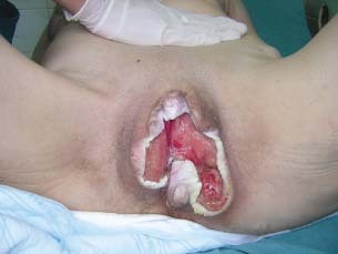
Over time, the wound continued to spread, encompassing on both sides of labia majora, labia minora, clitoris, introitus vaginae – a section of the perineum (Figure 2). At admission, the wound was 15 × 15 cm, permeated by a thick layer of fibrous tissue and covered in granulations. On the right side, in the lowest section of labia majora, a recessus, 3 cm deep, was found. In the introitus vagina was a clear delineation between fibrous lesion tissue covered in granulations and vaginal mucosa with normal appearance, moisture and elasticity, free from infection. The external urethral opening was on the front wall of the vagina. The external urethral opening moved from its usual position during the spread of the pressure ulceration accompanied by fibrosis and tissue deformation. The anus and the surrounding skin contained no lesions. After a detailed examination, an indication for surgery was set.
Figure 2.

Preoperative look of pressure sore in a vulvar region.
During preoperative preparation, a chest X‐ray, biochemical blood tests and internist examination were performed. Preoperative Hg values were 120 g/l which was a sufficient condition to start the operation.
SURGICAL TECHNIQUE
The patient was operated under analgosedation because general anaesthesia was not required. From the aspect of anaesthesiology, the patient required intensive monitoring because of arrhythmia, and previously treated hyposideraemic anaemia.
From the surgical aspect, the biggest intraoperative problem was profuse bleeding from the surgical wound. Ten centimetres of altered scar tissue was removed from the healthy tissue, along the borders of the wound and in depth. During excision, we continuously protected the urethra to prevent injury. As a result of profuse bleeding, excision was performed partially to reduce blood loss. By careful haemostasis using Thermocauter and haemostatic subcutaneous suturing (Vicryl 3‐0), we managed to stop bleeding.
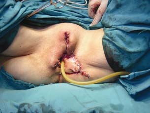
Using surrounding tissue (of the labia majora and ingvinal region), we reconstructed labia majora and introitus vaginae. Advanced skin slices were sutured to the mucosa of the vaginal introitus using absorbable thread Vicryl 3‐0. The junction between advancement skin slices in the pubic region was sutured using non absorbable thread Silk 4‐0 (Figure 3); no donor‐site morbidity.
Figure 3.

Direct postoperative result after vulvar reconstruction.
In the immediate postoperative course, the patient received three doses of blood, after which Hg was 93 g/l. There were no complications. Two antibiotics were prescribed parenterally: Cephtriaxon 2 g × 1 i.v. and Metronidazole 500 mg 2 × 1 i.v. Regular toilet of the surgical wound was performed and Gentamicin ointment was applied according to the swab from the wound and the antibiogram.
RESULTS
The patient was discharged after 12 days. The wound healed completely (per primam), sutures were removed 15 days after the operation. In the subsequent postoperative course there were no complications. An expected complication was scar formation and narrowing of introitus vaginae.
Follow‐up
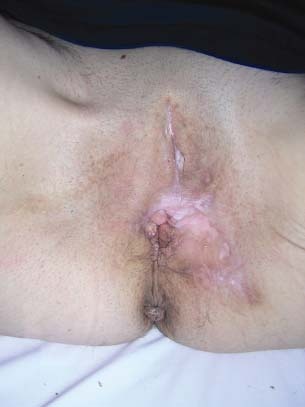
Three months after the operation (Figure 4), the urethral and vaginal openings are functional, there is no vaginal infection, no pronounced scar, the patient suffers no handicaps and she is satisfied with the achieved result. In the area of the reconstructed labia minora (which we made from the rest of the labia majora), there was atrophy of the hair root, which is an unexpected, but a welcomed effect. Postoperatively, the use of an antidecubital mattress for the wheelchair with a 20‐cm perforation in the middle was recommended. The patient was trained to perform daily checks of her external genitals, adequate toilet and care. Now, 2 years after surgery, she has no pains, no wound from the pressure on the vulva or in any other part of the body.
Figure 4.

Postoperative result 4 months after the operation.
DISCUSSION
The localisation of the pressure ulcus in this patient is very rare. The issue is how this pressure ulcus was formed, and if this is in fact pressure ulcus?
The established opinion is that pressure ulcus results only in places where bones exert high pressure on the skin and soft tissues. This is only partially true. Pressure exists not only at these characteristic prominent spots, therefore pressure ulcus is not formed only there. Pressure spreads in concentric circles around these known prominent spots, therefore the entire region exerts, that is, it is subject to certain pressure. Lindan et al. (4) investigated pressure distribution with the patient in the sitting and the reclining position. The highest pressure (100 mmHg) is present in the region of tubera ischiadicum, whereas in the region of the perineum and vulva, the pressure is 10–40 mmHg. Therefore, pressure as the most important cause quite apparently exists. Naturally, the mentioned pressure will not lead to the development of pressure ulcus quickly, but it will lead to even greater tissue damage than would be caused by higher pressure lasting a shorter period of time (5). Pressure is only the initial factor in our patient, because during the subsequent development of tissue damage, oedema plays the leading role. When the venous end of capillaries is subjected to pressure above 12 mmHg, capillary blood pressure in that section grows. As pressure grows, it surpasses blood pressure of 32 mmHg present in the arterial end of the capillary, leading to vasodilatation, plasma extravasation and oedema formation. In patients with denervation, oedema additionally develops because of non functional skeletal muscles, that is, incompetent lymphatic pump. In addition, due to the release of mediators of inflammation (resulting as a response to the pressure trauma), there is additional swelling. At that point, accumulation of interstitial liquid leads to dilution of the concentration of sebum which is present to protect the skin against infection.
Superposition of infection (especially in the anogenital region) is prompted by impeded lymphatic drainage, ischaemia, denervation as well as reduced immune function.
CONCLUSIONS
All three aetiological factors for onset of pressure ulcus – prolonged pressure, swelling and infection – have been proven in the patient. For this reason, we can claim that we were in fact dealing with a pressure ulceration of the vulva. The pressure ulcus was surgically removed and external female genitals were reconstructed. Reconstruction was performed simply without complications. As a result, normal function and natural appearance of organs were achieved without donor‐site morbidity.
ACKNOWLEDGEMENT
The author thanks Dr Maja J. Nikolic for assistance with the preparation of the article.
REFERENCES
- 1. Mancoll JS, Phillips LG. Pressure sores. In: Achauer BM, Erikson E, Guzuron B, Coleman J III, Russell R, Vander Kolk C, editors. Plastic surgery. Vol. 1 , St. Louis, Missouri: Mosby Inc, 2000:447–62. [Google Scholar]
- 2. Rathore FA, New PW, Waheed A. Pressure ulcers in spinal cord injury: unusual site and etiology. Am J Phys Med Rehabil 2009;88:587. [DOI] [PubMed] [Google Scholar]
- 3. Bergstrom N, Horn SD, Smout RJ, Bender SA, Ferguson ML, Taler G, Sauer AC, Sharkey SS, Voss AC. The National Pressure Ulcer Long‐Term Care Study: outcomes of pressure ulcer treatments in long‐term care. Am Geriatr Soc 2006;54:859. [DOI] [PubMed] [Google Scholar]
- 4. Lindan O, Greenway RM, Piazza JM. Pressure distribution of the surface of the human body. I. Evaluation of lying and sitting positions using a bed of springs and nails. Arch Phys Med Rehabil 1965;46:378. [PubMed] [Google Scholar]
- 5. Husain T. An experimental study of some pressure effects on tissues with reference to the bedsore problem. J Pathol Bacteriol 1953;66:347. [DOI] [PubMed] [Google Scholar]


