

Abstract
Stenotrophomonas maltophilia is a recently described organism which was mainly reported either in nosocomial setup, or in immunosuppresed individuals. This was rarely reported as cutaneous pathogenic organism causing cellulitis‐like lesion, paronychia, mucocutaneous ulcers and ecthyma gangrenosum in immunocompromised individuals. Here we describe a case of leg ulcer caused by S. maltophilia in an immuno‐competent patient. The infection was possibly community acquired as the patient had no exposure to hospital environment. The bacillus was sensitive to cotrimoxazole and levofloxacin, and the patient was successfully treated with cotrimoxazole. Our case is unique not only because it is probably the first ever case of leg ulcer caused by S. maltophilia, but also because of its unusual occurrence in immunocompetent patient.
Keywords: Leg ulcer, Stenotrophomonas
Introduction
Stenotrophomonas maltophilia, though newly described, has emerged as an important cause of nosocomial morbidity and mortality. This organism was originally classified as Pseudomonas maltophilia but was transferred to the genus Xanthomonas in 1993 and subsequently became the sole member to the genus Stenotrophomonas (1). The genus was proposed in 1993 by Palleroni and Bradbury (2).
Case report
A 45‐year‐old milkman presented to our outpatient department with non healing right leg ulcer for 2 months of duration. The patient attributed the onset to an injury inflicted by a cow. Before presenting to us, it was treated with different antibiotics without much improvement. The floor of the ulcer had crusted appearance with purulent discharge. The surrounding skin was erythematous and inflamed giving rise to cellulitis‐like appearance (Figure 1). The ulcer was gradually increasing in size and there was fever for 1 week.
Figure 1.
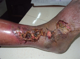
Infected ulcer on presentation.
The patient also had pedal oedema in right side and bilateral mild varicosity below knee joint. He did not have any history of prolonged standing, claudication, smoking, diabetes or drug intake. All peripheral pulses were palpable. There was no history of hospital admission and chemotherapeutic drug intake. Thorough clinical examination and routine investigation ruled out possibility of underlying malignancy or immunosuppression.
Artery–venous colour Doppler study of lower limbs was insignificant apart from mild bilateral superficial venous dilation below the knee joints. Blood and urine cultures were sterile.
However, pus culture showed moderate growth of S. maltophilia. Cultures grew non lactose‐fermenting colonies on McConkey's agar (Figure 2) and glistening colonies with greenish pigment on 5% sheep blood agar plate. The organism was catalase‐positive, oxidase‐negative, motile, non fermenting gram‐negative rods, which oxidised glucose and maltose and was lysine positive. The bacilli were identified as S. maltophilia.
Figure 2.

Growth on McConkey's agar.
It was sensitive to cotrimoxazole, levofloxacin, gentamycin and chloramphenicol and resistant to piperacillin+tazobactam, cefotaxim, ceftazidime, ceftriaxone, cefepime, imipenem, meropenem and aztreonem. Antimicrobial susceptibility test was performed by Kirby‐Bauer method as per Clinical and Laboratory Standards Institute guidelines (Figure 3). As culture sensitivity report was still pending, we started on oral amoxicillin and clavulonic acid for the patient. But even after 5 days of treatment there was no improvement. Later, as per the culture sensitivity report, we shifted to cotrimoxazole. Patient showed excellent response with fever subsiding in 2 days, and pain and erythema within 1 week. We continued cotrimoxazole for 2 weeks with complete healing of ulcer (Figure 4).
Figure 3.
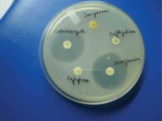
Cotrimoxazole and levofloxacin sensitivity.
Figure 4.
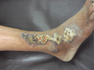
Ulcer healing after 1 week.
Discussion
S. maltophilia is an ubiquitous organism which has been isolated from water, soil, plant material (2) and hospital equipment (such as dressing materials, nebulizers, dialysis machines, intravenous fluids, etc.) (3). It has been emerged as an important opportunistic nosocomial infection 1, 2, 3 and primarily seen in malignancy 3, 4 during severe neutropenia (4) and in other immunocompromised condition 5, 6. In our patient, ulcer was infected with S. maltophilia, acquired from the environmental sources. Although S. maltophilia is regarded as being primarily a nosocomial pathogen, community acquired infection may occur more frequently than was previously recognised 3, 7, 8.
Although the infection usually occurs in immunocompromised host, routine investigation of our patient did not show any immunocompromise status.
Skin and soft tissue infections of S. maltophilia are increasing (4) and are most frequently associated with post‐traumatic, post‐surgical or burn‐related wounds and chronic cutaneous ulcers. Clinical manifestations include cellulitis, cellulitis‐like skin lesions, infected mucocutaneous ulcers, ecthyma gangrenosum and paronychia 3, 6.
There was a possibility of colonisation only (3) without infection by S. maltophilia. But chronicity of ulcer in spite of multiple therapy and quick relief from fever and cellulitis with starting of S. maltophilia‐sensitive antibiotic (i.e. cotrimoxazole) is more in favour of infection rather than a simple colonisation. Cotrimoxazole is recommended as the agent of choice for therapy, although resistance is increasing (4).
So from the above discussion it is obvious that S. maltophilia skin infection should be included as a differential diagnoses for skin lesions (especially when pus culture shows S. maltophilia) to start an appropriate antibiotic therapy.
Our present case is unique, not only because it is the first ever case of leg ulcer caused by community acquired, S. maltophilia infection, but also because of its unusual occurrence in immunocompetent patient.
References
- 1. Mukhopadhyay C, Bhargava A, Ayyagari A. Novel nosocomial infections by Stenotrophomonas maltophilia: first reported case from Lucknow, North India. J Clin Microbiol 2003;41:3989–90. [DOI] [PMC free article] [PubMed] [Google Scholar]
- 2. Denton M, Kerr KG. Microbiological and clinical aspects of infection associated with Stenotrophomonas maltophilia . Clin Microbiol Rev 1998; 11:57–80. [DOI] [PMC free article] [PubMed] [Google Scholar]
- 3. Son YM, Na SY, Lee HY, Baek JO, Lee JR, Roh JY. Ecthyma gangrenosum: a rare cutaneous manifestation caused by Stenotrophomonas maltophilia in a leukemic patient. Ann Dermatol 2009;21:389–92 [DOI] [PMC free article] [PubMed] [Google Scholar]
- 4. Teo WY, Chan MY, Lam CM, Chong CY. Skin manifestation of Stenotrophomonas maltophilia infection – a case report and review article. Ann Acad Med Singapore 2006;35:897–900 [PubMed] [Google Scholar]
- 5. Vartivarian SE, Papadakis KA, Palacios JA, Manning JT Jr, Anaissie EJ. Mucocutaneous and soft tissue infections caused by Xanthomonas maltophilia: a new spectrum. Ann Intern Med 1994;121:969–73 [DOI] [PubMed] [Google Scholar]
- 6. Lee GY, Choi YW, Choi HY, Myung KB. A case of onychia and paronychia by Sternotrophomonas maltophilia . Korean J Dermatol 2001;39:89–90. [Google Scholar]
- 7. Dyte PH, Gillians JA. Pseudomonas maltophilia infection in an abattoir worker. Med J Aust 1977;1: 444–5. [DOI] [PubMed] [Google Scholar]
- 8. Heath T, Currie B. Nosocomial and community acquired Xanthomonas maltophilia infection in tropical Australia. J Hosp Infect 1995;30:309–13. [DOI] [PubMed] [Google Scholar]