Abstract
Non‐invasive ventilation (NIV) provides an effective ventilatory support in patients with respiratory failure without endotracheal intubation. However, there are potential problems with its clinical application and the development of pressure ulcers represents a common complication. Often several intensive care units treat facial skin breakdown related to NIV. In this article, we report our experience in treatment and prevention of these lesions, emphasising the higher risk of certain age groups to develop them, such as preterm infants and elderly patients with comorbidities. We performed daily disinfection of the lesions followed by application of topical cream containing hyaluronic acid (HA) sodium salt. In addition, in order to prevent worsening of injury, we applied a cushion made of gauze pad containing HA sodium salt between the skin and the masks, so as to reduce friction between the NIV devices and the skin. Local medical treatment allowed complete reepithelialisation of the injured skin areas. Systematic monitoring of patients' faces is essential to detect early damages and to intervene with appropriate therapy, especially in preterm infants and elderly. Moreover, refining the devices with the proposed protective cushion can reduce pressure ulcers and increase comfort for the patients.
Keywords: Face mask, Hyaluronic acid, Non‐invasive ventilation, Pressure ulcers, Skin necrosis
Introduction
Non‐invasive ventilation (NIV) includes all methods of artificial ventilation that do not use an endotracheal tube but nasal or facial masks, mouthpiece or perithoracic devices 1. NIV represents one of the major technical advances in respiratory care over the last decade 2; in fact, in acute or chronic respiratory failure it helps to decrease the effort of breathing and also mortality by 36–75% 3, reducing intubation rates and exposure to the potentially lung‐damaging effects of mechanical ventilation.
NIV includes two types of ventilation 4: (i) non‐invasive positive pressure ventilation in which ventilation is associated with a positive pressure using nasal, facial or buccal masks; (ii) ventilation with negative pressure, which is not usually used because of its technological complexity. Nasal continuous positive airway pressure (NCPAP) belongs to the first NIV category and it is becoming increasingly popular as a method of respiratory support in the newborn 5, 6.
The choice of the interface and correct placement of mask are essential steps for realisation of an effective and well‐tolerated NIV. Most non‐invasive mask ventilation failures are due to technical problems such as air leaks, mask discomfort and skin lesions 7. Among the adverse effects of mask ventilation, skin breakdown, which occurs at the site of mask contact even after only a few hours of ventilation, is a frequent complication, ranging from 2% up to a maximum of 70% 8, 9, 10, 11, 12, 13, 14, 15, 16, 17, 18, 19, 20, 21. In most cases, masks were fitted tightly in order to reduce air leaks, which often caused skin lesions, especially where there is very little subcutaneous tissue, such as at the level of nasal bridge or columella 14, 22.
The Department of Plastic and Reconstructive and Aesthetic Surgery frequently receives consulting requests from several intensive care units to treat pressure ulcers associated with NIV devices. We report a review of the literature and our experience in treatment and prevention of facial skin breakdown related to the use of several NIV masks. This complication can affect patients of any age group, but we focus on higher risk of certain age groups such as preterm infants and elderly patients with comorbidities because of their poor general health conditions and/or their status of immunosuppression. Therefore, we present two selected clinical cases, first describing a case of nasal injury secondary to nasal continuous positive airway pressure in a preterm infant and then a case of skin necrosis caused by facial mask in an elderly patient.
Methods
Case 1
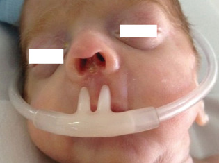
A 28‐week preterm male infant, admitted to the neonatal intensive care unit of our hospital, manifested severe respiratory insufficiency because of his immature lung function. Hence from birth, a NCPAP device (24 hours/day) was initiated. Four weeks later the infant developed nasal ulceration at the level of contact site with nasal prongs; he showed a loss of cutaneous substance of nasal columella with partial exposure of the underlying cartilaginous structures (Figure 1).
Figure 1.

Front view of a 28‐week preterm male infant showing nasal injury due to nasal continuous positive airway pressure (NCPAP): loss of cutaneous substance of the columella with partial exposure of the underlying cartilaginous structures at the level of contact site with nasal prongs.
Case 2
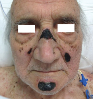
A 71‐year‐old gentleman with multiorgan failure was admitted to the medical sub‐intensive care unit for elderly patients of our hospital. The availability of technological equipment in this ward (such as monitors for cardiac and respiratory function, non‐invasive mechanical ventilators, peristaltic and volumetric pumps for i.v. therapy and enteral nutrition) allowed non‐invasive monitoring of the vital signs of the patient and intensive interventions. Once blood gases deteriorated, to manage the respiratory failure, a facial NIV mask (24 hours/day) was placed. Unfortunately, the prolonged use of this device resulted in increased pressure and consequent trauma at the points of contact between the facial skin and rigid parts of the mask frame. The patient showed four areas of skin necrosis located precisely at the level of nasal bridge, in the nasolabial region bilaterally and in the chin region (Figure 2). The time interval between the initiation of NIV and the onset of injury was 11 days.
Figure 2.

Front view of a 71‐year‐old gentleman with respiratory failure showing facial skin breakdown related to the use of facial non‐invasive ventilation (NIV) mask 24 hours/day. The image shows four areas of skin necrosis located at the points of contact between the facial skin and rigid parts of the mask frame, precisely at the level of nasal bridge, in the nasolabial region bilaterally and in the chin region.
In both cases, the use of NIV devices was absolutely necessary; when we moved them away from nares/face of patients, there was a sudden decrease in PO2. To manage these NIV complications, we performed daily disinfection of the lesions with a highly diluted solution of sodium hypochlorite (0·5% w/v) with topical anti‐infective activity (Dakin's solution) followed by accurate cleansing with normal saline in order to avoid hystotoxicity, and application of topical cream containing hyaluronic acid (HA) sodium salt. Moreover, in order to prevent worsening of injury, we applied a cushion made of gauze pad containing HA sodium salt around the nasal cannula for oxygen therapy in case 1, as well as under the frame of facial NIV mask in case 2, thereby reducing friction between the devices and the skin.
All patients were followed up for at least a month after the onset of skin breakdown. The condition of the facial skin was documented systematically by the same plastic and reconstructive surgeon who administered the cushion.
Results
The outcomes of the local medical treatment have been totally satisfactory leading to complete reepithelialisation of the injured skin areas.
Discussion
Until now, most of the results with NIV usage have been beneficial, and it provides a safe and effective ventilatory support in patients with respiratory failure. Nevertheless, there are potential problems with its clinical application that need to be addressed 23, 24, 25.
A common complication correlated with the use of NIV is the development of pressure ulcers on the nasal bridge, in the columella region and around the mask 8, 9, 10, 11, 12, 13, 14, 15, 16, 17, 18, 19, 20, 21, 26, 27, 28, 29, 30, 31, 32, as reported in the above two cases.
A pressure ulcer can be defined as an area of local tissue damage caused by pressure, shear or friction 33; in fact, the major underlying mechanism of nasal and/or facial injury related to NIV appears to be pressure generated on the skin by the rigid parts of these devices. Previous literature on pressure sore has shown that despite the ability of the skin to autoregulate its flow, pressure sustained at 35 mmHg or greater for longer than 2‐hour duration leads to irreversible ischaemia and subsequent tissue necrosis 24, 26, 34.
Although the type of damage is the same, the sites of injury differ. In patients with nasal mask or prongs, injuries occur primarily at the base of the nasal septum at the junction between the nasal septum and the philtrum (case 1). This suggests that this is the area where the mask exerts the greatest pressure, as prolonged pressure leads to impairment of tissue perfusion with resultant skin trauma. Instead, in patients with facial mask, lesions are usually multifocal and they are located all around the nose and mouth regions (case 2).
The choice of NIV device should be based on some important parameters: clinical age of patient, severity of respiratory failure, degree of vigilance, morphology of the face, patient compliance and subjective tolerance towards the various available devices. If the patient is able to ventilate for a long time effectively through the nose, then a nasal mask should be prescribed. Otherwise, a face mask should be used in the first instance. In selected cases it is also possible to alternate between several types of masks 4.
However, regardless of the type of chosen mask, the most important practice is to periodically check the facial skin of these patients in order to detect early damages and to intervene with appropriate therapy. Although pressure ulcers due to NIV can occur in a variety of age groups, some groups are at increased risk, such as preterm infants and elderly patients with comorbidities.
In fact, these two categories of patients are usually very debilitated, and have two important conditions that predispose to tissue injury related to NIV devices: prolonged immobility during long hospitalisations and immunosuppression. The prolonged immobility causes a reduction of tissue blood flow and consequently results in alteration of the skin trophism. The condition of immunosuppression, due to immature immune system in preterm infants or its deterioration by several comorbidities such as in elderly patients, hinders the regular mechanism of wound repair. Thus, the combination of these adverse conditions makes preterm infants and the elderly with NIV devices particularly vulnerable to rapid and early mechanisms of damage 35.
Therefore, in the application of NIV, it is important to pay close attention not only to the general health conditions of each patient but also to the trophic status of the skin, especially considering the delicate skin of children and the thin, fragile and inelastic skin of elderly patients 36.
Various attempts have been made to improve the design and performance of NIV devices; some have concentrated on making the materials used soft and malleable, whereas other have changed the shape of the mask to facilitate usage 25. Meduri et al. 14 recommend using a patch of wound care dressing on the nasal bridge to reduce skin lesions. Weng 29 found that refining the materials of the masks with polyurethane hydrocolloid dressings that are applied to prevent pressure ulcers can increase tolerance of NIV. Günlemez et al. 30 proposed nasal silicon shield application to achieve the same goal.
According to our experience in the management of skin breakdown associated with NIV devices, we suggest an application a protective cushion—made of gauze pad containing HA sodium salt—around the nasal prongs for oxygen therapy and under the frame of facial NIV masks. We found that the use of this protection not only reduced the pressure ulcer rate significantly but also decreased the severity of injuries, reducing friction between the device and the skin as demonstrated in our clinic. In addition, HA generates a microenvironment stimulating the secretion of growth factors, proliferation and migration of fibroblasts, endothelial cells, keratinocytes and angiogenesis, and it has a positive effect on inflammatory response: the essential conditions for wound healing 37.
In conclusion, we found that refining NIV devices with a protective cushion made of gauze pad containing HA sodium salt can reduce pressure ulcers and increase comfort for patients. It is a safe, simple, reliable, reproducible and versatile method. Thus, we propose it as an alternative approach in the management of facial skin breakdown associated with NIV. In addition, we emphasise the importance of performing systematic monitoring of the facial skin in these patients at the points of contact between the skin and the rigid parts of the mask frame, recommending a close follow‐up (checks every 3–4 hours) especially in preterm infants and elderly, who represent classes of patients with increased risk of development of these common complications.
Acknowledgements
All authors hereby declare not to have any potential conflict of interests and not to have received funding for this work from any of the following organisations: National Institutes of Health (NIH), Wellcome Trust, Howard Hughes Medical Institute (HHMI) and other(s). Each author participated sufficiently in the work to take public responsibility for the content.
References
- 1. Mehta S, McCool FD, Hill NS. Leak compensation in positive pressure ventilators: a lung model study. Eur Respir J 2001;17:259–67. [DOI] [PubMed] [Google Scholar]
- 2.Royal College of Physicians, British Thoracic Society, Intensive Care Society. Chronic obstructive pulmonary disease: non‐invasive ventilation with bi‐phasic positive airways pressure in the management of patients with acute type 2 respiratory failure. Concise Guidance to Good Practice series, No 11. London: RCP, 2008, p. 2.
- 3. Landefeld CS, Palmer RM, Kresevic DM, Fortinsky RH, Kowal J. A randomized trial of care in a hospital medical unit especially designed to improve the functional outcomes of acutely ill older patients. N Engl J Med 1995;332:1338–44. [DOI] [PubMed] [Google Scholar]
- 4. Cuvelier A, Benhamou D, Muir JF. Non‐invasive ventilation of elderly patients in the intensive care unit. Rev Mal Respir 2004;21(5 pt 3):8S139–50 Review. French. [PubMed] [Google Scholar]
- 5. Wiswell TE, Srinivasan P. Continuous distending pressure. In: Goldsmith JP, Karotkin EH, editors. Assisted ventilation of the neonate. Philadelphia: Saunders, 2003:127–47. [Google Scholar]
- 6. Bancalari E, Claure N. Non‐invasive ventilation of the preterm infant. Early Hum Dev 2008;84:815–9 Review. [DOI] [PubMed] [Google Scholar]
- 7. Soo Hoo GW, Santiago S, Williams JW. Nasal mechanical ventilation for hypercapnic respiratory failure in chronic obstructive pulmonary disease: determinants of success and failure. Crit Care Med 1994;22:1253–61. [DOI] [PubMed] [Google Scholar]
- 8. Benhamou D, Girault C, Faure C, Portier F, Muir JF. Nasal mask ventilation in acute respiratory failure. Experience in elderly patients. Chest 1992;102:9912–7. [DOI] [PubMed] [Google Scholar]
- 9. Vitacca M, Rubini F, Foglio K, Scalvini S, Nana S, Ambrosino N. Non‐invasive modalities of positive of pressure ventilation improve the outcome of acute exacerbation of COLD patients. Intensive Care Med 1993;19:450–5. [DOI] [PubMed] [Google Scholar]
- 10. Wysocki M, Tric L, Wolff MA, Gertner J, Millet H, Herman B. Noninvasive pressure support ventilation in patients with acute respiratory failure. Chest 1993;103:907–13. [DOI] [PubMed] [Google Scholar]
- 11. Kramer N, Mayer TJ, Meharg J, Cece RD, Hill NS. Randomized, prospective trial of noninvasive positive pressure ventilation in acute respiratory failure. Am J Respir Crit Care Med 1995;151:1799–806. [DOI] [PubMed] [Google Scholar]
- 12. Brochard L, Mancebo J, Wysocki M, Lofaso F, Conti G, Rauss A, Simmoneau G, Benito S, Gasparetto A, Lemaire F, Isabey D, Harf A. Noninvasive ventilation for acute exacerbations of chronic obstructive pulmonary disease. N Engl J Med 1995;333:817–22. [DOI] [PubMed] [Google Scholar]
- 13. Wysocki M, Tric L, Wolff MA, Millet H, Herman B. Noninvasive pressure support ventilation in patients with acute respiratory failure. Randomized comparison with conventional therapy. Chest 1995;107:761–8. [DOI] [PubMed] [Google Scholar]
- 14. Meduri GU, Turner RE, Abou‐Shala N, Wunderink R, Tolley E. Noninvasive positive pressure ventilation via face mask: first line intervention in patients with acute hypercapnic and hypoxemic respiratory failure. Chest 1996;109:179–92. [DOI] [PubMed] [Google Scholar]
- 15. Ambrosino N. Noninvasive mechanical ventilation in acute respiratory. Eur Respir J 1996;9:795–807. [DOI] [PubMed] [Google Scholar]
- 16. Nava S, Ambrosino N, Clini E, Prato M, Orlando G, Vitacca M, Brigada P, Fracchia C, Rubini F. Noninvasive mechanical ventilation in the weaning of patients with respiratory failure due to chronic obstructive pulmonary disease. A randomized, controlled study. Ann Intern Med 1998;128:721–8. [DOI] [PubMed] [Google Scholar]
- 17. Celikel T, Sungur M, Ceyhan B, Karakart S. Comparison of noninvasive positive pressure ventilation with standard medical therapy in hypercapnic acute respiratory failure. Chest 1998;114:1636–42. [DOI] [PubMed] [Google Scholar]
- 18. Antonelli M, Conti G, Rocco M, Bufi M, De Blasi RA, Vivino G, Gasparetto A, Meduri GU. A comparison of noninvasive ventilation and conventional mechanical ventilation in patients with acute respiratory failure. N Engl J Med 1998;339:429–35. [DOI] [PubMed] [Google Scholar]
- 19. Gregoretti C, Beltrame F, Lucangelo U, Burbi L, Conti G, Turello M, Gregori D. Physiologic evaluation of non invasive pressure support ventilation in trauma patients with acute respiratory failure. Intensive Care Med 1998;24:785–90. [DOI] [PubMed] [Google Scholar]
- 20. Confalonieri M, Potena A, Carbone G, Della Porta R, Tolley EA, Meduri GU. Acute respiratory failure in patients with severe community‐acquired pneumonia: a prospective randomized evaluation of noninvasive ventilation. Am J Respir Crit Care Med 1999;160:1585–91. [DOI] [PubMed] [Google Scholar]
- 21. Beltrame F, Lucangelo U, Gregori D, Gregoretti C. Noninvasive positive pressure ventilation in trauma patients with acute respiratory failure. Monaldi Arch Chest Dis 1999;54:109–14. [PubMed] [Google Scholar]
- 22. Maruccia M, Fanelli B, Ruggieri M, Onesti MG. Necrosis of the columella associated with nasal continuous positive airway pressure in a preterm infant. Int Wound J 2012. DOI: 10.1111/j.1742-481X.2012.01121.x Epub ahead of print. [DOI] [PMC free article] [PubMed] [Google Scholar]
- 23. Demoule A, Girou E, Richard JC, Taille S, Brochard L. Benefits and risks of success or failure of noninvasive ventilation. Intensive Care Med 2006;32:1756–65. [DOI] [PubMed] [Google Scholar]
- 24. Racca F, Appendini L, Gregoretti C, Stra E, Patessio A, Donner CF, Ranieri VM. Effectiveness of mask and helmet interfaces to deliver noninvasive ventilation in a human model of resistive breathing. J Appl Physiol 2005;99:1262–71. [DOI] [PubMed] [Google Scholar]
- 25. Racca F, Appendini L, Berta G, Barberis L, Vittone F, Gregoretti C, Ferreyra G, Urbino R, Ranieri VM. Helmet ventilation for acute respiratory failure and nasal skin breakdown in neuromuscular disorders. Anesth Analg 2009;109:164–7. [DOI] [PubMed] [Google Scholar]
- 26. Ahmad Z, Venus M, Kisku W, Rayatt S. A case series of skin necrosis following use of non invasive ventilation pressure masks. Int Wound J 2013;10:87–90. [DOI] [PMC free article] [PubMed] [Google Scholar]
- 27. Callaghan S, Trapp M, Callaghan S, Trapp M. Evaluating two dressings for the prevention of nasal bridge pressure sores. Prof Nurse 1998;13:361–4. [PubMed] [Google Scholar]
- 28. Layfield C. Non‐invasive BiPAP—implementation of a new service. Intensive Crit Care Nurs 2002;18:310–9. [DOI] [PubMed] [Google Scholar]
- 29. Weng MH. The effect of protective treatment in reducing pressure ulcers for non‐invasive ventilation patients. Intensive Crit Care Nurs 2008;24:295–9. [DOI] [PubMed] [Google Scholar]
- 30. Günlemez A, Isken T, Gökalp AS, Türker G, Arisoy EA. Effect of silicon gel sheeting in nasal injury associated with nasal CPAP in preterm infants. Indian Pediatr 2010;47:265–7. [DOI] [PubMed] [Google Scholar]
- 31. Yong SC, Chen SJ, Boo NY. Incidence of nasal trauma associated with nasal prong versus nasal mask during continuous positive airway pressure treatment in very low birthweight infants: a randomised control study. Arch Dis Child Fetal Neonatal Ed 2005;90:F480–3. [DOI] [PMC free article] [PubMed] [Google Scholar]
- 32. Gregoretti C, Confalonieri M, Navalesi P, Squadrone V, Frigerio P, Beltrame F, Carbone G, Conti G, Gamna F, Nava S, Calderini E, Skrobik Y, Antonelli M. Evaluation of patient skin breakdown and comfort with a new face mask for non‐invasive ventilation: a multi‐centre study. Intensive Care Med 2002;28:278–84. [DOI] [PubMed] [Google Scholar]
- 33. Theaker C. Pressure sore prevention in the critically ill: what you don't know, what you should know and why it's important. Intensive Crit Care Nurs 2003;19:163–8. [DOI] [PubMed] [Google Scholar]
- 34. Thompson D. A critical review of the literature on pressure ulcer aetiology. J Wound Care 2005;14:87–90. [DOI] [PubMed] [Google Scholar]
- 35. Jaul E. Assessment and management of pressure ulcers in the elderly: current strategies. Drugs Aging 2010;27:311–25 Review. [DOI] [PubMed] [Google Scholar]
- 36. Worley CA, Worley CA. Aging skin and wound healing. Dermatol Nurs 2006;18:265–6. [PubMed] [Google Scholar]
- 37. Hollander D, Schmandra T, Windolf J. Using an esterified hyaluronan fleece to promote healing in difficult‐to‐treat wounds. J Wound Care 2000;9:463–6. [DOI] [PubMed] [Google Scholar]


