Abstract
Chronic leg ulcers in patients with rheumatological diseases can cause significant morbidity. We performed a retrospective case review to describe the epidemiology, clinical features and outcome of chronic leg ulcers in this group of patients. Twenty‐nine patients with underlying rheumatological conditions, such as, rheumatoid arthritis (15 patients), systemic lupus erythematosus (8 patients), overlap syndromes (3 patients), systemic sclerosis (1 patient) and ankylosing spondylitis (1 patient) were included. The ulcers were mostly located around the ankle (55·2%) and calves (37·9%). The predominant aetiology of the ulcers, in decreasing order of frequency, was venous disease, multifactorial, vasculitis or vasculopathy, infective, pyoderma gangrenosum, ischaemic microangiopathy and iatrogenic. Treatment modalities included aggressive wound bed preparation, compression therapy (17 patients), changes in immunosuppressive therapy (15 patients), hyperbaric oxygen therapy (4 patients) and cellular skin grafting (2 patients). Management of chronic leg ulcers in rheumatological patients is challenging and the importance of careful clinicopathological correlation and treatment of the underlying cause cannot be overemphasised.
Keywords: Chronic ulcer, Rheumatological diseases
Introduction
The prevalence of chronic leg ulcers in patients with an underlying rheumatological disease has been reported to range from 5·6% to 10% in Western literature 1, 2. The aetiology of these leg ulcers is often multifactorial and can be the result of underlying venous disease, peripheral artery disease, vasculitis and vasculopathy, trauma or neuropathy 3, 4, 5. There is a paucity of data regarding chronic leg ulcers in Asian patients with rheumatological diseases. The aim of this study was to describe the epidemiological data, clinical features, underlying aetiology, treatment and outcome of chronic leg ulcers in adult patients with rheumatological diseases, seen at a tertiary dermatological centre in Singapore.
Methods
This was a 7‐year retrospective case review of all patients with leg ulcers seen at the National Skin Centre in Singapore from August 2003 to August 2010. Patients with leg ulcers of more than 6 weeks duration and a history of a concomitant rheumatological disease were included. Patients below the age of 21 years or who had ulcers that healed within 6 weeks were excluded. This study was approved by the country's research ethics committee.
Results
Patient demographics
There was a total of 29 patients, with a mean age of 53·9 years (range 23–78 years). The majority were female (82·8%). The racial distribution was Chinese (65·5%), Malay (17·2%), Indian (10·3%) and others (7·0%).
Approximately half of the patients (52%) had rheumatoid arthritis (RA). There were eight patients with systemic lupus erythematosus (SLE), two with systemic sclerosis (SSc), one with ankylosing spondylitis (AS) and three patients with overlap syndromes. The mean duration of the rheumatological disease was 13·6 years (range 0–30 years) and majority were on immunosuppressive therapy (75·8%) at the time of presentation of their chronic leg ulcers. There were two patients with diabetes mellitus (Table 1).
Table 1.
Demographics and clinical characteristics of the study patients
| RA | SLE | SSc | AS | Overlap syndromes | |
|---|---|---|---|---|---|
| Number of patients | 15 | 8 | 2 | 1 | 3 |
| Mean age, years (range) | 56·3 (23–78) | 51·4 (31–70) | 52·5 (35–70) | 65 | 46 (38–54) |
| Gender | |||||
| Male | 2 | 1 | 0 | 1 | 0 |
| Female | 13 | 7 | 2 | 0 | 2 |
| Race | |||||
| Chinese | 10 | 4 | 1 | 1 | 3 |
| Malay | 1 | 3 | 1 | 0 | 0 |
| Indian | 2 | 1 | 0 | 0 | 0 |
| Others | 2 | 0 | 0 | 0 | 0 |
| Mean duration of disease, years (range) | 14·3 (2–25) | 16·9 (12–36) | 4 (2–6) | 30 | 4·3 (0–8) |
| On immunosuppressives at presentation | 13 | 8 | 0 | 0 | 1 |
| No. patients with diabetes mellitus | 0 | 2 | 0 | 0 | 0 |
AS, ankylosing spondylitis; RA, rheumatoid arthritis; SLE, systemic lupus erythematosus; SSc, systemic sclerosis.
Clinical characteristics of ulcers
The mean duration of the leg ulcers was 11·1 months (range 0·1–60 months) prior to presentation. The majority of patients were presented with a single ulcer (65·5%) and unilateral involvement (86·2%). The maximum diameter of ulcers ranged from 2 to 16.2 cm. The location of the ulcers, in descending frequency, was around the ankle (55·2%), lower leg (37·9%), foot (24·1%) and thigh (3·4%) (Table 2).
Table 2.
Clinical characteristics of the ulcers
| Number of ulcers | |
| 1 | 19 (65·5%) |
| ≥2 | 10 (34·5%) |
| Unilateral | 86·2% |
| Location | |
| Foot | 7 (24·1%) |
| Around ankle | 16 (55·2%) |
| Between ankle and knee | 11 (37·9%) |
| Thigh | 1 (3·4%) |
| Size (greatest diameter of ulcers, cm) | Range, 2–16·2 cm |
Predominant aetiology
Skin biopsies performed on 18 patients (62·1%) showed vasculitis in 50% of cases. Other histological diagnosis included chronic ulcer, granulomatous dermatitis caused by mycobacterial infection, dermal mucinosis with pseudomembranous fat alteration, stasis changes, fibrosis and scar tissue. Venous doppler ultrasound studies were performed in nine patients (31·0%), which showed deep venous incompetence in three patients, superficial venous incompetence in four patients and incompetence in the perforators in two patients. Arterial studies were performed in 12 patients (41·4%) that showed arterial occlusion in 3 patients.
After clinicopathological correlation, the predominant aetiology of the chronic leg ulcers was attributed to venous disease (34·5%), multifactorial (24·1%), vasculitis or vasculopathy (20·7%), atypical mycobacterial infection (7·0%), pyoderma gangrenosum (7·0%), ischaemic microangiopathy (3·4%) and iatrogenic (3·4%).
Treatment, complications and outcome
Pain was the most common complication, occurring in 20 of 22 patients (69·0%). Of the 22 patients, 20 had positive wound swabs for qualitative bacterial cultures. Of these 20 patients with positive pyogenic wound cultures, 19 had concomitant clinical signs of local wound infection or cellulitis (65·5%), supporting the diagnosis of wound infection. Staphylococcus aureus was the most commonly isolated organism, found in 14 patients (48·3%), followed by Pseudomonas aeruginosa in 10 patients (34·5%). Other isolated organisms include Escherichia coli, Klebsiella species, Streptococcus milleri group, Acinetobacter baumannii, coagulase negative Staphylococcus, Enterobacter species, coliforms, mixed gram negative bacilli and mixed flora. All the patients received antibiotic treatment, either topically, orally or intravenously. Topical antibiotics used include 23 mupirocin 2% ointment and tetracycline 3% ointment.
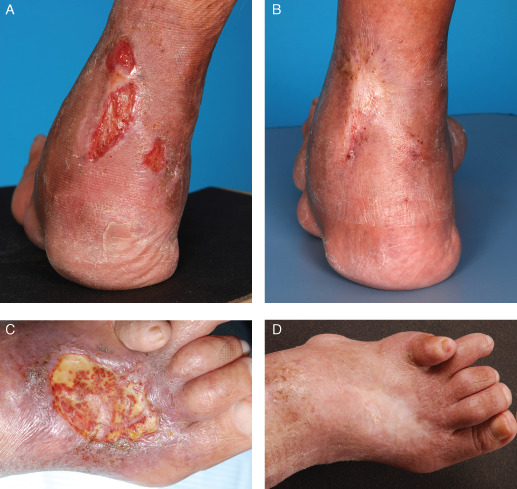
Treatment was directed at the underlying cause. Fifteen patients (51·7%) required an addition, increase or change in immunosuppressive agents for the treatment of their chronic ulcers. All patients received aggressive wound bed preparation, 17 patients (58·6%) were treated with compression therapy, 4 patients (13·8%) received hyperbaric oxygen therapy and 2 patients (6·9%) underwent cellular skin grafting, of which 1 patient (3·4%) also received two doses of intravenous anti‐CD20 rituximab 500 mg 2 weeks apart for recurrent, recalcitrant ulcers (Figure 1).
Figure 1.

Chronic ulcers in a patient with rheumatoid arthritis, (A) before and (B) after cellular skin grafting, with reepithelisation; (C) before and (D) after IV rituximab, with reepithelisation.
Follow‐up data were available for 20 ulcers among 15 patients. The mean time for ulcer reepithelisation was 8·6 months (range 3–42 months). Of the 15 patients, 11 developed ulcers either concomitantly or after their initial ulcers had healed.
Discussion
Chronic leg ulcers in patients with rheumatological diseases can cause significant morbidity and its point prevalence has been reported to be about 9% in patients with RA 1 and 5·6% in patients with systemic lupus erythematosus 2. In this study, chronic leg ulcers were most commonly associated with RA (52%) among patients with rheumatological disease.
The aetiology of chronic leg ulcers in patients with rheumatological disease is often multifactorial 3. In this study, venous disease was the most common cause of chronic leg ulceration (34·5%), in general. However, for the cohort of 15 patients with RA, we found that the predominant aetiology in decreasing order of frequency was vasculitis or vasculopathy (33·3%), venous disease (20%), mixed venous disease and vasculitis (20%), pyoderma gangrenosum (13·3%), ischaemic microangiopathy (6·7%) and infective (6·7%). The high number of patients with ulcers of a vasculitic or vasculopathic origin is comparable to previous published studies of 18–50% 6, 7. This is in contrast to a recent study which reported that only 8% of RA patients had ulcers of a vasculitic origin 5. It is important to ensure careful clinicopathological correlation to distinguish between primary and secondary vasculitis, occurring because of infection or inflammation, which is common in RA patients 8.
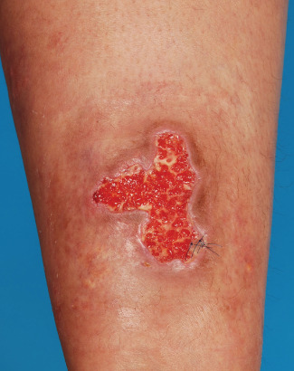
An interesting case which also highlighted the need for careful clinicopathological correlation was a 38‐year‐old female patient with overlap syndrome that had recurrent atypical mycobacterial infection and ulceration of her right lower leg (Figure 2). She had been on long‐term immunosuppressive agents including Prednisolone and Cyclophosphamide. During the first episode of ulceration, histology showed a septolobular pannicultitis with granulomatous inflammation. The initial clinical impression was that of a panniculitis, possibly secondary to lupus erythematosus or underlying atypical mycobacterial infection. A repeat histology was performed when ulcers and papules recurred after 1 year, which showed a granulomatous dermatitis with necrosis. The polymerase chain reaction for non‐tuberculous mycobacterium was negative in the first episode, but positive in the second episode, although the specific strain could not be identified as tissue cultures for both MTB and NMTB were negative. The patient was treated with Clarithromycin and Ciprofloxacin over 14 and 27 weeks, respectively, for the first and second episodes, with full resolution of the ulcers. This case also highlights the need for a high index of suspicion for atypical causes or infective agents, especially in immunosuppressed hosts.
Figure 2.

Chronic ulcer secondary to atypical mycobacterial infection in a patient with overlap syndrome.
The management of chronic leg ulcers in this group of patients is challenging and treatment should be directed at the underlying cause. Published systematic reviews showed no additional benefit of one dressing type over another for the healing of use of venous ulcers 9, 10, whereas compression therapy is effective in increasing the healing rates of venous ulcers 11.
Other treatment options for recalcitrant lower limb ulcers include hyperbaric oxygen and skin grafting. A recent Cochrane review reported the short‐term benefit of hyperbaric oxygen therapy in the healing of diabetic foot ulcers, although no definitive conclusions regarding the effects of hyperbaric oxygen therapy for chronic wounds with other underlying pathologies can be made 12. Skin grafts may be autografts, allografts or xenografts and various surgical techniques (for example pinch grafts, punch grafts, full‐thickness skin grafts, split‐thickness skin grafts) have been tried. In our series of patients, two patients underwent autologous cellular skin grafting for recalcitrant chronic ulcers as part of a research study to facilitate ulcer healing. One patient with chronic refractory RA and recurrent, treatment‐refractory vasculitic ulcers at her left lower leg had reepithelisation of her leg ulcers 10 weeks after cellular skin grafting. The second patient had systemic lupus erythematosus and a persistent chronic lower limb ulcer after surgery for necrotising faciitis. Her ulcer reepithelised 11 weeks after cellular skin grafting. Unfortunately, in the first patient, new lower limb ulcers appeared a month after and in the second patient, there was a relapse of her ulcer 1 week after it had reepithelised.
The need for continued monitoring of these patients is vital as these patients can experience recurrent leg ulcerations as exemplified in the three patients mentioned. In our cohort, 73·3% developed concomitant or recurrent leg ulceration.
In summary, the importance of careful clinicopathological correlation to determine and treat the underlying cause of chronic leg ulceration in patients with associated rheumatological disease cannot be overemphasised. Key strategies include sustained immunosuppression, broad spectrum antibiotics, compression therapy and aggressive wound bed preparation.
Acknowledgements
The authors have no conflict of interests to declare.
References
- 1. Thurtle OA, Cawley MI. The frequency of leg ulceration in rheumatoid arthritis: a survey. J Rheumatol 1983;10:507–9. [PubMed] [Google Scholar]
- 2. Tuffaneilli DL, Dubois EL. Cutaneous manifestations of systemic lupus erythematosus. Arch Dermatol 1964;90:377–86. [DOI] [PubMed] [Google Scholar]
- 3. Hafner J, Schneider E, Burg G, Cassina PC. Management of leg ulcers in patients with rheumatoid arthritis or systemic sclerosis: the importance of concomitant arterial and venous disease. J Vasc Surg 2000;32:322–9. [DOI] [PubMed] [Google Scholar]
- 4. Shanmugam VK, Steen VD, Cupps TR. Lower extremity ulcers in connective tissue disease. Isr Med Assoc J 2008;10:534–6. [PubMed] [Google Scholar]
- 5. Seitz CS, Berens N, Bröcker EB, Trautmann A. Leg ulceration in rheumatoid arthritis—an underreported multicausal complication with considerable morbidity: analysis of thirty‐six patients and review of the literature. Dermatology 2010;220:268–73. [DOI] [PubMed] [Google Scholar]
- 6. Pun YL, Barraclough DR, Muirden KD. Leg ulcers in rheumatoid arthritis. Med J Aust 1990;153:585–7. [DOI] [PubMed] [Google Scholar]
- 7. Oien RF, Håkansson A, Hansen BU. Leg ulcers in patients with rheumatoid arthritis— a prospective study of aetiology, wound healing and pain reduction after pinch grafting. Rheumatology (Oxford) 2001;40:816–20. [DOI] [PubMed] [Google Scholar]
- 8. Cawley MI. Vasculitis and ulceration in rheumatic diseases of the foot. Baillieres Clin Rheumatol 1987;1:315–33. [DOI] [PubMed] [Google Scholar]
- 9. Brölmann FE, Ubbink DT, Nelson EA, Munte K, van der Horst CM, Vermeulen H. Evidence‐based decisions for local and systemic wound care. Br J Surg 2012;99:1172–83. [DOI] [PubMed] [Google Scholar]
- 10. Palfreyman SJ, Nelson EA, Lochiel R, Michaels JA. Dressings for healing venous leg ulcers. Cochrane Database Syst Rev 2006;CD001103. [DOI] [PubMed] [Google Scholar]
- 11. O'Meara S, Cullum NA, Nelson EA. Compression for venous leg ulcers. Cochrane Database Syst Rev 2009;CD000265. [DOI] [PubMed] [Google Scholar]
- 12. Kranke P, Bennett MH, Martyn‐St James M, Schnabel A, Debus SE. Hyperbaric oxygen therapy for chronic wounds. Cochrane Database Syst Rev 2012;4:CD004123. [DOI] [PubMed] [Google Scholar]


