Abstract
Chronic venous ulcer can often be associated with asymptomatic peripheral arterial disease (PAD), which usually remains undiagnosed adding significantly to the morbidity of these patients. The Ankle‐Brachial Pressure Index (ABPI) is suggested for PAD evaluation. Many PAD studies were conducted in western countries, but there is a scarcity of data on the prevalence of PAD in clinical venous ulcer patient in developing countries. We conducted a study in a tertiary care hospital of eastern part of India to find out the prevalence of PAD in venous ulcer patients, and also to find the sensitivity of ABPI as a diagnostic tool in these patients. We evaluated clinically diagnosed patients with venous ulcer using ABPI and Colour Doppler study for the presence of PAD. Possible associations such as age, sex, body mass index (BMI), smoking, hypertension and atherosclerosis were studied. All results were analysed using the software Statistica version 6. PAD was present in 23 (27·71%) patients. Older age, longer duration, smoking, high BMI and hypertension were found to be significantly associated with PAD. A very strong level of agreement was found between venous Doppler and ABPI. Assessment for the presence of PAD is important in all clinically diagnosed venous ulcer patients. ABPI being a simple, non‐invasive outpatient department (OPD)‐based procedure, can be routinely used in cases of venous ulcer to find out the hidden cases of PAD even in developing countries.
Keywords: ABPI, Peripheral arterial disease, Venous ulcer
Introduction
Chronic venous ulcer in the lower leg is a common cause of considerable physical and social morbidity 1. Recent studies showed that a significant proportion of venous ulcer may also be associated with peripheral artery diseases (PAD) 2, 3. Compression therapy is the most widely used treatment in the management of venous leg ulcers 3, 4. This may not only be ineffective but can also be harmful by further compromising the arterial circulation in the settings of coexisting PAD 5. Most clinicians diagnose PAD in patients with symptoms of intermittent claudication, rest pain and signs of diminished peripheral pulses, digital ulceration or gangrene, although majority of the patients with PAD may remain asymptomatic 6. So a simple low‐cost outpatient department (OPD)‐based test was the need of the day to find out these ‘hidden cases' of PAD amongst venous ulcer patients especially in developing countries where the patient load is more than the infrastructure. The Ankle‐Brachial Pressure Index (ABPI) had gained popularity in recent times in western countries as a simple non‐invasive tool to detect PAD 7, 8. We conducted an analytical observational (cross‐sectional) study to evaluate the prevalence of PAD by a simple, non‐invasive, OPD‐based ABPI measurement in clinical venous ulcer patients.
Methods
Patients
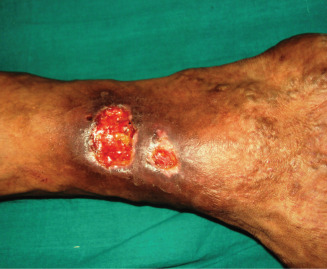
Patients, who were clinically diagnosed with venous ulcer, attending the dermatology OPD (Figure 1) were included after duly signed inform consent form. Clinical diagnosis was based on the site of ulcer (gaiter area), signs of chronic venous insufficiency (oedema, varicosity, lipodermatosclerosis, pigmentation and eczema) and characteristics of ulcer (shallow margin, granulation tissue in ulcer bed). Factors such as age, sex, body mass index (BMI), duration of ulcer, smoking history, hypertension and dyslipidemia were noted.
Figure 1.

Venous leg ulcer.
ABPI measurement
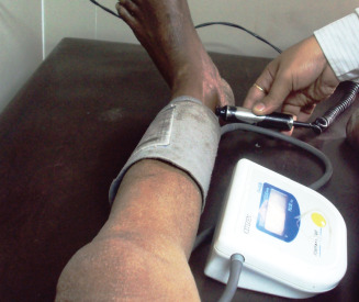
Measurement were performed with the patient in supine position by a hand‐held Doppler (8 MHz) device (Figure 2). ABPI was calculated by dividing the systolic blood pressure (SBP) obtained at ankle level of one leg with the SBP of brachial artery.
Figure 2.

Ankle‐Brachial Pressure Index measurement by hand‐held Doppler (8 MHz).
ABPI assessment
PAD was defined as ABPI ≤ 0·9 9 as per the criteria of the American College of Cardiology (ACC) and American Heart Association (AHA) for the diagnosis of PAD 10.
ABPI of the leg with lower value was taken to determine the presence of PAD 11.
Colour Doppler study
We also conducted arterial‐venous colour Doppler study on all the patients of this study.
Statistical analysis
Data were entered into a Microsoft Excel spreadsheet and then analysed by Statistica version 6 (StatSoft Inc., 2001, Tulsa, OK) software. Data have been summarised as mean and standard deviation for numerical variables (also median and interquartile range where applicable) and counts and percentages for categorical variables. Numerical variables have been compared between groups by Student's independent samples t‐test, if normally distributed, and by Mann–Whitney U‐test if otherwise. The Fisher's exact test was used to compare categorical variables between groups. Extent of agreement between ABPI and Doppler as diagnostic tool for PAD in venous ulcer cases was quantified by Kappa statistic. All analyses have been two‐tailed and P < 0·05 was considered statistically significant.
Results
After 1 year of study a total of 83 patients were evaluated. Of these, 24 patients (28·9%) had ulcer on the right side, 32 patients (38·5%) on the left side and, in 27 patients (32·5%), both sided ulcer.
Prevalence of PAD
Of the 83 patients studied, prevalence of concomitant PAD as calculated by ABPI was 23 [27·71% (95% CI: 18·08–37·34%)].
Among the 23 PAD patients, 14 patients (60·8%) were asymptomatic. History of intermittent claudication was found in eight (34·7%) patients, and signs of diminished peripheral pulses in one patient. None of the patients complained of rest pain.
The arterial‐venous colour Doppler study showed venous abnormality in the form of valve incompetence, deep vein thrombosis or varicosities, or perforator defects in all the patients of this study. That points out very high quality of clinical diagnosis of chronic venous leg ulcer.
Arterial Doppler showed that 22 of our the patients had PAD in the form of atherosclerosis, narrowing or block. Extent of agreement between ABPI and Doppler as diagnostic tool for PAD in venous ulcer cases by Kappa statistic was 0·906 (95% CI: 0·691–1·121) indicating a strong agreement.
Age
Mean age of presentation was 45·0 ± 14·5 years in clinical venous ulcer associated with PAD patient and 36·6 ± 11·4 years in non‐PAD patient. Older age patients were found to be more prone to developing PAD with the P value being 0·007.
Sex
Of 83 patients, 73 (87·95%) were male and, among the PAD patients, 22 (95·65%) were male.
Body mass index (BMI)
Average BMI in PAD patient was 25·5 ± 2·1 and in non‐PAD patient 24·1 ± 2·3. A statistically significant association was found between obesity and PAD.
Duration of ulcer
A very strong association could be found between longer duration (7 ± 3·4 months) of ulcer and PAD.
Smoking, hypertension and dyslipidemia
Although a strong association was observed for smoking and hypertension with PAD, the same was lacking for elevated serum triglyceride and cholesterol (Table 1).
Table 1.
Clinical characteristics of the study group*
| Risk factor | PAD (n = 23) | Non‐PAD (n = 60) | P value† |
|---|---|---|---|
| Age (Years) | 45·0 ± 14·5 | 36·6 ± 11·5 | 0.007 |
| Sex (Male/Female) | 22 (95·6%)/1 (4·3%) | 51(85%)/9(15%) | 0.270 |
| BMI | 25·5 ± 2·1 | 24·1 ± 2·3 | 0.011 |
| Duration (Months) | 7 ± 3·4 | 4·4 ± 2·3 | <0.001 |
| Smoking | 19 (82·61%) | 28 (46·67%) | 0.003 |
| Hypertension | 7 (30·4%) | 5 (8·3%) | 0.017 |
| Dyslipidemia | 3 (13%) | 4 (6·7%) | 0.390 |
| ABPI | Median 0·9 | 4 (6·67%) | <0.001 |
| Lower quartile 0·8 | Lower quartile 1·0 | ||
| Upper quartile 0·9 | Upper quartile 1·2 |
PAD, peripheral arterial disease; BMI, Body mass index; ABPI, Ankle‐Brachial pressure index.
Values are presented as the mean ± standard deviation, number (%) or median with lower and upper quartile.
P < 0·05 was considered significant.
Discussion
The prevalence of PAD in general population is variable 11 and was found to be 3·2% in a study from south India 12. PAD was associated with 21% of chronic leg ulcer irrespective of aetiology 5. In another study, among the venous ulcer patients 17% had PAD 13.
A study from Portugal observed that the diagnosis of leg ulcer was performed on clinical basis in 56% cases and only 8% had undergone PAD evaluation by ABPI 14. We evaluated 83 clinically diagnosed venous ulcer patients and PAD was found to be associated in 23 (27·71%) patients.
Majority of PAD patients (14; 60·8%) were asymptomatic, which supports the finding of earlier studies 6. Compression therapies that are routinely used in cases of venous ulcers can actually compromise the blood supply in these patients, furthermore adding to the morbidity and complications. Necrosis, limb lost by amputation following compression therapy, is not an unusual complication even with recently available superior compression stockings 5. So the results of this study clearly suggest that careful assessment for PAD in clinical venous ulcer patients is mandatory even in the absence of symptoms of PAD before treating them with compression therapy. Ulcer present in the classical venous site (gaiter area) should not lead to the assumption that arterial disease is not present 5.
Risk of PAD increases with age, obesity, diabetes and smoking 5, 12 male patients 15. In this study, PAD was significantly more with older age, obesity, hypertension and smoking. Mean age of presentation (45 years) was lower than western studies.
PAD of the leg is an important manifestation of systemic atherosclerosis 16, 17. Studies suggest that both symptomatic and asymptomatic PAD are associated with an increased risk of morbidity and mortality including cardiovascular disease, stroke 18, increase in serum creatinine level 19 and dementia 20. However, in this study no significant association of PAD with dyslipidemia was observed.
We found that ABPI is a simple, low‐cost and yet reliable tool for the diagnosis of PAD with a very strong level of agreement with a more conventional Doppler Studies. International guidelines on leg ulcers also recommend ABPI to assess and diagnose PAD 13.
We suggest that every patient with clinical diagnosis of venous ulcer should be carefully evaluated for PAD by a simple, non‐invasive OPD‐based ABPI measurement even in the absence of symptoms. It not only helps to arrive at a correct diagnosis and management, but also prevents dreaded complications arising from the indiscriminate use of compression therapies.
References
- 1. Graham ID, Harrison MB, Nelson EA, Lorimer K, Fisher A. Prevalence of lower‐limb ulceration: a systematic review of prevalence studies. Adv Skin Wound Care 2003;16:305–16. [PubMed ID 14652517]. [DOI] [PubMed] [Google Scholar]
- 2. Mekkes JR, Loots MA, Van Der Wal AC, Bos JD. Causes, investigation and treatment of leg ulceration. Br J Dermatol 2003;148: 388–401. [PubMed ID 12653729]. [DOI] [PubMed] [Google Scholar]
- 3. Humphreys ML, Stewart AH, Gohel MS, Taylor M, Whyman MR, Poskitt KR. Management of mixed arterial and venous leg ulcers. Br J Surg 2007;94:1104–7. [PubMed ID 17497654]. [DOI] [PubMed] [Google Scholar]
- 4. Milic DJ, Zivic SS, Bogdanovic DC, Karanovic ND, Golubovic ZV. Risk factors related to the failure of venous leg ulcers to heal with compression treatment. J Vasc Surg 2009;49:1242–7. [PubMed ID 19233601]. [DOI] [PubMed] [Google Scholar]
- 5. Callam MJ, Harper DR, Dale JJ, Ruckley CV. Arterial disease in chronic leg ulceration: Lothian and Forth Valley leg ulcer study. BMJ 1987;294:929–31. [PubMed ID 3107659]. [DOI] [PMC free article] [PubMed] [Google Scholar]
- 6. Sillesen H, Falk E. Peripheral artery disease (PAD) screening in the asymptomatic population: why, how, and who? Curr Atheroscler Rep 2011;13:390–5. [PubMed ID 21811798]. [DOI] [PubMed] [Google Scholar]
- 7. Keen D. Critical evaluation of the reliability and validity of ABPI measurement in leg ulcer assessment. J Wound Care 2008;17:530–3. [PubMed ID 19052517]. [DOI] [PubMed] [Google Scholar]
- 8. Lazarides MK. Giannoukas AD. The role of hemodynamic measurements in the management of venous and ischemic ulcers. Int J Low Extrem Wounds 2007;6:254–61. [PubMed ID 18048871]. [DOI] [PubMed] [Google Scholar]
- 9. Guo X, Li J, Pang W, Zhao M, Luo Y, Sun Y, Hu D. Sensitivity and specificity of ankle‐brachial index for detecting angiographic stenosis of peripheral arteries. Circ J 2008;72:605–10. [PubMed ID 18362433]. [DOI] [PubMed] [Google Scholar]
- 10. Hirsch AT, Criqui MH, Treat‐Jacobson D, Regensteiner JG, Creager MA, Olin JW, Krook SH, Hunninghake DB, Comerota AJ, Walsh ME, McDermott MM, Hiatt WR. Peripheral arterial disease detection, awareness, and treatment in primary care. JAMA 2001;286:1317–24. [PubMed ID 11560536]. [DOI] [PubMed] [Google Scholar]
- 11. Yu JH, Hwang JY, Shin MS, Jung CH, Kim EH, Lee SA, Koh EH, Lee WJ, Kim MS, Park JY, Lee KU. The prevalence of peripheral arterial disease in Korean patients with type 2 diabetes mellitus attending a university hospital. Diabetes Metab J 2011;35:543–50. [PubMed ID 22111047]. [DOI] [PMC free article] [PubMed] [Google Scholar]
- 12. Premalatha G, Shanthirani S, Deepa R, Markovitz J, Mohan V. Prevalence and risk factors of peripheral vascular disease in a selected South Indian population: the Chennai Urban Population Study. Diabetes Care 2000;23:1295–300. [PubMed ID 10977021]. [DOI] [PubMed] [Google Scholar]
- 13. Lazareth I, Taieb JC, Michon‐Pasturel U, Priollet P. Ease of use, feasibility and performance of ankle arm index measurement in patients with chronic leg ulcers. Study of 100 consecutive patients. J Mal Vasc 2009;34:264–71. [PubMed ID 19539439]. [DOI] [PubMed] [Google Scholar]
- 14. Pina E, Furtado K, Franks PJ, Moffatt CJ. Leg ulceration in Portugal: prevalence and clinical history. Eur J Vasc Endovasc Surg 2005;29:549–53. [PubMed ID 15966097]. [DOI] [PubMed] [Google Scholar]
- 15. Diehm C, Schuster A, Allenberg JR, Darius H, Haberl R, Lange S, Pittrow D, von Stritzky B, Tepohl G, Trampisch HJ. High prevalence of peripheral arterial disease and co‐morbidity in 6880 primary care patients: cross‐sectional study. Atherosclerosis 2004;172:95–105. [PubMed ID 14709362]. [DOI] [PubMed] [Google Scholar]
- 16. Hiatt WR. Medical treatment of peripheral arterial disease and claudication. N Engl J Med 2001;344:1608–21. [PubMed ID 11372014]. [DOI] [PubMed] [Google Scholar]
- 17. Murabito JM, Evans JC, Nieto K, Larson MG, Levy D, Wilson PW. Prevalence and clinical correlates of peripheral arterial disease in the Framingham Offspring Study. Am Heart J 2002;143:961–5. [PubMed ID 12075249]. [DOI] [PubMed] [Google Scholar]
- 18. Ding YM, Wang Y, Li Y, Yang P, Liu MY, Liu L, Zhu P, Li XY. Association of ankle‐brachial index with clinical coronary heart disease, stroke in aged Chinese hypertensive men. Zhongguo Ying Yong Sheng Li Xue Za Zhi 2011;27:129–33. [PubMed ID 21845851]. [PubMed] [Google Scholar]
- 19. O’Hare AM, Rodriguez RA, Bacchetti P. Low ankle‐brachial index associated with rise in creatinine level over time: results from the atherosclerosis risk in communities study. Arch Intern Med 2005;165:1481–5. [PubMed ID 16009862]. [DOI] [PubMed] [Google Scholar]
- 20. Laurin D, Masaki KH, White LR, Launer LJ. Ankle‐to‐brachial index and dementia: the Honolulu‐Asia Aging Study. Circulation 2007;116:2269–74. [PubMed ID 17967779]. [DOI] [PubMed] [Google Scholar]


