Abstract
Necrotizing fasciitis is a destructive invasive infection of skin, subcutaneous tissue and deep fascia, with relative sparing of muscle. It is a life‐threatening condition.
Here we report two cases of necrotizing fasciitis, which were not responding to conventional antibiotic therapy and local wound care after aggressive debridement. These two cases were treated simply by local application of 3% citric acid. Thus, citric acid was used to compliment wound management following surgical treatment with antibiotics.
Keywords: Citric acid treatment, Necrotizing fasciitis
INTRODUCTION
Necrotizing fasciitis is a rapidly spreading severe soft‐tissue infection characterised by rapidly progressive necrosis, involving subcutaneous tissue, including fascia, muscle or both. Surgical procedure, trauma, scratch or insect bites are the predisposing factors that favour development of necrotizing fasciitis (1).
It is a life‐threatening surgical emergency that needs early diagnosis, prompt and aggressive debridement and definitive therapy 2, 3 that is intravenous fluids, injectable antibiotics, inotropic support and oxygenation, if needed. Immediate institution of appropriate treatment (aggressive debridement and definitive therapy) improves the outcome.
We reported two cases of necrotizing fasciitis, which were not responding to conventional therapy after aggressive debridement, treated simply by local application of 3% citric acid. This study was approved by institutional ethical committee and informed consent from both the patients was obtained before application of citric acid to their wounds.
Case 1
A 40‐year‐old non diabetic male with a history of above knee amputation of left leg 10 years back presented with complains of fever, pain in back and extremities, and swelling of right thigh since 24 hours. On examination, his general condition was found to be very poor – the patient was in septic shock, his body temperature was 97°F, systolic blood pressure (BP) was 60 mm Hg, pulse was 146, respiratory rate was 28 breathes per minute and oxygen saturation was 86%. The abnormal haematological and biochemical parameters were as follows:
Total leucocytes count – 2100/mm3
Platelets – 37 000/mm3
Blood urea nitrogen – 27 mg/dl
Serum creatinine – 16 mg/dl
Patient showed mild cyanosis and cold clammy extremities. Local examination showed generalised swelling over right thigh and tenderness over the medial aspect of thigh with indurations. Based on clinical findings, the case was provisionally diagnosed as septic shock with soft‐tissue infection and subjected to various investigations. The patient was immediately given intravenous (iv) fluids and antibiotics, ceftizoxime 2 gm BD, netromycin 300 mg OD, crystalline penicillin 2 000 000 units every 6 hours and metronidazole 500 mg TDS for 7 days.
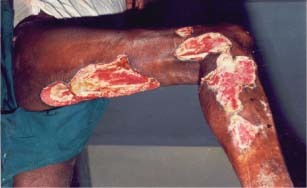
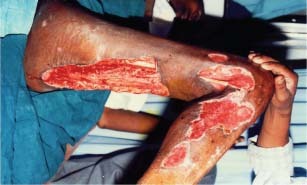
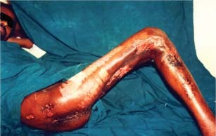
The infection extended up to anterior‐ and posterior‐abdominal wall of right side and blebs were developed on thigh. With the treatment given, swelling over thigh and abdominal wall was reduced but there was necrosis of the superficial skin of posterior aspect of right thigh and gluteal region. So the decision of surgical debridement was taken. Under general anaesthesia, incision was taken for drainage of anterolateral aspect of thigh opposite to greater trochanter. The dead necrosed skin and underlying fascial sheaths, found to be necrosed, were removed from posterior and anterior aspect of thigh. The necrosed material was sent for culture. The culture yielded Staphylococcus aureus and Escherichia coli. S. aureus was moderately susceptible to ampicillin and vancomycin, and resistant to cotrimoxazole, erythromycin, gentamicin, amikacin, netromycin, tetracycline, cloxacillin, piperacillin, ceftazidime, ceftriaxone, cephotaxime, cefuraxime and ciprofloxacin. E. coli was susceptible to amikacin and netromycin, and resistant to ampicillin, gentamicin, cephalexcin and pefloxacin. As the patient was not in position to afford costly antibiotic therapy, with written consent from patient, application of 3% citric acid was started. After through debridement of wound and removal of necrotic tissue, citric acid dressing was started on alternate day in the form of wound irrigation and wound wash and pads soaked in 3% citric acid were placed on raw area. This treatment was continued for 20 days and during this period, a total of 11 dressings with citric acid were performed. This treatment resulted in the formation of healthy granulation tissue. Because of large raw area, wound over lateral aspect of knee and leg was covered by skin graft (1, 2, 3).
Figure 1.

Necrotizing fasciitis – before application of citric acid.
Figure 2.

Necrotizing fasciitis – after 11 applications of citric acid.
Figure 3.

Necrotizing fasciitis – final photograph after skin grafting.
Case 2
A 60‐year‐old non diabetic female presented with fever and chills, soft‐tissue infection of left leg with blebs and skin discolouration. She was given ciprofloxacin (100 ml) twice daily, metronidazole (100 ml) every 8 hours and injection amoxycillin + clavulanic acid 1.2 g every 8 hours for 5 days but in vain. On surgical intervention, infection was found to be extended up to deep fascia, particularly the medial aspect of thigh, calf, ankle and dorsum. It was diagnosed as necrotizing fasciitis. Culture of necrosed material yielded Streptococcus pyogenes susceptible to amikacin, ceftazidime, cefepime, ciprofloxacin and resistant to cefazolin, gatifloxacin, and Pseudomonas aeruginosa susceptible to amikacin and resistant to ceftazidime, cefepime, imipenem, azlocillin, ciprofloxacin and cefazolin. As the patient was not in position to afford costly antibiotic therapy, with written consent from patient, application of 3% citric acid after thorough surgical debridement was started. Citric acid dressing was applied daily once in the form of wound irrigation and wound wash and pads soaked in 3% citric acid were placed on raw area. Thorough surgical debridement followed by local application of 3% citric acid resulted in the formation of healthy granulation after 20 applications. The wound closure was performed by skin grafting. Skin graft was accepted and wound was successfully closed.
DISCUSSION
Necrotizing fasciitis is a rapidly spreading soft‐tissue infection. It is a life‐threatening condition. Early diagnosis followed by surgical and medical management influences prognosis. The conventional treatment includes aggressive debridement, appropriate antibiotic therapy and meticulous local care. Sometimes, it does not respond to conventional therapy and spreads rapidly in spite of meticulous care and threatens the life and affected limb of patient. In the present study also, patients were non responsive to conventional antibiotic therapy and local wound care after aggressive debridement.
Use of citric acid for the effective treatment of a variety of chronic wound infections has been reported 4, 5, 6. In a histopathologic study also, citric acid has been found to actuate the wound healing process by boosting fibroblastic growth and neovascularisation that in turn increases microcirculation of wound enabling the formation of healthy granulation tissue, thereby leading to faster healing of the wound (7).
In the present study, an attempt was made to treat necrotizing fasciitis with 3% citric acid. The use of citric acid successfully controlled the infection and promoted formation of healthy granulation tissue making wound closure suitable by skin grafting. It is noteworthy that in case 1, we could successfully save the affected limb of the person who had lost his left leg before 10 years for the same reason.
From these results, it is concluded that when the management of necrotizing fasciitis is a matter of great concern by conventional treatment strategy (conventional antibiotic therapy and local wound care after aggressive debridement), citric acid treatment could be the reliable and effective option for the clinical management of necrotizing fasciitis.
REFERENCES
- 1. Singh G, Sinha SK, Adhikary S, Babu KS, Ray P, Khanna SK. Necrotizing infection of the soft tissue: a clinical profiles. Eur J Sur 2002;168: 366–71. [DOI] [PubMed] [Google Scholar]
- 2. Naqvi GA, Malik SA, Jan W. Necrotizing fasciitis of the lower extremity: a case report and current concept of diagnosis and management. Scandinavian J Trauma, Resuscitation Emergency Med 2009;17:28–40. [DOI] [PMC free article] [PubMed] [Google Scholar]
- 3. Sarani B, Strong M, Pascual J, Schwab CW. Necrotizing fasciitis: current concepts and review of literature. J Am Coll Surg 2009;208:279–88. [DOI] [PubMed] [Google Scholar]
- 4. Nagoba BS, Wadher BJ, Deshmukh SR, Mahabaleshwar L, Gandhi RC, Kulkarni PB, Mane VA, Deshmukh JS. Treatment of superficial pseudomonal infections with citric acid – an effective and economical approach. J Hosp Infect 1998;40:155–7. [DOI] [PubMed] [Google Scholar]
- 5. Nagoba BS, Wadher BJ, Chandorkar AG. Citric acid treatment of non‐healing ulcers in leprosy patients. Br J Dermatol 2002;146:1101. [DOI] [PubMed] [Google Scholar]
- 6. Nagoba BS, Wadher BJ, Rao AK, Kore GD, Gomashe AV, Ingle AB. Simple and effective approach for the treatment of chronic wound infections caused by multiple antibiotic resistant Escherichia coli . J Hosp Infect 2008;69:177–80. [DOI] [PubMed] [Google Scholar]
- 7. Nagoba BS, Gandhi RC, Wadher BJ, Potekar RM, Kolhe SM. Microbiological, histopathological and clinical changes in chronic wounds after citric acid treatment. J Med Microbiol 2008;57:681–2. [DOI] [PubMed] [Google Scholar]


