Abstract
Aims
A comprehensive classification for coronal lower limb alignment with predictive capabilities for knee balance would be beneficial in total knee arthroplasty (TKA). This paper describes the Coronal Plane Alignment of the Knee (CPAK) classification and examines its utility in preoperative soft tissue balance prediction, comparing kinematic alignment (KA) to mechanical alignment (MA).
Methods
A radiological analysis of 500 healthy and 500 osteoarthritic (OA) knees was used to assess the applicability of the CPAK classification. CPAK comprises nine phenotypes based on the arithmetic HKA (aHKA) that estimates constitutional limb alignment and joint line obliquity (JLO). Intraoperative balance was compared within each phenotype in a cohort of 138 computer-assisted TKAs randomized to KA or MA. Primary outcomes included descriptive analyses of healthy and OA groups per CPAK type, and comparison of balance at 10° of flexion within each type. Secondary outcomes assessed balance at 45° and 90° and bone recuts required to achieve final knee balance within each CPAK type.
Results
There was similar frequency distribution between healthy and arthritic groups across all CPAK types. The most common categories were Type II (39.2% healthy vs 32.2% OA), Type I (26.4% healthy vs 19.4% OA) and Type V (15.4% healthy vs 14.6% OA). CPAK Types VII, VIII, and IX were rare in both populations. Across all CPAK types, a greater proportion of KA TKAs achieved optimal balance compared to MA. This effect was largest, and statistically significant, in CPAK Types I (100% KA vs 15% MA; p < 0.001), Type II (78% KA vs 46% MA; p = 0.018). and Type IV (89% KA vs 0% MA; p < 0.001).
Conclusion
CPAK is a pragmatic, comprehensive classification for coronal knee alignment, based on constitutional alignment and JLO, that can be used in healthy and arthritic knees. CPAK identifies which knee phenotypes may benefit most from KA when optimization of soft tissue balance is prioritized. Further, it will allow for consistency of reporting in future studies.
Cite this article: Bone Joint J 2021;103-B(2):329–337.
Keywords: Coronal Plane Alignment Knee classification, Arithmetic HKA, Joint line obliquity, Knee alignment, Constitutional alignment, Kinematic alignment, CPAK
Introduction
Determining the ideal coronal alignment for individuals undergoing total knee arthroplasty (TKA) is one of the great challenges in reconstructive knee surgery. The ‘mechanical alignment’ (MA) method1 has been the gold-standard technique since early in TKA development, with good historic long-term survivorship.2-4 MA results in a horizontal joint line and a neutral mechanical axis, which has long been believed to provide the best mechanical environment for prosthetic longevity.5 MA, however, disregards the significant inherent variability in coronal alignment that exists across individuals6-10 and the biomechanical sequelae that may result from this ‘one-size-fits-all’ approach.11-16
The pursuit of improvement in patient satisfaction has led some to suggest a shift in technique favouring recreation of a patient’s constitutional (prearthritic) alignment, possibly resulting in more natural knee movements11-13 and improved soft tissue balance.16-20 Commonly termed the ‘kinematic alignment’ (KA) method,21 this approach attempts to restore the constitutional knee joint by replicating the original movement around the three kinematic axes that make up normal knee motion.17,21,22 However, there remains uncertainty about alignment targets, optimal kinematic surgical techniques, and choice of suitable patients.17,18,20,22-26
The existing nomenclature for coronal alignment (varus, valgus, or neutral) is inadequate as it only describes the patient’s alignment at a static moment in time and does not take the joint line into consideration.27 Once arthritic deformity commences, the lower limb mechanical axis shifts, commonly accentuating the original alignment (e.g. increasing varus deformity from initial constitutional varus). Other times, however, the constitutional alignment that was present at skeletal maturity reverses with disease progression (e.g. constitutional varus shifting to valgus deformity due to lateral joint space loss). Without knowing an individual’s constitutional alignment, replication of native anatomy with KA techniques is not easily achieved. Similarly, joint line obliquity (JLO) of the knee has not been clearly defined, an element that may be just as important as limb alignment to restoring natural kinematics of the prosthetic joint.28
Multiple methods to classify coronal alignment have been proposed, but these are complex and have not quantified constitutional limb alignment and JLO.8,29 Another unresolved question in kinematic alignment is which patients are most likely to benefit from restoration of constitutional alignment. Such ambiguity in the literature highlights the need for a clear, simple, and universal classification system for the coronal alignment of the knee. In addition, a system that has the capacity to determine which types of knees are more amenable to which alignment strategies would be of value.
The primary aim of this paper is to propose a new classification system for the Coronal Plane Alignment of the Knee (CPAK). We also aim to determine the CPAK types for which KA may provide a greater benefit than MA in optimizing soft tissue balance. The study’s first primary outcome was to examine the universal applicability of the CPAK classification using descriptive analyses of large, population-based, cross-sectional radiological datasets from healthy volunteers and osteoarthritic (OA) patients undergoing TKA. The second primary outcome was to assess the relative proportion of balanced knees at 10° of flexion for each CPAK type using KA versus MA techniques. Secondary outcomes included the quantitative mean intercompartmental pressure difference (ICPD) at 10°, 45°, and 90°, and the need for major knee balancing procedures comparing KA and MA for each CPAK type. Data from this study will provide a framework for classifying coronal plane alignment of the knee and, furthermore, will allow preoperative identification of the patients most likely to benefit from kinematic TKA according to CPAK-type.
Methods
Study design
Part 1 of the study outlines a stepwise methodological description of the CPAK classification by undertaking a cross-sectional radiological descriptive analysis of healthy and arthritic cohorts. Part 2, CPAK Surgical Validation, was a retrospective analysis of soft tissue balance based on CPAK type. Data were obtained from a convenience sample of patients from a randomized controlled trial (RCT) that compared intraoperative soft tissue balance in TKAs positioned with KA versus MA, the methodology and findings of which were described previously.16
Ethical approval for the overarching RCT was provided by Bellberry Limited (approval #2017-12-911) and was prospectively registered with the Australian New Zealand Clinical Trials Registry (#ACTRN12617001627347p). Approval for the current study was provided by Hunter New England Local Health District (approvals #EX201905-03 and #EX201905-04).
Study groups
The study group used to validate the CPAK classification for Part 1 comprised two cohorts. The healthy population consisted of 250 young adults aged between 20 and 27 years from a previous cross-sectional study of knee alignment by one of the authors (JB).7 Both limbs were imaged, providing data from a total of 500 knees. Participants were recruited at high school and university campuses, cinemas, and job recruitment bureaux in Leuven, Belgium between October 2009 and March 2010. In all, 50% (n = 125) of the volunteers were female. Only asymptomatic volunteers with no history of orthopaedic injury or disease were included. The arthritic population consisted of 500 consecutive patients scheduled for primary total or unicompartmental knee arthroplasty by two of the authors (SJM, DBC) at a private hospital in Sydney, Australia between October 2016 and March 2018. Only the limb undergoing surgery was included. The patients’ mean age was 66 years (44 to 88). Overall, 62% (n = 310) of patients were female. Patients were included regardless of underlying diagnosis and any history of lower limb surgery or trauma.
The study group for Part 2 consisted of a separate cohort of patients scheduled for primary unilateral or bilateral TKA. Two authors (SJM, DBC) performed all operations at a single institution in Sydney, Australia. There were 125 patients included in the study, with 13 bilateral procedures; 138 knees received the allocated intervention and were analyzed—68 in the MA group and 70 in the KA group. The mean age was 67.4 years (36 to 89) with a mean body mass index of 30.1 kg/m2 (21.5 to 54.8). There were 74 females and 51 males.
Radiological measurements
All participants underwent digital long leg radiographs (LLRs) as per Paley and Pfeil.30 Measurements were taken by a single observer in the healthy group and by two observers in the arthritic group (WGJ), using the same methodology (described below), which has been shown to have high inter- and intraobserver reliability.31 The mechanical hip-knee-ankle (mHKA) angle was the angle subtended by the mechanical axes of the femur and tibia. The mechanical lateral distal femoral angle (LDFA) was defined as the lateral angle formed between the femoral mechanical axis and the joint line of the distal femur. The mechanical medial proximal tibial angle (MPTA) was defined as the medial angle formed between the tibial mechanical axis and the joint line of the proximal tibia.
As an assessment of reproducibility of these measurements, the correlations were calculated using Pearson’s r in a subgroup of 25 arthritic LLRs among three surgeons (SJM, DBC, WGJ) and one trainee for the mHKA. The interobserver r was near-perfect (0.99; p < 0.001) for measures between all four observers, and the intraobserver r was near-perfect (0.99; p < 0.001) for measures among observers at a one-week interval.
Part 1: CPAK classification
The arithmetic HKA
With the unicompartmental joint space narrowing that occurs as part of the arthritic process, the overall alignment of the limb (mHKA) can change significantly with time (Figure 1a and b). 32 In the absence of arthritic bone loss at the central compartmental contact points, the constitutional alignment of the lower limb can be determined using “the arithmetic HKA”, by identifying bony landmarks and applying the algorithm: aHKA = MPTA - LDFA. This algorithm has been previously validated in a matched-pairs radiological study by our group to predict constitutional alignment.27 A negative aHKA indicates varus, and a positive aHKA indicates valgus constitutional limb alignment (Figure 2). The aHKA is not affected by joint space narrowing or tibiofemoral subluxation. It disregards the joint line convergence angle, which has been shown in our prior study of normal knees to be approximately -0.5°, and its contribution to prediction of constitutional knee alignment has minimal clinical significance.7 The method makes the assumption that when the distal femoral and proximal tibial joint lines are parallel, the aHKA equals the mHKA. Hence, the aHKA can be used to estimate constitutional alignment.
Fig. 1.
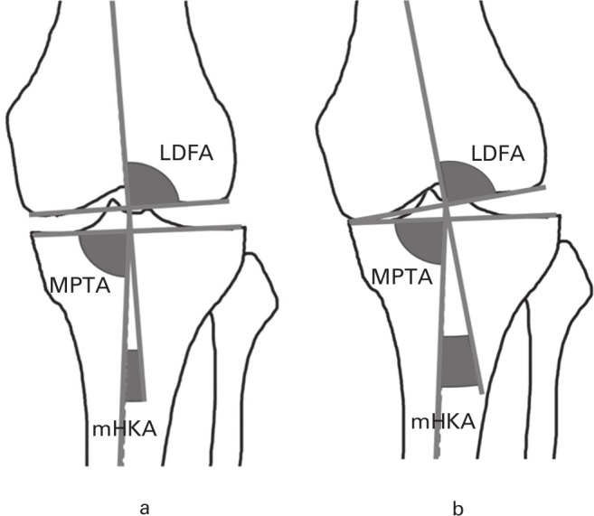
a) Lateral distal femoral angle (LDFA), medial proximal tibial angle (MPTA), and mechanical hip-knee-ankle angle (mHKA) in a knee with preserved joint space and mild constitutional varus alignment. b) The same knee following degenerative loss of medial joint space, showing a change in mHKA (with shift to further varus) but no change to LDFA and MPTA.
Fig. 2.
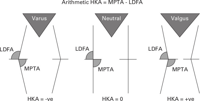
Relationship between the lateral distal femoral angle (LDFA) and medial proximal tibial angle (MPTA) in varus, neutral, and valgus lower limb alignment with the arithmetic hip-knee-ankle angle (aHKA).
Joint line obliquity
JLO of the knee is independent of the mechanical axis of the lower limb. Several studies have previously described the native JLO, but no consistent methodology has been universally adopted.28,33 For example, the JLO commonly referred to as ‘varus’ is the product of tibial varus and femoral valgus in the neutrally aligned lower limb. Similarly, distal femoral valgus and a neutral proximal tibia can combine to create ‘valgus’ lower limb alignment and a JLO commonly referred to as ‘varus’. To use the same terms (‘varus’ and ‘valgus’) for these two independent variables in the coronal plane of the knee creates ambiguity. For this reason, CPAK describes the direction of the JLO as ‘apex distal’, ‘neutral’, and ‘apex proximal’. This terminology clearly specifies whether the joint lines of both knees when extended to the midline is either below, level with, or above the level of a horizontal joint line (Figure 3).
Fig. 3.
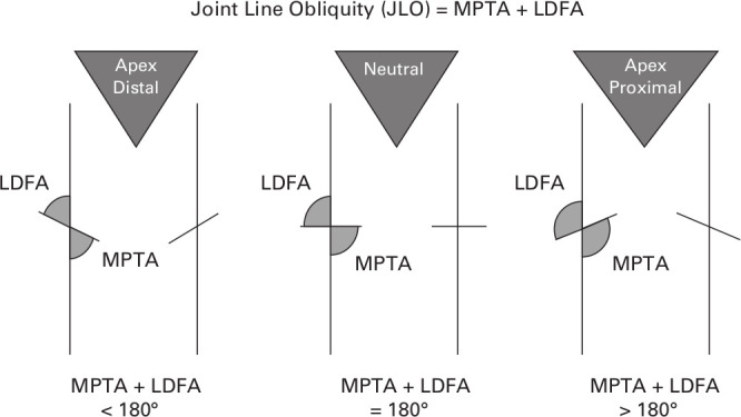
Use of medial proximal tibial angle (MPTA) and lateral distal femoral angle (LDFA) to indicate joint line obliquity (JLO).
Calculation of the JLO is derived from the same two variables used to calculate the aHKA (JLO = MPTA + LDFA) and defines its obliquity relative to the floor in double leg stance. If the sum of these two angles is 180°, the joint line is approximately neutral. A sum of greater than 180° indicates an apex proximal joint line, while a sum of less than 180° indicates that the joint line is apex distal.
CPAK classification matrix
The CPAK classification incorporates the two independent variables of aHKA (with varus, neutral, and valgus subgroups) and JLO (with apex distal, neutral, and apex proximal subgroups). The three subgroups of aHKA are set against the three subgroups of JLO in a matrix to create nine different phenotypes of knees (Figure 4).
Fig. 4.
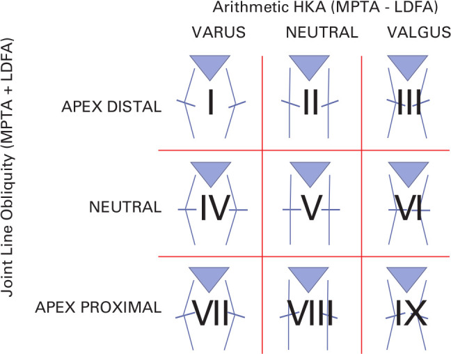
Coronal Plane Alignment of the Knee classification (CPAK) with nine theoretical types of knee. Arithmetic HKA, arithmetic hip-knee-ankle angle.
CPAK type boundaries were determined to be one standard deviation (SD) (rounded to the nearest whole number) for the mean aHKA and JLO of the combined dataset of all 1,000 knees. CPAK boundaries for neutral aHKA are 0° ± 2°, inclusive (SD 1.80°). A varus aHKA is less than -2°, while a valgus aHKA is greater than +2°. CPAK boundaries for a neutral JLO are 180° ± 3°, inclusive (SD 2.90°). An apex distal JLO is less than 177°, while an apex proximal JLO is greater than 183°.
Part 2: CPAK surgical validation
Surgical planning
The mHKA, LDFA, and MPTA were measured in the surgical validation group of 138 knees. This allowed calculation of the aHKA, JLO, CPAK type, and distal femoral and proximal tibial resection angles.
In the MA group, bone resections were made perpendicular to the mechanical axis of the femur and tibia, with the aim of restoring a neutral (0°) mHKA. Femoral rotation was set parallel to the surgical transepicondylar axis, with secondary referencing perpendicular to the AP femoral axis and 3° externally rotated from the posterior condylar axis.
In the KA group, coronal bone resections were undertaken within a restricted alignment safe zone, with the aim of restoring constitutional LDFA, MPTA, and aHKA for each patient. The restricted safe zone was defined as 86° to 93° for recreation of both the LDFA and the MPTA, and -5° varus to +4° valgus for the final mHKA. If the aHKA was outside the final mHKA safe zone, the femoral and tibial resections were incrementally reduced to be within the safe zone. For patients who were older than 80 years or who had a history of osteoporosis, the safe zones for LFDA and MPTA were narrowed to 87° to 93°, and the final HKA was narrowed to -4° to +3° due to concern about greater risk of implant subsidence in those patients with alignment deviations further from neutral. Femoral rotation was initially planned parallel to the posterior condylar bone but adjusted if the tibial resection had to be reduced to fall within the safe zone.
Surgical technique
All procedures were performed using optical navigation (OrthoMap Precision Navigation, Stryker, Mahwah, New Jersey, USA) to ensure accurate restoration of target alignments. A posterior-stabilized, fully cemented total knee prosthesis was used with patellar resurfacing in all cases (Legion, Smith & Nephew, Memphis, Tennessee, USA). After trial implant insertion, but prior to any soft tissue releases, a wireless pressure sensor (VERASENSE, OrthoSensor, Dania Beach, Florida, USA) was inserted, and medial and lateral compartmental pressures recorded at 10°, 45°, and 90° of knee flexion with the arthrotomy closed. Pressures were recorded by both the operating surgeon and an assistant, with the mean of the two readings used. The intercompartmental pressure difference (ICPD) was calculated as the absolute pressure difference between medial and lateral compartments at each flexion angle. An ICPD of 15 psi or less at each flexion angle was considered to be balanced based on prior studies showing improved patient-reported outcomes using this definition.34,35 If the ICPD was between 16 and 40 psi, a soft tissue release was performed.36 Bone recuts were performed if an ICPD was greater than 40 psi or if the absolute pressure in one compartment was greater than 60 psi.
Outcome measures
The primary outcome for Part 1 of the study was frequencies for each CPAK type in the healthy and arthritic populations. The primary outcome for Part 2 was a comparison of the relative proportion of balanced knees (KA versus MA) among CPAK types. Near-full extension (10°) was chosen as the angle for this primary outcome because the methodology for calculation of constitutional alignment is based on coronal alignment, which restores extension gap balance. Secondary outcomes included the mean ICPD for each CPAK type at 10°, 45°, and 90° of flexion as a quantitative comparison of knee balance of KA versus MA. Additionally, numbers of bone recuts required in each CPAK type were analyzed for MA and KA as an estimate of severe imbalance requiring major balancing procedures.
Statistical analysis
Scatterplots for each population were created to demonstrate alignment distributions for healthy and arthritic groups. Normality of data distribution was assessed for continuous variables using Shapiro-Wilk test and Q-Q plots. An independent-samples t-test was used to compare differences in means for normally distributed data and Mann-Whitney U test for non-parametric data. The chi-squared test and Fisher’s exact test were used for categorical data analysis. Statistical significance was set at a p-value ≤ 0.05. Statistical analyses were performed using XLSTAT v22.3.1 (Addinsoft, New York, New York, USA) and SPSS Statistics Package v.25 (IBM, Armonk, New York, USA).
Results
The mean MPTAs of the healthy and arthritic groups were 87.0° (SD 2.1°) and 87.3° (SD 2.1°) respectively. The mean LDFAs of the healthy and arthritic groups were 87.9° (SD 1.7°) and 88.1° (SD 2.1°) respectively. The mean and variance for mHKA were different between the healthy and arthritic groups (-1.3° (SD 2.3°) vs -2.9° (SD 7.4°)), but the mean and variance for aHKA were similar (-0.9° (SD 2.5°) vs -0.8° (SD 2.8°)).
CPAK classification
The frequencies of individuals representing all CPAK types were similar when comparing the two populations (Figures 5 and 6). The commonest CPAK types in order were Type II (neutral aHKA, apex distal JLO; 39.2% (n = 196) healthy vs 32.2% (n = 161) OA), Type I (varus aHKA, apex distal JLO; 26.4% (n = 132) healthy vs 19.4% (n = 97) OA), and Type V (neutral aHKA, neutral JLO; 15.4% (n = 77) healthy vs 14.6% (n = 73) OA). CPAK Types VII, VIII, and IX were rare in both populations.
Fig. 5.
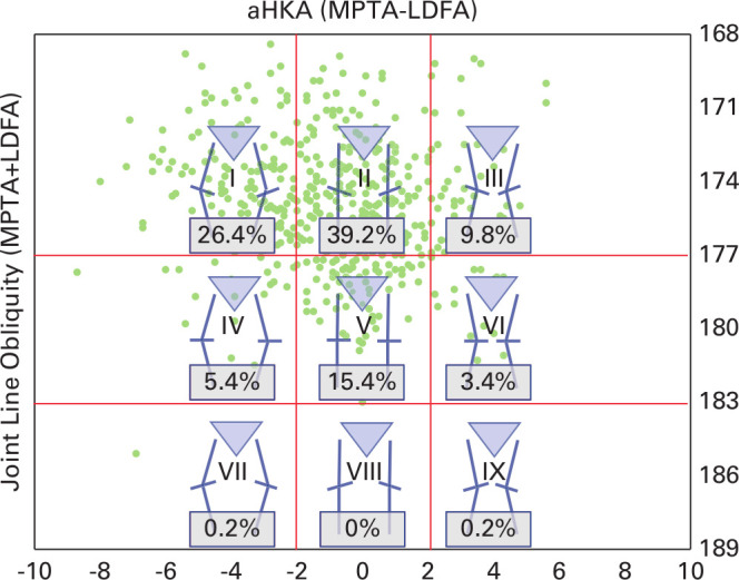
Plot of arithmetic hip-knee-ankle angle (aHKA) against joint line obliquity for a healthy population showing distribution by percentage in the nine Coronal Plane Alignment of the Knee (CPAK) types. LDFA, lateral distal femoral angle; MPTA, medial proximal tibial angle.
Fig. 6.
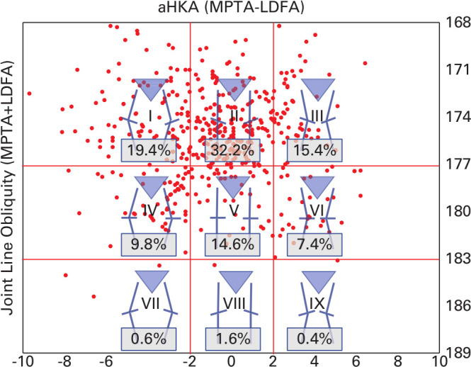
Plot of arithmetic hip-knee-ankle angle (aHKA) against joint line obliquity for an arthritic population showing distribution by percentage in the nine Coronal Plane Alignment of the Knee (CPAK) types. LDFA, lateral distal femoral angle; MPTA, medial proximal tibial angle.
CPAK surgical validation
A higher proportion of KA TKAs were balanced compared to MA TKAs for all CPAK types (Table I and Figure 7). Types I, II, and IV with KA had a significantly higher likelihood of having optimal balance and the largest effect sizes compared to MA (Type I, 100% KA vs 15% MA; p < 0.001, chi-squared test; Type II, 78% KA vs 46% MA; p = 0.018, chi-squared test; Type IV, 89% KA versus 0% MA; p < 0.001, Fisher's exact test).
Table I.
Coronal Plane Alignment Knee type and balance at 10° knee flexion with kinematic alignment and mechanical alignment.
| CPAK type |
Knees, n | KA balanced, % (balanced/total) | MA balanced, % (balanced/total) | p-value |
|---|---|---|---|---|
| I | 23 | 100 (10/10) | 15 (2/13) | < 0.001* † |
| II | 53 | 78 (21/27) | 46 (12/26) | 0.018* † |
| III | 28 | 62 (8/13) | 40 (6/15) | 0.290† |
| IV | 15 | 89 (8/9) | 0 (0/6) | < 0.001* ‡ |
| V | 12 | 100 (7/7) | 60 3/5) | 0.152‡ |
| VI | 7 | 50 (2/4) | 33 (1/3) | 1.000‡ |
Statistically significant.
Chi-squared test.
Fisher's exact test.
CPAK, Coronal Plane Alignment Knee; KA, kinematic alignment; MA, mechanical alignment.
Fig. 7.
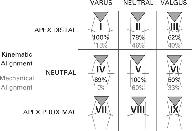
Probability of achieving knee balance based on Coronal Plane Alignment Knee type for kinematic alignment and mechanical alignment at 10° of knee flexion.
Secondary outcome measures
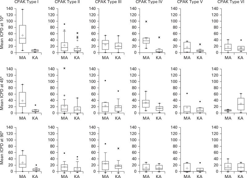
There was a significant ICPD at all three flexion angles for CPAK Type I, at 90° for CPAK Type II, and at 10° and 45° for CPAK Type IV, with MA having a greater difference (worse) than KA (Table II and Figure 8). There was a higher proportion of TKAs requiring bone recuts to achieve knee balance in CPAK Types I, II and III when MA was performed compared to KA (Table III).
Table II.
Descriptive statistics for intercompartmental pressure differences at 10°, 45°, and 90° of knee flexion for kinematic alignment and mechanical alignment.
| CPAK type | Knee angle, ° | Mean KA ICPD (SD; range) |
Mean MA ICPD (SD; range) |
p-value |
|---|---|---|---|---|
| I | 10 | 6.5 (3.2; 1 to 10) | 55.9 (39.0; 7 to 138) | 0.001* ‡ |
| 45 | 8.7 (7.0; 2 to 26) | 39.7 (28.4; 2 to 91) | 0.004* † | |
| 90 | 7.3 (6.0; 0 to 21) | 28.2 (21.9; 1 to 66) | 0.008* ‡ | |
| II | 10 | 13.7 (16.5; 1 to 63) | 24.0 (24.1; 2 to 92) | 0.065† |
| 45 | 16.9 (14.6; 2 to 51) | 26.0 (28.1; 3 to 141) | 0.157† | |
| 90 | 12.4 (13.3; 2 to 50) | 18.5 (13.3; 0 to 61) | 0.039* † | |
| III | 10 | 22.5 (16.2; 1 to 54) | 25.8 (17.3; 1 to 55) | 0.612‡ |
| 45 | 25.1 (18.3; 7 to 72) | 26.4 (25.3; 1 to 104) | 0.990† | |
| 90 | 22.3 (18.6; 0 to 75) | 28.6 (23.9; 1 to 89) | 0.316† | |
| IV | 10 | 11.6 (14.9; 3 to 50) | 45.6 (28.6; 16 to 99) | 0.006* † |
| 45 | 15.2 (9.8; 3 to 32) | 33.6 (21.5; 9 to 69) | 0.041* ‡ | |
| 90 | 15.3 (8.2; 4 to 28) | 16.1 (11.9; 3 to 32) | 0.887‡ | |
| V | 10 | 10.3 (8.4; 4 to 28) | 19.0 (16.2; 4 to 38) | 0.606† |
| 45 | 16.1 (12.4; 5 to 40) | 22.0 (25.6; 5 to 65) | 0.606‡ | |
| 90 | 11.4 (9.9) | 14.5 (14.7) | 0.965† | |
| VI | 10 | 18.3 (15.7) | 19.8 (20.2) | 0.911‡ |
| 45 | 32.6 (26.5) | 12.2 (3.8) | 0.251‡ | |
| 90 | 16.5 (14.3) | 19.8 (20.2) | 0.807‡ |
Statistically significant.
Mann-Whitney U test.
Independent-samples t-test.
CPAK, Coronal Plane Alignment Knee; ICPD, intercompartmental pressure difference; KA, kinematic alignment; MA, mechanical alignment.
Fig. 8.
Box plot comparison of mean intercompartmental pressure differences at 10°, 45°, and 90° of knee flexion for kinematic alignment (KA) and mechanical alignment (MA). CPAK, Coronal Plane Alignment of the Knee; ICPD, intercompartmental pressure difference.
Table III.
Requirements for bone recuts for each Coronal Plane Alignment Knee type.
| CPAK type | KA group, n (total) |
KA recuts, % | MA group, n (total) |
MA recuts, % | p-value |
|---|---|---|---|---|---|
| I | 0 (10) | 0 | 9 (13) | 69 | 0.001* † |
| II | 4 (27) | 15 | 11 (26) | 42 | 0.026* |
| III | 0 (13) | 0 | 7 (15) | 47 | 0.004* † |
| IV | 1 (9) | 11 | 3 (6) | 50 | 0.235‡ |
| V | 0 (7) | 0 | 2 (5) | 40 | 0.152‡ |
| VI | 1 (3) | 25 | 1 (3) | 33 | 1.000‡ |
Bone recuts performed when absolute pressure in either compartment was greater than 60 psi, or an intercompartmental pressure difference was greater than 40 psi.
Statistically significant.
Chi-squared test.
Fisher’s exact test.
CPAK, Coronal Plane Alignment Knee; KA, kinematic alignment; MA, mechanical alignment.
Discussion
We describe a straightforward, pragmatic, and comprehensive classification system incorporating algorithms for constitutional lower limb alignment and JLO. When comparing healthy and arthritic populations, there were similar frequencies among all CPAK types, suggesting that this classification can be used in healthy and arthritic knees. Although there are nine knee types, CPAK Types VII, VIII, and IX are rare. We believe their inclusion for completeness is important as they provide a visual aid to understanding the CPAK matrix and can also be used in postoperative TKA assessment where the JLO has been inadvertently altered. The study also demonstrates that for each CPAK type, a greater proportion of knees implanted in KA achieved balance compared to those implanted in MA.
Despite CPAK Type V (neutral aHKA, neutral JLO) being the target for MA, only 15% of both populations fell within the classification boundaries. A greater proportion of Type V knees were objectively balanced when undertaking KA versus MA (100% versus 60% respectively). Although not statistically significant, it is likely that subtle knee alignment changes with KA to both the aHKA and JLO within 2° of its boundaries may increase the likelihood of achieving knee balance. This technique of altering alignment around the neutral resections is referred to as ‘adjusted MA’ or ‘modified MA.’37
CPAK Type II knees (neutral aHKA and apex distal JLO) are the commonest knee type, comprising nearly 40% of knees in the normal population. This CPAK type is the foundation for which Hungerford et al38 described the anatomical alignment (AA) method. Despite this technique alig ning the joint line based on mean population values of 3° femoral valgus and 3° tibial varus, precisely replicating these resection targets with conventional instrumentation was difficult and largely abandoned. In this CPAK type, where mechanical axis (aHKA) is neutral, the current study found a significant difference in the proportion of knees balanced at 10° and a trend for lower ICPD differences in favour of KA. At 90°, there was significantly improved balance in favour of KA (Table II). This suggests that JLO, as a separate variable to coronal limb alignment (aHKA), independently improves knee balance in flexion.
The distributions by CPAK type (Figures 5 and 6) show that 32% of normal and 30% of arthritic patients have constitutional varus and 76% of normal and 67% of arthritic patients have an apex distal JLO. This challenges the common philosophy of aligning the knee into a neutral mechanical alignment with neutral JLO, as this combination only represents normal for a small proportion of the population. The effect size for achieving a balanced knee for KA when compared to MA with constitutional varus was large for CPAK Type I (varus aHKA, apex distal JLO) and CPAK Type IV knees (varus aHKA, neutral JLO). Approximately 90% or more of CPAK Types I and IV were balanced at 10° if randomized to KA, compared to 15% or fewer when MA was used. When analyzing ICPDs, Type I knees had better balance at 10°, 45°, and 90°, supporting the proposition that restoring the apex distal JLO with KA has an impact on flexion balance as well, while creating a non-physiological neutral JLO with MA is unfavourable in this group. Type IV knees in the KA cohort had better balance at 10° and 45°, but both were equivalent, with normal balance, at 90°. Conceptually, this illustrates that while MA does not restore the varus aHKA and extension balance in the Type IV knee, it does restore the neutral JLO of this group; this is the reason KA and MA are both balanced in flexion. It is our opinion that CPAK Types I and IV are better aligned with a kinematic approach from the commencement of surgery. Otherwise, if MA is undertaken, significant interventions to restore balance will most likely be required, with either recuts into varus or extensive releasing of the medial collateral ligament.
CPAK Types III and VI are constitutional valgus knees, with an apex distal and neutral JLO respectively. In these types, complex morphological factors beyond coronal plane alignment may drive alterations in soft tissue balance.39 These include lateral femoral and tibial bone deficiencies, external rotation deformities of the femur and tibia, and secondary femoral metaphyseal remodelling. Soft tissue alterations may occur, particularly contractures of the lateral soft tissues, and as arthritic deformity increases, so may secondary attenuation of the medial collateral ligament.40 CPAK Types III and VI represent a more complex reconstructive solution beyond restoring constitutional alignment. A proportion of patients in these CPAK types had constitutional alignment and lateral distal femoral angles outside the restricted safe zones defined in this study. Further, there were fewer patients in these groups, which may have had an impact on whether a true difference existed when undertaking KA in these knees. Despite having a similar mean ICPD at 10° for MA and KA, significantly more Type III knees required bone recuts into valgus when MA was applied (47% MA vs 0% KA), suggesting that in this group, normalization of JLO is an important component for restoration of complex 3D kinematics.
In 2018, Lin et al8 described a classification system using LLRs of 214 healthy Taiwanese individuals aged 20 to 70 years. This classification had 27 possible combinations, although only five were described as clinically relevant. The following year, Hirschmann et al9 proposed a classification based on CT imaging from 160 nonarthritic individuals aged 16 to 44 years (308 knees), with 125 theoretical ‘functional phenotypes’ and 43 described as clinically relevant. Both classifications utilized three variables: mechanical limb alignment, distal femoral angle, and proximal tibial angle. In contrast, CPAK combines the anatomical joint line measures of the LDFA and MPTA, resulting in only two critical variables: aHKA (constitutional limb alignment) and JLO. In this way, CPAK classification simplifies categorization into nine knee phenotypes. Also, because CPAK also incorporates aHKA, it can be used in both healthy subjects and arthritic patients.
The CPAK classification allows for customization of preoperative alignment planning based on individualized, surgeon-defined restricted boundaries for aHKA and JLO. Secondly, CPAK determines whether a patient should be considered for a MA TKA (CPAK Type V), AA TKA (CPAK Type II), or KA TKA (including but not restricted to other CPAK Types I, III, IV, VI). Further, it allows for consideration of a ‘functional alignment’ strategy, where bone resections are performed within traditionally accepted boundaries, with the aim to limit alteration to the native soft tissue envelope.37
This research has several limitations. First, the arthritic cohort included all patients on whom knee arthroplasty was conducted irrespective of significant arthritic bone loss, extra-articular bone deformity, or prior osteotomy. Second, the two populations studied were from different continents, and racial background was not analyzed. Racial differences have been shown in prior studies to influence alignment.8,41 Third, the groups studied did not have an equal sex distribution: the OA group had a predominance of females, which is typical of a TKA population. Fourth, in the second part of the study, small sample sizes (particularly in CPAK Types V and VI) make comprehensive comparison of balance among groups less reliable, as this study was not powered to detect differences among all CPAK types. Fifth, radiological measurement errors related to rotational malpositioning and fixed flexion contractures could not be excluded, and as such, other advanced imaging methods may provide greater accuracy. Bone loss related to advanced OA will also contribute to measurement errors for determination of alignment parameters and resection angle calculations. Further research into methods that can account for bone loss is warranted. Sixth, because we used a restricted safe zone for KA surgery, it is possible that widening of alignment boundaries may have resulted in an even higher proportion of knees being balanced in the KA group. Seventh, although this classification has demonstrated utility in prediction of soft tissue balance, future research is required to correlate knee phenotype and patient outcomes. We believe that CPAK phenotype should be considered when assessing outcomes in kinematically aligned surgery. And finally, CPAK does not address axial or sagittal alignment, which also contribute to knee balance. Future research that addresses our understanding of 3D alignment and balance is warranted.
In summary, the new CPAK classification provides a simple and comprehensive system for describing knee alignment in the arthritic and healthy knee.In addition, CPAK allows determination of which patients are most likely to benefit from kinematic alignment when optimization of soft tissue balance is prioritized. With a greater understanding of the knee phenotypes, surgeons now have a preoperative method to determine which alignment strategy is best suited for each patient.
Take home message
- The new coronal plane alignment of the knee (CPAK) classification provides a simple and comprehensive system for describing knee alignment in the arthritic and healthy knee and provides categorical data that will aid communication and stimulate further research.
- CPAK allows determination of which patients are most likely to benefit from kinematic alignment when optimization of soft tissue balance is prioritized.
- With a greater understanding of knee phenotypes, surgeons now have a preoperative method to determine which alignment strategy is best suited for each patient.
Author contributions
S. J. MacDessi: Conceptualized and designed the study, Produced the manuscript, Analyzed the data.
W. Griffiths-Jones: Conceptualized and designed the study, Produced the manuscript, Analyzed the data.
I. A. Harris: Interpreted the data, Produced the manuscript.
J. Bellemans: Produced the manuscript.
D. B. Chen: Designed the study, Produced the manuscript.
Funding statement
The author or one of more of the authors have received or will receive benefits for personal or professional use from a commercial party related directly or indirectly to the subject of this article. In addition, benefits have been or will be directed to a research fund, foundation, educational institution, or other non-profit organization with which one or more of the authors are associated.
ICMJE COI statement
J. Bellemans reports personal fees from Stryker, outside the submitted work. W. Griffiths-Jones reports personal fees from Stryker, outside the submitted work, and a patent pending (PCT/AU2018/000241). D. B. Chen reports fellowship funding from Smith and Nephew and Zimmer Biomet, consulting fees from Amplitude SAS, personal fees from Stryker, a patent pending (PCT/AU2018/000241), all outside the submitted work. S. J. MacDessi reports fellowship funding from Smith and Nephew and Zimmer Biomet, consulting fees from Amplitude SAS, personal fees from Stryker, a patent pending (PCT/AU2018/000241), all outside the submitted work.
Acknowledgements
We are sincerely appreciative of the efforts of Ms Jil Wood, MSN, Clinical Research Manager at Sydney Knee Specialists, for her assistance with editing the manuscript and oversight of the study. We also wish to acknowledge Dr William Colyn, Consulting Surgeon at the General Hospital of Turnhout, Belgium, and Dr Sol Han and Dr Nikolas Fountas, Orthopaedic Registrars at The Canterbury Hospital, for performing radiographic measurements in this study.
Open access statement
This is an open-access article distributed under the terms of the Creative Commons Attribution Non-Commercial No Derivatives (CC BY-NC-ND 4.0) licence, which permits the copying and redistribution of the work only, and provided the original author and source are credited. See https://creativecommons.org/licenses/by-nc-nd/4.0/
This article was primary edited by A. Wood and first proof edited by G. Scott.
References
- 1.Insall JN, Binazzi R, Soudry M, Mestriner LA. Total knee arthroplasty. Clin Orthop Relat Res. 1985;192:13–22. [PubMed] [Google Scholar]
- 2.Evans JT, Walker RW, Evans JP, Blom AW, Sayers A, Whitehouse MR. How long does a knee replacement last? A systematic review and meta-analysis of case series and national registry reports with more than 15 years of follow-up. Lancet. 2019;393(10172):655–663. [DOI] [PMC free article] [PubMed] [Google Scholar]
- 3.Australian Orthopaedic Association National Joint Replacement Registry (AOANJRR) . Hip, knee & shoulder arthroplasty: 2019 Annual Report. AOA: Adelaide. 2019. https://aoanjrr.sahmri.com/documents/10180/668596/Hip%2C+Knee+%26+Shoulder+Arthroplasty/c287d2a3-22df-a3bb-37a2-91e6c00bfcf0
- 4.Healthcare Quality Improvement Partnership . National joint Registry for England, Wales, Northern Ireland and the Isle of man. 15th annual report. 2018. https://www.hqip.org.uk/resource/national-joint-registry-15th-annual-report-2018/#.XnQemC1L00o (date last accessed 15 January 2019).
- 5.Hadi M, Barlow T, Ahmed I, Dunbar M, McCulloch P, Griffin D. Does malalignment affect revision rate in total knee replacements: a systematic review of the literature. Springerplus. 2015;4:835. [DOI] [PMC free article] [PubMed] [Google Scholar]
- 6.Cooke D, Scudamore A, Li J, Wyss U, Bryant T, Costigan P. Axial lower-limb alignment: comparison of knee geometry in normal volunteers and osteoarthritis patients. Osteoarthritis Cartilage. 1997;5(1):39–47. [DOI] [PubMed] [Google Scholar]
- 7.Bellemans J, Colyn W, Vandenneucker H, Victor J. The Chitranjan Ranawat Award: is neutral mechanical alignment normal for all patients? the concept of constitutional varus. Clin Orthop Relat Res. 2012;470(1):45–53. [DOI] [PMC free article] [PubMed] [Google Scholar]
- 8.Lin Y-H, Chang F-S, Chen K-H, Huang K-C, Su K-C. Mismatch between femur and tibia coronal alignment in the knee joint: classification of five lower limb types according to femoral and tibial mechanical alignment. BMC Musculoskelet Disord. 2018;19(1):411. [DOI] [PMC free article] [PubMed] [Google Scholar]
- 9.Hirschmann MT, Hess S, Behrend H, Amsler F, Leclercq V, Moser LB. Phenotyping of hip-knee-ankle angle in young non-osteoarthritic knees provides better understanding of native alignment variability. Knee Surg Sports Traumatol Arthrosc. 2019;27(5):1378–1384. [DOI] [PubMed] [Google Scholar]
- 10.Moreland JR, Bassett LW, Hanker GJ. Radiographic analysis of the axial alignment of the lower extremity. J Bone Joint Surg Am. 1987;69(5):745–749. [PubMed] [Google Scholar]
- 11.Blakeney W, Clément J, Desmeules F, Hagemeister N, Rivière C, Vendittoli P-A. Kinematic alignment in total knee arthroplasty better reproduces normal gait than mechanical alignment. Knee Surg Sports Traumatol Arthrosc. 2019;27(5):1410–1417. [DOI] [PubMed] [Google Scholar]
- 12.Maderbacher G, Keshmiri A, Krieg B, Greimel F, Grifka J, Baier C. Kinematic component alignment in total knee arthroplasty leads to better restoration of natural tibiofemoral kinematics compared to mechanic alignment. Knee Surg Sports Traumatol Arthrosc. 2019;27(5):1427–1433. [DOI] [PubMed] [Google Scholar]
- 13.Niki Y, Nagura T, Nagai K, Kobayashi S, Harato K. Kinematically aligned total knee arthroplasty reduces knee adduction moment more than mechanically aligned total knee arthroplasty. Knee Surg Sports Traumatol Arthrosc. 2018;26(6):1629–1635. [DOI] [PubMed] [Google Scholar]
- 14.Blakeney W, Beaulieu Y, Kiss M-O, Rivière C, Vendittoli P-A. Less gap imbalance with restricted kinematic alignment than with mechanically aligned total knee arthroplasty: simulations on 3-D bone models created from CT-scans. Acta Orthop. 2019;90(6):602–609. [DOI] [PMC free article] [PubMed] [Google Scholar]
- 15.Blakeney W, Beaulieu Y, Puliero B, Kiss M-O, Vendittoli P-A. Bone resection for mechanically aligned total knee arthroplasty creates frequent gap modifications and imbalances. Knee Surg Sports Traumatol Arthrosc. 2020;28(5):1532–1541. [DOI] [PubMed] [Google Scholar]
- 16.MacDessi SJ, Griffiths-Jones W, Chen DB, et al. Restoring the constitutional alignment with a restrictive kinematic protocol improves quantitative soft-tissue balance in total knee arthroplasty: a randomized controlled trial. Bone Joint J. 2020;102-B(1):117–124. [DOI] [PMC free article] [PubMed] [Google Scholar]
- 17.Howell SM, Howell SJ, Kuznik KT, Cohen J, Hull ML. Does a kinematically aligned total knee arthroplasty restore function without failure regardless of alignment category? Clin Orthop Relat Res. 2013;471(3):1000–1007. [DOI] [PMC free article] [PubMed] [Google Scholar]
- 18.Calliess T, Bauer K, Stukenborg-Colsman C, Windhagen H, Budde S, Ettinger M. Psi kinematic versus non-PSI mechanical alignment in total knee arthroplasty: a prospective, randomized study. Knee Surg Sports Traumatol Arthrosc. 2017;25(6):1743–1748. [DOI] [PubMed] [Google Scholar]
- 19.Dossett HG, Estrada NA, Swartz GJ, LeFevre GW, Kwasman BG. A randomised controlled trial of kinematically and mechanically aligned total knee replacements: two-year clinical results. Bone Joint J. 2014;96-B(7):907–913. [DOI] [PubMed] [Google Scholar]
- 20.Hutt JRB, LeBlanc M-A, Massé V, Lavigne M, Vendittoli P-A. Kinematic TKA using navigation: surgical technique and initial results. Orthop Traumatol Surg Res. 2016;102(1):99–104. [DOI] [PubMed] [Google Scholar]
- 21.Howell SM, Kuznik K, Hull ML, Siston RA. Results of an initial experience with custom-fit positioning total knee arthroplasty in a series of 48 patients. Orthopedics. 2008;31(9):857–863. [DOI] [PubMed] [Google Scholar]
- 22.Howell SM, Papadopoulos S, Kuznik KT, Hull ML. Accurate alignment and high function after kinematically aligned TKA performed with generic instruments. Knee Surg Sports Traumatol Arthrosc. 2013;21(10):2271–2280. [DOI] [PubMed] [Google Scholar]
- 23.Almaawi AM, Hutt JRB, Masse V, Lavigne M, Vendittoli P-A. The impact of mechanical and restricted kinematic alignment on knee anatomy in total knee arthroplasty. J Arthroplasty. 2017;32(7):2133–2140. [DOI] [PubMed] [Google Scholar]
- 24.McEwen P, Balendra G, Doma K. Medial and lateral gap laxity differential in computer-assisted kinematic total knee arthroplasty. Bone Joint J. 2019;101-B(3):331–339. [DOI] [PubMed] [Google Scholar]
- 25.Waterson HB, Clement ND, Eyres KS, Mandalia VI, Toms AD. The early outcome of kinematic versus mechanical alignment in total knee arthroplasty: a prospective randomised control trial. Bone Joint J. 2016;98-B(10):1360–1368. [DOI] [PubMed] [Google Scholar]
- 26.Young SW, Walker ML, Bayan A, Briant-Evans T, Pavlou P, Farrington B. The Chitranjan S. Ranawat Award : No difference in 2-year functional outcomes using kinematic versus mechanical alignment in TKA: A randomized controlled clinical trial. Clin Orthop Relat Res. 2017;475(1):9–20. [DOI] [PMC free article] [PubMed] [Google Scholar]
- 27.MacDessi SJ, Griffiths-Jones W, Harris IA, Bellemans J, Chen DB. The arithmetic HKA (aHKA) predicts the constitutional alignment of the arthritic knee compared to the normal contralateral knee. Bone & Joint Open. 2020;1(7):339–345. [DOI] [PMC free article] [PubMed] [Google Scholar]
- 28.Victor JMK, Bassens D, Bellemans J, Gürsu S, Dhollander AAM, Verdonk PCM. Constitutional varus does not affect joint line orientation in the coronal plane. Clin Orthop Relat Res. 2014;472(1):98–104. [DOI] [PMC free article] [PubMed] [Google Scholar]
- 29.Hirschmann MT, Moser LB, Amsler F, Behrend H, Leclerq V, Hess S. Functional knee phenotypes: a novel classification for phenotyping the coronal lower limb alignment based on the native alignment in young non-osteoarthritic patients. Knee Surg Sports Traumatol Arthrosc. 2019;27(5):1394–1402. [DOI] [PubMed] [Google Scholar]
- 30.Paley D, Pfeil J. [Principles of deformity correction around the knee]. Orthopade. 2000;29(1):18–38. [Article in German] [DOI] [PubMed] [Google Scholar]
- 31.Abu-Rajab RB, Deakin AH, Kandasami M, McGlynn J, Picard F, Kinninmonth AWG. Hip-knee-ankle radiographs are more appropriate for assessment of post-operative mechanical alignment of total knee arthroplasties than standard AP knee radiographs. J Arthroplasty. 2015;30(4):695–700. [DOI] [PubMed] [Google Scholar]
- 32.Cerejo R, Dunlop DD, Cahue S, Channin D, Song J, Sharma L. The influence of alignment on risk of knee osteoarthritis progression according to baseline stage of disease. Arthritis Rheum. 2002;46(10):2632–2636. [DOI] [PubMed] [Google Scholar]
- 33.Hutt J, Massé V, Lavigne M, Vendittoli P-A. Functional joint line obliquity after kinematic total knee arthroplasty. Int Orthop. 2016;40(1):29–34. [DOI] [PubMed] [Google Scholar]
- 34.Chow JC, Breslauer L. The use of intraoperative sensors significantly increases the patient-reported rate of improvement in primary total knee arthroplasty. Orthopedics. 2017;40(4):e648–e651. [DOI] [PubMed] [Google Scholar]
- 35.Gustke KA, Golladay GJ, Roche MW, Elson LC, Anderson CR. Primary TKA patients with quantifiably balanced soft-tissue achieve significant clinical gains Sooner than unbalanced patients. Adv Orthop. 2014;2014:1–6. [DOI] [PMC free article] [PubMed] [Google Scholar]
- 36.Roche M, Elson L, Anderson C. Dynamic soft tissue balancing in total knee arthroplasty. Orthop Clin North Am. 2014;45(2):157–165. [DOI] [PubMed] [Google Scholar]
- 37.Oussedik S, Abdel MP, Victor J, Pagnano MW, Haddad FS. Alignment in total knee arthroplasty. Bone Joint J. 2020;102-B(3):276–279. [DOI] [PubMed] [Google Scholar]
- 38.Hungerford DS, Kenna RV, Krackow KA. The porous-coated anatomic total knee. Orthop Clin North Am. 1982;13(1):103–122. [PubMed] [Google Scholar]
- 39.Ranawat AS, Ranawat CS, Elkus M, Rasquinha VJ, Rossi R, Babhulkar S. Total knee arthroplasty for severe valgus deformity. J Bone Joint Surg Am. 2005;87 Suppl 1(Pt 2):271–284. [DOI] [PubMed] [Google Scholar]
- 40.Lange J, Haas SB. Correcting severe valgus deformity: taking out the knock. Bone Joint J. 2017;99-B(1 Supple A):60–64. [DOI] [PubMed] [Google Scholar]
- 41.Song M-H, Yoo S-H, Kang S-W, Kim Y-J, Park G-T, Pyeun Y-S. Coronal alignment of the lower limb and the incidence of constitutional varus knee in Korean females. Knee Surg Relat Res. 2015;27(1):49–55. [DOI] [PMC free article] [PubMed] [Google Scholar]


