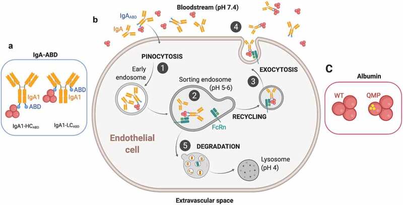
Figure 1.

Recycling model of IgAABD through binding to albumin and indirect rescue by FcRn. A) Illustration of the two ABD-fused human IgA1 formats studied; IgA1-LCABD and IgA1-HCABD. B) (1) IgA1-LCABD and IgA1-HCABD will be taken up in complex with albumin via fluid-phase pinocytosis and enter early endosomes. (2) FcRn predominantly resides within acidified endosomes, where the lower pH will trigger binding of albumin bound IgA1ABD to the receptor. As such, the IgA1 fusions will be indirectly bound to FcRn. (3–4) The ligands bound to the receptor will then be recycled back to the cell membrane, where the physiological pH of blood (pH 7.4) results in release of the ligands back into the circulation. (5) Proteins that do not bind FcRn in the sorting endosomes, such as naked IgA, will end in lysosomes for degradation. C) Illustrations of wild-type (WT) and engineered (QMP) human albumin variants. The figure was made with BioRender
