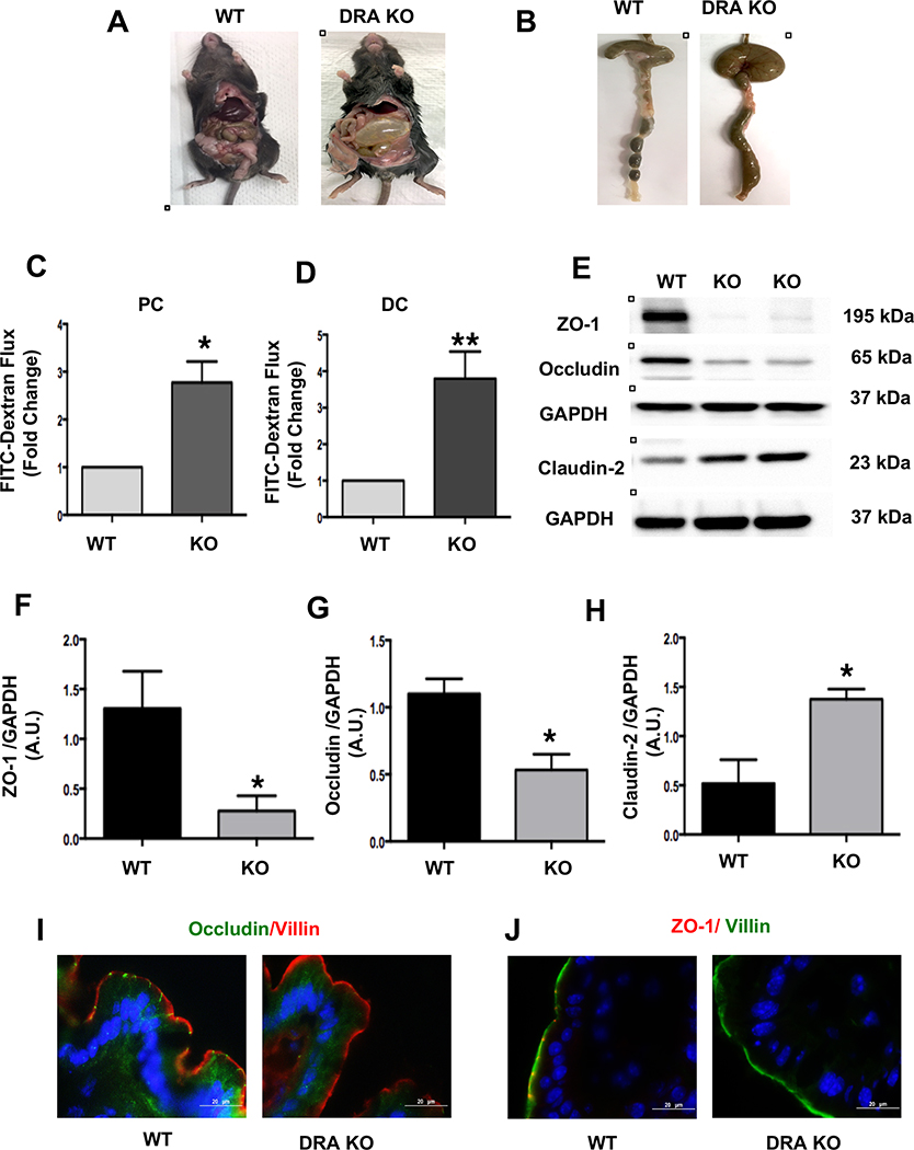
Figure 1: Loss of DRA results in increased paracellular permeability and decreased TJ protein expression in mouse colon:
DRA KO mice exhibit severe diarrhea, a distended abdomen and enlarged cecum (Figures A and B). DRA KO mice showed increased paracellular permeability in proximal colon (PC) and distal colon (DC) (Figures C and D). Intestinal permeability was determined by comparing mucosal-to-serosal flux of 4 kDa-FITC dextran (FD4) fluorescence over 120 minutes (n=3–4). Effects of DRA knockdown on protein levels of TJ components in wild type and DRA KO mice distal colon by immunoblotting are shown (Figure E) (n=3–6). GAPDH was used as the internal control. Densitometric analyses of band intensities are shown (Figures F, G, H). Values are means ± SEM. Data was analyzed by unpaired t-test (*p <0.05 vs. wild type; **p <0.01 vs. wild type). Immunofluorescence staining of distal colon sections of wild type and DRA KO mice for occludin (green), villin (red), and DAPI (blue) (Figure I) and ZO-1 (red), villin (green), and DAPI (blue) (Figure J). Scale bar: 20 μm.

