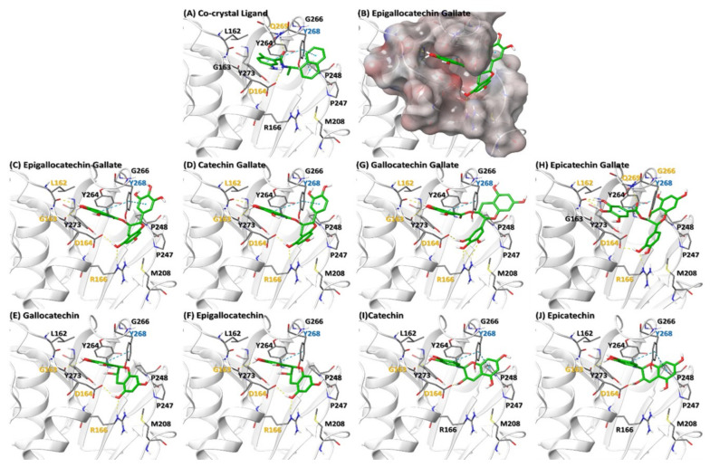
Figure 5.
Binding of various catechins to SARS-CoV-2 PLPro protein. (A) Ligand (GRL0617) Co-crystal of PLPro (PDB ID: 7CMD). (B) Electrostatic surface of PLPro binding site (C–J) represent interaction of various catechins with the ligand binding site of PLPro. The residues in orange, cyan and black color represent H-bond, Cation-pi and hydrophobic interaction respectively. Orange dotted line represents H-bond whereas cyan dotted lines represent cation-pi interaction.

