Abstract
Background
Rectus abdominis is the main core muscle. Weakness or any alteration in it may increase the pressure over the lower back, in obese women diastasis of rectus abdominis muscle found to be very common condition. Therefore, there may be a correlation between diastasis of rectus abdominis muscle and low back pain in obese women that needs to be explored, as there is no literature available.
Methods
In this study, 72 female subjects with Body Mass Index <30 kg/m2 were recruited by snowball sampling method. Demographic (name, age) and anthropometric characteristics (height, weight and body mass index) were recorded. The separation in the rectus abdominis muscle was assessed with vernier calliper.
Results
Total subjects were included in the study; all the subjects were Female without any recent abdominal surgical history. The subjects included in the study with age of 30 years to 55 years old with body mass index of the included females must be (30-30.9) kg/m2 i.e. women must come under obese category. Diastasis of rectus abdominis muscle was another variable used that must be present in each women. Low back pain was also used as the variable that may be present or may not be present in the women with diastasis of rectus abdominis muscle. The collected data were analysed by the appropriate statistical analysis tools. The p-value was found more than 0.05 (the alpha level set was less than 0.05) which is non-significant.
Conclusion
The study concluded a non-significant correlation between the diastasis of rectus abdominis muscle and low back pain in obese women. The present study concludes that it is not necessary that all obese women with low back pain always propose to have diastasis of rectus abdominis muscle.
Keywords: Diastasis of rectus abdominis muscle, Low back pain, Vernier calliper, Inter-rectus distance, Obesity
INTRODUCTION
Diastasis Recti also referred as Diastasis of Rectus Abdominis Muscle (DRAM) is determined by reduced compactness of the Linea Alba amalgamated with indulgence of the ventral abdominal musculature. It’s after effect, leads to bulging of midline when there is a rise in intra-abdominal pressure. Beer classification explains it as an inter-rectus distance of 22 mm, in relaxed state which is measured 3 cm above the umbilicus [1].
DRAM develops anywhere above Linea Alba, from pubic bone to Xiphoid Process. There are six layers in the antero-lateral wall of abdomen from in to outward i.e. parietal peritoneum, transversalis fascia, abdominal muscles, fat, superficial fascia and the skin [2]. Four muscles in the muscular layer whose fibres aligned perpendicularly are of Rectus Abdominis, diagonal fibres of External and Internal oblique and parallel fibres of Transverse Abdominis muscle, through thoracolumbar fascia these muscles are attached to spine, pelvis and rib cage [3].
It has been reported by researchers, that the prevalence of DRAM in postpartum female around was 60.0% at 6 weeks, 32.6% at 12 months, 45.5% at 6 months and 33.1% at gestation week. Studies report the most common occurrence point of DRAM was at the umbilicus [4]. The stimulant activities of the abdominal muscles hold up and shield the viscera, these muscles are important for maintaining correct posture and equilibrium of the lumbar and pelvic spine. Throughout pregnancy, the symmetry of abdominal muscles changes but still they continue to function. The inter-rectus space settle progressively with in the postpartum phase with characteristic inconsistency commonly gets resolves in almost 8 weeks postpartum [5]. The presence of the separation of abdominal muscles was acknowledged in 1858, when Gray explained the “Deviating from each other in their conquest, becoming of significant thickness following considerable expansion of the abdomen after the pregnancy”. Poor posture, Back pain, Pelvic floor problems and gastro intestinal disturbances like bloating and constipation are all the manifestations that occurs due to DRAM [6].
When studied in a population, it persisted in 100% postpartum cases. It can also be seen during pregnancy because of hormones like oestrogen, progesterone and relaxin level increases which leads to decreased strength of Linea alba and connective tissue [7]. So, the prolonged raised stretch when combined with hormonal changes, whose consequences leads to splitting of Linea Alba which leads to this condition i.e. DRAM. Other common causes for Diastasis Recti is Obesity [5]. Obesity being a chronic metabolic disease leads to ill health and a common cause of disability and death as there is increased fat stores of body. Risk factors involving cardiovascular, metabolic, cancer and respiratory disorders. Body can be evaluated by Body Mass Index (BMI) calculated by body weight in (kilograms) divided by height in m2. Overweight patients have a BMI of 25 kg/m2 or more, whereas obese patients are classified into 3 grades i.e. Grade 1 includes BMI of (30.0-34.9) kg/m2, Grade 2 includes (35.0-39.9) kg/m2 and Grade 3 equal to or more than 40 kg/m2 [8].
It has been reported by researchers, that the prevalence of DRAM in postpartum female around was 60.0% at 6 weeks, 32.6% at 12 months, 45.5% at 6months and 33.1% at gestation week. Studies report the most common occurrence point of DRAM was at the umbilicus [4]. Vernier calliper has been used as an instrument to record short lengths. By means of a screw it can be fixed at any position. A Vernier calliper consists of metallic bar which has inches on one end and centimetres & millimetres on the other. The zero of Vernier should meet zero of main scale, when both the junctions are pressed together. It has two upper jaws which are used for measuring internal diameter of pipes and hollow cylinder [9]. In this study Digital Vernier calliper has been used for measuring the Inter Rectus Distance (IRD).
DRAM decreases strength and soundness of abdominal wall muscles, which further increase pelvic instability and low back pain (LBP) [9]. As the obesity is the other factor which is increasing the chances of DRAM occurrence, so it is important to find out that is there any connection between the LBP and DRAM in obese women.
MATERIALS AND METHODS
For this correlational study a cross sectional (analytical) study design was incorporated. A number of (N = 72) obese female patients were taken through snowball sampling method and were palpated to check whether they are having Diastasis Recti or not. Obese females with diastasis were included with applicable inclusion and exclusion selection criteria. Women within age group of 35 to 55 years, BMI must be (30-30.9) kg/m2 and must have DRAM were included in the inclusion criteria. Whereas, exclusion criteria comprised psychological issues, recent abdominal surgery, spinal surgery and BMI < 30 kg/m2. An information sheet expressing details of the researcher and the ongoing project was provided as leaflet to individuals. Following which, subjects willing to participate in the study were asked to sign an informed consent form for their voluntary participation. Patient were given a calm and relaxed environment. They were asked to lie in supine position with arm abducted at 90 degree with elbow flexed, hands in supination and fingers of both hands entangled with each other under the occiput. In addition to the above patient’s knees were maintained in flexed position throughout the evaluation. After putting the patient in required position, they were asked to elevate the upper thorax till the spine of scapula clears the couch. The patient was advised not to hold breath during the process, rather inhale from nose and exhale through mouth.
Therapist stood on either side of the patient with bilateral straight elbow. The therapist used three fingers (index, middle and ring) of the right hand for palpating Rectus Abdominis muscle. This act was performed 4 centimetre above and below the umbilicus. At this instinct, the patient was asked to elevate the upper thorax till the scapula clears the couch for palpating DRAM, after which with the help of Vernier calliper accurate distance was measured. After making sure that subject is having DRAM, subject was asked “Do you have low back pain?”
The main researcher examined and entered the collected data. Data collected was statistically analysed through the statistical software Statistical Package for Social Sciences (SPSS), version 16. Kolmogorov-Smirmov test was used to verify normal data distribution. As the data did not follow the normal distribution, non-parametric tests were used. The significance or alpha level was set less than 0.05 i.e. p < 0.05. Chi square correlation coefficient was used to rule out association between BMI and Inter Rectus Distance Lower (IRDL), BMI and IRDU and association between Inter Rectus Distance Upper and Lower was established with LBP.
RESULTS
72 subjects were involved in the present study. All subjects were female with BMI (30-30.9) kg/m2 and age from 35 years to 55 years old. All subjects posed with DRAM but wasn’t necessary to pose with Low Back Pain. Demographic characteristics of subjects have been described through descriptive data whereas correlation of DRAM with low back pain in women with BMI (obese). The demographic findings have been described in the form of Table 1. As follows are the results:
Table 1.
Demographic characteristics
| Demographic characteristics | Median (inter quartile range) | Range |
|---|---|---|
| Age (years) | 43.50 (35, 51.75) | 30-55 |
| Height (cm) | 162 (78, 88) | 151-174 |
| Weight (kg) | 81.50 (1.57, 1.67) | 69-95 |
| Body mass index (BMI) kg/m2 | 30.90 (30.40, 31.67) | 30-34.50 |
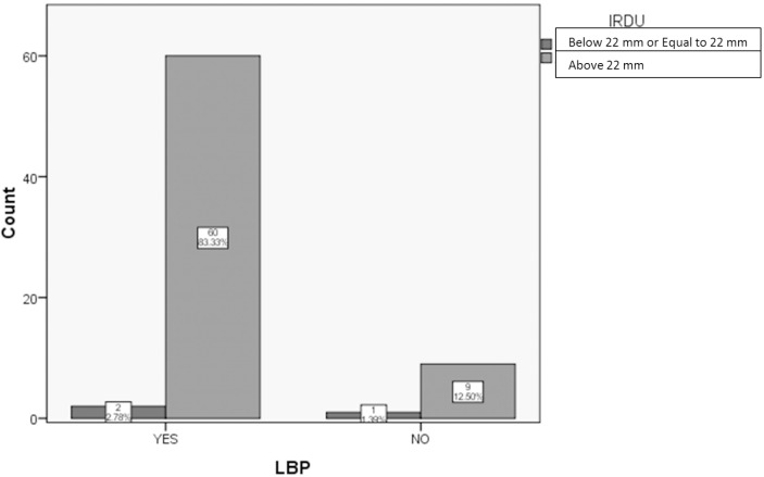
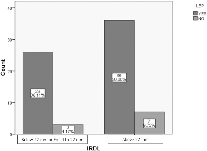
As it is a correlation study after the result the correlational graph was built between IRDU and LBP in Fig. 1 and another graph was made between IRDL and LBP in Fig. 2, showing how many obese females with DRAM having LBP or not.
Fig. 1.
Bar graph displaying correlation between IRDU and LBP in patients having inter rectus distance ≤ 22 mm and > 22 mm.
Fig. 2.
Bar graph showing relationship between IRDL and LBP in patients with inter-rectus ≤ 22 mm and > 22 mm.
DISCUSSION
The purpose of the study was to observe the relation between DRAM and LBP. A total of 72 subjects were recruited for the study. To determine the normality of all the collected data, Kolmogrov-Smirnov test was used. As the data was categorical, chi-square test was used. The correlation coefficient’s value is an evaluation of strength of alliance between the 2 variables. The result of the present study showing non-significant relationship between LBP and IRDU as well as between LBP and IRDL respectively.
In 2016, Khandale SR et al. has done the study on the outcome of abdominal exercises on lowering DRAM in postnatal women. This study revealed that diastasis of rectus abdominis cause alteration in abdominal musculature. Thus, interfering the mechanics of abdomen and its function leading to physical disquiet such as LBP [7]. The outcome of the present study depicts though obese women with DRAM have low back pain but still there are some women who don’t have low back pain even with a DRAM.
CONCLUSION
The study concludes a non-significant correlation between the DRAM and LBP in obese women. The present study concludes that it is not necessary that all obese women with low back pain always propose to have Diastasis of Rectus Abdominis.
REFERENCES
- 1.Mommer EHH, Ponten JEH, Al Omar AK, Reilingh TS, Bouvy ND, Nienhuijs SW. The general surgeon's perspective of rectus diastasis. A systematic review of treatment options. Surg Endosc. 2017;31(12):4934–49. doi: 10.1007/s00464-017-5607-9. [DOI] [PMC free article] [PubMed] [Google Scholar]
- 2.Mota P, Pascoal AG. Diastasis recti abdominis in pregnancy and postpartum period. Risk factors, functional implications and resolution. Curr Women's Health Rev. 2015;11(1):59–67. doi: 10.2174/157340481101150914201735. [DOI] [Google Scholar]
- 3.Tomlinson DJ, Erskine RM, Morse CI, Winwood K, Pearson GO. The impact of obesity on skeletal muscle strength and structure through adolescence to old age. Biogerontology. 2016;17(3):467–83. doi: 10.1007/s10522-015-9626-4. [DOI] [PMC free article] [PubMed] [Google Scholar]
- 4.Sperstad JB, Tennfjord MK, Hilde G. Diastasis recti abdominis during pregnancy and 12 months after childbirth: prevalence, risk factors and report of lumbopelvic pain. Br J Sports Med. 2016;50:1092–6. doi: 10.1136/bjsports-2016-096065. [DOI] [PMC free article] [PubMed] [Google Scholar]
- 5.Michalska A, Rokita W, Wolder D, Pogorzelska J, Kaczmarczyk K. Diastasis recti abdominis - a review of treatment methods. Ginekologia Polska. 2018;89(2):97–101. doi: 10.5603/GP.a2018.0016. [DOI] [PubMed] [Google Scholar]
- 6.Adkitte RG, Yeole U, Gawali P, Gharote GM. Prevalence of Diastasis of Rectus Abdominis Muscle in Immediate Post- Partum Women of Urban and rural areas. Eur J Pharm Med Res. 2016;3(5):460–2. [Google Scholar]
- 7.Khandale SR, Hande D. Effects of Abdominal Exercises on Reduction of Diastasis Recti in Postnatal Women. IJSR. 2016;6(6):182–91. [Google Scholar]
- 8.Akhtar N, Qureshi NK. Obesity: A Review of Pathogenesis and Management Strategies in Adult. Delta Med Col J. 2017;5(1):35–48. doi: 10.3329/dmcj.v5i1.31436. [DOI] [Google Scholar]
- 9.Giridharan GV. Effectiveness of Exercise in Treating Rectus Abdominis Diastasis. Biomedicine (India) 2019;38(4):147–51. [Google Scholar]


