Abstract
Background
Rapid identification of pathogenic Mycobacterium species is critical for a successful treatment. However, traditional method is time-consuming and cannot discriminate isolated non-tuberculosis mycobacteria (NTM) at species level. In the retrospective study, we evaluated the clinical applicability of PCR-reverse blot hybridization assay (PCR-REBA Myco-ID) with clinical specimens for rapid detection and differentiation of mycobacterial species.
Methods
A total of 334 sputum and 362 bronchial alveolar lavage fluids (BALF) from 696 patients with mycobacterium pulmonary disease (MPD) and 210 patients with non-mycobacterium pulmonary disease used as controls were analyzed. Sputum or BALF were obtained for MGIT 960-TBc ID test and PCR-REBA Myco-ID assay. High resolution melt analysis (HRM) was used to resolve inconsistent results of MGIT 960-TBc ID test and PCR-REBA Myco-ID assay.
Results
A total of 334 sputum and 362 BALF specimens from 696 MPD patients (292 MTB and 404 NTM) were eventually analyzed. In total, 292 MTBC and 436 NTM isolates (mixed infection of two species in 32 specimens) across 10 Mycobacterium species were identified. The most frequently isolated NTM species were M. intracellulare (n = 236, 54.1%), followed by M. abscessus (n = 106, 24.3%), M. kansasii (n = 46, 10.6%), M. avium (n = 36, 8.3%). Twenty-two cases had M. intracellulare and M. abscessus mixed infection and ten cases had M. avium and M. abscessus mixed infection. A high level of agreement (n = 696; 94.5%) was found between MGIT 960-TBc ID and PCR-REBA Myco-ID (k = 0.845, P = 0.000). PCR-REBA Myco-ID assay had higher AUC for both MTBC and NTM than MGIT 960-TBc ID test.
Conclusion
PCR-REBA Myco-ID has the advantages of rapid, comparatively easy to perform, relatively low cost and superior accuracy in mycobacterial species identification compared with MGIT 960-TBc ID. We recommend it into workflow of mycobacterial laboratories especially in source-limited countries.
Keywords: Mycobacterium tuberculosis (MTB), Nontuberculous mycobacteria (NTM), Identification, PCR-reverse blot hybridization assay (PCR-REBA), Molecular diagnosis
Background
The genus Mycobacterium contains a number of acid-fast bacilli (AFB), including Mycobacterium tuberculosis complex (MTBC), Mycobacterium leprae, and non-tuberculosis mycobacteria (NTM) [1]. NTM are ubiquitous environmental organisms that incidentally cause opportunistic NTM pulmonary disease (NTM-PD) and extrapulmonary infections in immunocompromised individuals [2]. With the prevalence of acquired immunodeficiency syndrome (AIDS), the recognition of clinical importance of NTM is also growing [3]. Recently, many countries have reported a dramatically increased incidence of NTM-PD. [4–7] NTM infections constitutes 0.5–35% of all human mycobacterial infections [8]. For patients with risk factors, such as bronchiectasis, cystic fibrosis, and immune deficiency, the figure was higher still, over 50% [9]. However, a high percentage of patients have no risk factors [10]. Infections of most NTM species have been traditionally considered from environmental sources rather than person-to-person transmission like MTBC [11]. In addition, many NTM strains are insensitive to most anti-tuberculosis drugs [12, 13]. Therefore, quality and timely identification of mycobacterium species is necessary for better patient management and appropriate chemotherapy. However, most clinical features (cough, sputum production, fatigue, hemoptysis and fever) and abnormalities on chest radiography (centrilobular nodules, tree-in-bud opacity and cavitation) generally associated with NTM-PD are atypical, which may lead to false diagnoses as PTB, and hence to overtreatment of patients who do not have TB [14, 15]. The traditional methods used in mycobacterial laboratories require cultivated isolates and the results are only obtained after weeks to months of incubation. Therefore, these culture-based methods are time-consuming and labor-intensive [16]. Moreover, the traditional methods could not discriminate NTM to the species level, or detect mixed infections in which two or more mycobacterial species were simultaneously detected in the same specimen [17]. All NTM isolates from respiratory samples should be identified at least to species level, since that optimal therapeutic regimen differs according to different species, especially between slow-growing and rapid-growing species. Therefore, current clinical methods are not conducive to guide clinical decisions in a timely manner. Clinicians sometimes refer only to interferon-γ release assays (IGRAs). IGRAs are based on M. tuberculosis specific antigens. These antigens are not present in NTM strains other than M. marinum, M. kansasii, M. gordonae and M. szulgai [18]. However, it is worth mentioning that a number of TB patients are also negative in IGRAs [19]. So, IGRAs could not make a reliably distinction between NTM and MTBC.
Advances in molecular assays have accelerated the diagnosis of mycobacteria.
infections over recent years. Among these advances, nucleic acid amplification (NAA)-based techniques permit for identifying mycobacteria at the species level [20–23]. Sequencing of 16S rRNA has been recommended as the gold standard method for definitive identification and discrimination of mycobacterial species [24]. Unfortunately, although fast, some of these techniques still require cultured strains and expensive equipment such as Hain line-probe and minion [22]. A commercial kit based on 16S rRNA sequencing and nucleic acid probes and reverse blot hybridization (REBA), PCR-REBA Myco-ID (Yaneng BioSciences, Shenzhen), was developed for simultaneous genotyping of 22 clinical important mycobacterial species including MTB, M. smegmatis, M. Intracellulare, M. kansasii, M. chelonae, M. marinum, M. fortuitum, M. terrae, M. nonchromogenicum, M. avium, M. scrofulaceum, M. abscessus, M. xenopi, M. gilvum, M. phlei, M. triviale, M. gordonae, M. gastri, M. vaccae, M. szulgai, M. diernhoferi, M. simiae. Moreover, as a major advantage, PCR-REBA Myco-ID can be performed with clinical specimens. Despite the fact that this technology has been used as a rapid, low cost and easy-to-use method in mycobacterial research works, it has not been incorporated into workflow of many mycobacterial laboratories. Therefore, the purpose of this study was to evaluate clinical applicability of PCR-REBA Myco-ID assay for prompt and accurate identification of NTM at species level directly from 696 clinical specimens in China.
Methods
Ethics statement and informed consent
This study was approved by The Ethics Committee of the Shanghai Pulmonary Hospital, Tongji University School of Medicine in China (Approval number K20–422). Each participant gave written informed consent before enrollment. The study was performed according to the Declaration of Helsinki with respect to ethical principles for research involving application of human specimens.
Clinical specimen collection
Clinically suspected NTM-PD patients admitted to Shanghai Pulmonary Hospital between January 3, 2018 and November 28, 2019 were eligible for screening if they met the following inclusion criteria: 1) HIV test negative; 2) providing two sputum specimens or bronchial alveolar lavage fluids (BALF) from sputum-scarce patients for MGIT 960-TBc ID test and PCR-REBA Myco-ID test. Samples should be processed within 24 h of collection.
Fiberoptic bronchoscopy
Bronchoscopy procedures were performed as previously described [25].
Routine identification methods
Nacetyl-L-cysteine (NALC)–NaOH method was used to decontaminate about 5 ml specimens [26]. The specimens were exposed to 4%NaOH for 15–20 min. The sediment was washed with sterile 0.9% NaCl solution and resuspended in 1.5 ml sterile 0.9% NaCl solution. Two separate 500-μl aliquots were prepared in 1.5 ml tubes for MGIT 960-TBc ID test and PCR-REBA Myco-ID test. Specimens were cultured by MGIT 960 (Becton Dickinson Diagnostic Systems, Sparks, MD) for 6 weeks following the standard procedure of the manufacturer [27]. In order to confirm the presence of mycobacteria and exclude contamination, samples from all positive MGIT 960 tubes were Ziehl-Neelsen (ZN) -stained and Gram-stained. TBc ID (Becton Dickinson, Sparks, MD) is an assay for the detection of MPT64 Ag, a mycobacterial protein secreted by MTBC and certain strains of M. bovis. 100-μl liquid media from the positive MGIT tubes was added to the TBc ID card and was incubated for 15 min at room temperature. The results were visually assessed. A positive test result indicated MTBC and a negative test result indicated NTM.
PCR-reverse blot hybridization assay (PCR-REBA Myco-ID)
a) DNA Isolation: 500 μl bacterial precipitation was treated with DNA Lysis Buffer (10 mmol/L NaCl, 1 mg/ml SDS, 0.15 g/ml Chelex-1 s00 glass beads, 1% Tween 20) at 50 °C for 1 h, then at 100 °C for 10 min, and centrifuged at 10000 r/min for 2 min. The supernatant containing genomic DNA was transferred to another tube and preserved at − 20 °C for further PCR. b) PCR: PCR was carried out in a 50 μl reaction mixture including 5 μl DNA template, 10 mM Tris/HCl (pH 8.3), 50 mM KCl, 2 mM MgCl2, 0.2 mM dNTP, 0.4 mM each primer, and 2 U AmpliTaq Gold polymerase. First, the mixture was incubated at 94 °C, to activate the Taq polymerase, followed by 40 cycles of amplification (94 °C for 1 min, annealing and extension for 30s at 65 °C, 72 °C for 1 min), and finally incubated at 72 °C for 10 min. 5 μl PCR product was electrophoresed on 6% polyacrylamide gel with silver staining. c) REBA: The amplified PCR products were REBA tested with Mycobacterium species identification detecting Kit (Yaneng BioSciences, Shenzhen) according to the manufacturer’s guidelines. In brief, biotinylated PCR products were denatured at 25 °C for 5 min and were added to the REBA membrane strip in the blotting tray provided. Denatured single-stranded PCR products were hybridized with the probes on the strip at 55 °C for 30 min. The strips were then cleaned twice with gentle shaking in 1.0 ml of washing solution for 10 min at 55 °C, incubated at 25 °C for 30 min, and cleaned twice with 1.0 ml of CDS at room temperature for 1 min. Finally, visualize the signals of colorimetric hybridization and read the band pattern.
PCR-high resolution melting (PCR-HRM) analysis
To confirm mycobacterium species inconsistently identified by the two different assays, fluorescence PCR-HRM Assay was carried out with Mycobacterium Identification Kit (Zeesan Biotech, Xiamen). The amplification of rpoB gene was performed on the following conditions: preincubation at 95 °C for 10 min, then denaturation at 95 °C for 10s, 45 cycles, annealing for 30s at 65 °C, and extension for 10s at 72 °C followed by the Tm analysis with increasing temperatures from 60 to 95 °C in a 0.2 °C s-1 slope increment for 10s. The HRM analysis was carried out with Gene Scanning Software Version 1.5.0 (Roche Instrument Centre, Switzerland). The aggregation of the melting curves was based on the regions of the melting curve corresponding to the pre-melting, melting, and post-melting regions. Distilled water was used as the non-template control (NTC).
Statistical analysis
Data analysis was performed using SPSS for Windows (Version 19.0, SPSS Inc., Chicago). All patients were followed up for at least 6 months. The sensitivity, specificity, positive predictive value (PPV), and negative predictive value (NPV) of PCR-REBA Myco-ID assay and MGIT 960-TBc ID test was calculated. The categorical variables were analyzed using Fisher exact or Pearson X2 tests where appropriate and 2-tailed tests were used. The concordance of agreement between MGIT 960-TBc ID test and PCR-REBA Myco-ID assay was evaluated using Cohen’s kappa test (k > 0.75, excellent agreement; 0.4 < k < 0.75, moderate agreement; and k < 0.4, poor agreement). Receiver operating characteristic (ROC) curve analysis was performed and the area under curve (AUC) with a 95% confidence interval (CI) was further calculated. P < 0.05 was considered statistically significant.
Results
Demographic and clinical characteristics of the participants
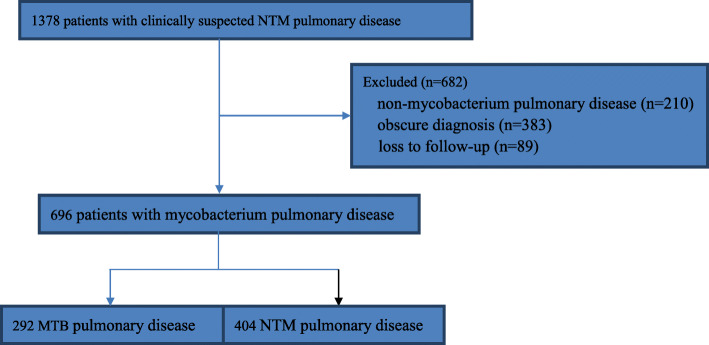
In all, 1378 patients (698 sputum and 680 BALF) with clinically suspected NTM-PD were enrolled into this study. Finally, 682 patients were excluded, including 383 patients with obscure diagnosis, 89 patients lost to follow up and 210 patients with non-mycobacterium pulmonary disease (Table 1). The remaining 696 patients (334 sputum and 362 BALF) with mycobacterium pulmonary disease (MPD) (292 MTB and 404 NTM) were eventually analyzed (Fig. 1). Baseline characteristics of the 696 patients were summarized in Table 2. In total, 292 MTBC and 436 NTM isolates (mixed infection of two species in 32 specimens) across 10 Mycobacterium species were identified. The most frequently isolated NTM species were M. intracellulare (n = 236, 54.1%), followed by M. abscessus (n = 106, 24.3%), M. kansasii (n = 46, 10.6%), M. avium (n = 36, 8.3%), M.scrofulaceum (n = 4, 0.9%), M. phlei (n = 2, 0.5%), M.chelonae (n = 2, 0.5%), M. xenopi (n = 2, 0.5%), and M. marinum (n = 2, 0.5%). Twenty-two patients had M. intracellulare and M. abscessus mixed infection and ten patients had M. avium and M. abscessus mixed infection (Table 3).
Table 1.
Baseline characteristics of patients with non-mycobacterium pulmonary disease
| Condition | Age ± SD (range) | Gender (male: female) | MGIT 960 (+) | PCR-REBA (+) | Total (210) |
|---|---|---|---|---|---|
| Lung cancer | 48 ± 16 (35–80) | 49:38 | 0 | 0 | 87 |
| Pneumonia | 49 ± 17 (21–78) | 35:30 | 1(NTM) | 0 | 65 |
| Pulmonary mycosis | 51 ± 17 (35–80) | 15:13 | 0 | 0 | 28 |
| Silicosis | 48 ± 8 (40–57) | 20:1 | 0 | 0 | 21 |
| Bronchogenic cyst | 49 ± 4 (45–51) | 1:1 | 0 | 0 | 2 |
| Interstitial lung disease | 50 ± 2 (48–52) | 1:1 | 0 | 0 | 2 |
| Pulmonary embolism | 50 ± 1 (49–51) | 1:0 | 1(MTBC) | 0 | 1 |
| Granulomatous vasculitis | 43 | 0:1 | 0 | 0 | 1 |
| Right lower lobe sequestration | 46 | 1:0 | 0 | 0 | 1 |
| Lung tissue X disease | 51 | 1:0 | 0 | 0 | 1 |
| Myelodysplastic syndrome | 45 | 0:1 | 0 | 0 | 1 |
Fig. 1.
Flow chart of the study
Table 2.
Baseline characteristics of patients with mycobacterium pulmonary disease
| MTB pulmonary disease (n = 292) | NTM pulmonary disease (n = 404) | |
|---|---|---|
| Age, SD (range) | 44 ± 15 (25–80) | 49 ± 17 (32–86) |
| Gender (male: female), n | 190:102 | 191:213 |
| Body mass index, median (range) | 19.3 (14–29) | 19.2 (13–28) |
| Diabetes mellitus, n | 32 | 21 |
| Autoimmune diseases, n | 18 | 25 |
| bronchiectasis or cystic fibrosis | 101 | 98 |
| Smear (+) / QFT-GITa (−) | 73 | 240 |
| No response to anti-TB medication | 118 | 66 |
aQFT-GIT, QuantiFERON-TB Gold In-Tube
Table 3.
Identification of clinical isolates using PCR-REBA
| Clinical organism | Number of strains (728) | Identified by PCR-REBA (718) |
|---|---|---|
| MTB | 292 (40.1%) | 286 (97.9%) |
| M. intracellulare | 236 (32.4%) | 232 (98.3%) |
| M. abscessus | 106 (14.6%) | 106 (100%) |
| M. kansasii | 46 (6.3%) | 46 (100%) |
| M. avium | 36 (4.9%) | 36 (100%) |
| M.scrofulaceum | 4 (0.5%) | 4 (100%) |
| M. xenopi | 2 (0.3%) | 2 (100%) |
| M. marinum | 2 (0.3%) | 2 (100%) |
| M. phlei | 2 (0.3%) | 2 (100%) |
| M.chelonae | 2 (0.3%) | 2 (100%) |
Identification of mycobacterial by MGIT 960-TBc ID
Of the 292 pulmonary tuberculosis (PTB) patients, 274 isolates (93.8%) were identified as MTBC and 10 were negative in MGIT 960-TBc ID. Of the 404 NTM-PD patients, 394 isolates (96.5%) were identified as NTM and 6 were negative in MGIT 960-TBc ID (Table 4). Since there were two false-positive cases in the non-TB group, the specificity of MGIT 960-TBc ID test for both MTBC and NTM was 99.5%. Eight MTBC were misidentified as NTM, and four NTM misidentified as MTBC.
Table 4.
Comparison of PCR-REBA and MGIT 960 for Detection of mycobacterium
| PCR-REBA | MGIT 960 | P | ||
|---|---|---|---|---|
| MTB | sensitivity | 97.9% | 93.8% | 0.012 |
| specificity | 100% | 99.5% | 0.317 | |
| PPV | 100% | 98.6% | 0.040 | |
| NPV | 98.0% | 96.7% | 0.323 | |
| NTM | sensitivity | 99.0% | 96.5% | 0.106 |
| specificity | 100% | 99.5% | 0.317 | |
| PPV | 100% | 98% | 0.004 | |
| NPV | 99.0% | 98.5% | 0.530 |
Identification of mycobacterial by PCR-REBA Myco-ID
Of the 292 PTB patients, 286 isolates (97.9%) were identified as MTBC and 6 were negative in PCR-REBA Myco-ID assay. Of the 404 NTM-PD patients, 400 isolates (99.0%) were identified as NTM and 4 were negative in PCR-REBA Myco-ID assay. The specificity and PPVs of PCR-REBA Myco-ID assay for both MTBC and NTM were 100%, which were significantly higher than those of MGIT 960-TBc ID (P = 0.04). The sensitivity of PCR-REBA Myco-ID to MTBC was also significantly higher than that of MGIT 960-TBc ID (P = 0.012).
Consistency between MGIT 960-TBc ID and PCR-REBA Myco-ID
MGIT 960-TBc ID and PCR-REBA Myco-ID were concordant except for 38 samples (5.5%) (Table 5). A high level of agreement (n = 696; 94.5%) was found between MGIT 960-TBc ID and PCR-REBA Myco-ID (k = 0.845, P < 0.0001), indicating an optimal consistency between the 2 tests. For the 38 strains with inconsistent results of the two methods, PCR-HRM analysis was used for further analysis.
Table 5.
Identification of clinical isolates using PCR-REBA, MGIT 960 and HRM
| PCR-REBA | MGIT 960 | HRM | Total (696) |
|---|---|---|---|
| MTB | (−) | MTB | 10 |
| MTB | NTM | MTB | 8 |
| (−) | MTB | MTB | 6 |
| M.scrofulaceum | MTB | M.scrofulaceum | 4 |
| (−) | NTM | M. intracellular | 4 |
| M. intracellular | (−) | M. intracellular | 2 |
| M. phlei | (−) | M. phlei | 2 |
| M.chelonae | (−) | M.chelonae | 2 |
| MTB | MTB | / | 268 |
| M. intracellulare | NTM | / | 208 |
| M. abscessus | NTM | / | 74 |
| M. kansasii | NTM | / | 46 |
| M. avium | NTM | / | 26 |
| M. xenopi | NTM | / | 2 |
| M. marinum | NTM | / | 2 |
| M. intracellulare-M. abscessus | NTM | / | 22 |
| M. avium-M. abscessus | NTM | / | 10 |
Establishment of ROC curve
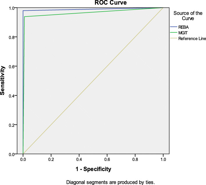
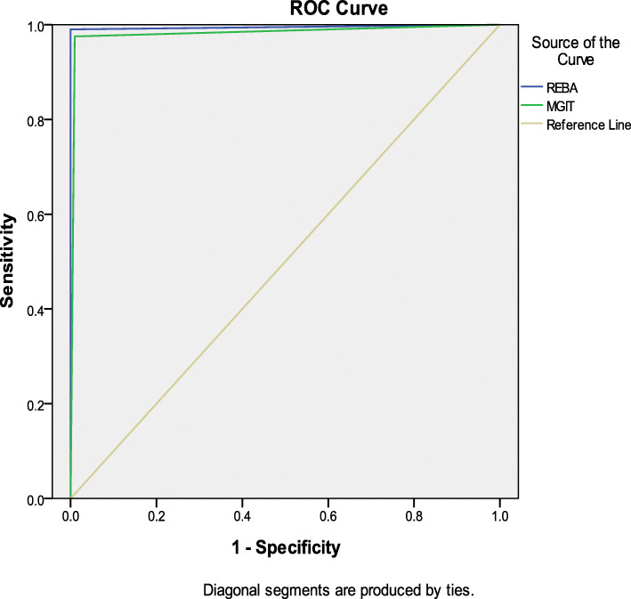
The AUC of PCR-REBA Myco-ID and MGIT 960-TBc ID for MTBC and NTM was 0.990 (95% CI 0.980–0.999) and 0.964 (95% CI 0.947–0.982), 0.995 (95% CI 0.988–1.0) and 0.983 (95% CI 0.971–0.994) respectively (Figs. 2, 3). PCR-REBA Myco-ID had the higher AUC for both MTBC and NTM than MGIT 960- TBc ID.
Fig. 2.
ROC curve of REBA and MGIT 960 for MTBC
Fig. 3.
ROC curve of REBA and MGIT 960 for NTM
Discussion
In current retrospective study, we evaluated the clinical applicability of PCR-REBA Myco-ID assay for rapid detection and differentiation of mycobacterial species directly from respiratory samples (sputum and BALF) in clinically suspected NTM-PD patients. Our results indicate that PCR-REBA Myco-ID assay has higher sensitivity and PPV than MGIT 960-TBc ID, which is consistent with the results of previous study by Lee et al. [28]. Because NTM are ubiquitous in the environment, a single isolated NTM strain from sputum without repeated isolation on culture, is not clinically significant, in general. Therefore, in the investigation of clinically suspected NTM-PD patients, sputum samples should be collected at least two other days for mycobacterium culture. Differentiation of Mycobacteria at the species level by phenotypic and biochemical testing is time-consuming because of the slow growth rate of mycobacteria. However, the major advantage of some direct molecular detection is to rapidly differentiate MTB from NTM in clinical samples. PCR-REBA Myco-ID assay has a short turnaround time of about 4 h. Timely intervention is also the key to the success of NTM treatment. Another limitation of MGIT 960-TBc ID test is that the accuracy is heavily affected by mycobacterial species and the quality of specimen. Relatively high contamination rate was reported [29]. Our results showed that the overall contamination rate was 1.7% (12/696) in this study. In addition, there were two false positive cultures in the non-TB group including one MTBC and one NTM isolate. Culture of NTM can be difficult even in mycobacterial reference laboratories. For example, while many NTM species grow best at 28 °C or 45 °C, conventional cultures are performed only at 35 °C. Other NTM species require prolonged incubation or special medium. Some NTM cultivars require supplementary media or extended culture cells. In our current study, six strains of NTM were not cultured, but were detected by PCR-REBA Myco-ID assay and confirmed by PCR-HRM analysis. Routinely culture also cannot discriminate NTM species. At present, more than 160 distinct Mycobacterium species have been validly published (http://www.bacterio.net/mycobacterium.html). The clinical relevance of isolated NTM differs strongly by species, from pathogenic species (e.g., Mycobacterium kansasii and Mycobacterium malmoense) to typical saprophytes (Mycobacterium gordonae and Mycobacterium phlei) [30]. In practice, treatment recommendations presented for NTM are based on the NTM species. NTM isolates species should be subspeciated. For example, the prognosis of diseases caused by M. intracellulare is better than that caused by M. avium, and the prognosis of diseases caused by M. abscessus is worse than that caused by M. massiliense [31, 32]. However, PCR-REBA Myco-ID assay cannot further classify M. abscessus as M. massiliense, M. bolletii and M. abscessus subspecies. If person-to-person transmission of M. abscessus is suspected, it is recommended to use whole genome sequencing (WGS), which has been developed in recent years [33]. Several methods based on WGS achieve subspecies-level resolution [34]. However, this technique is more sophisticated but not cost-effective and requires expensive equipment. It is too expensive to be used routinely in mycobacterial laboratories especially in source-limited countries. A limit of this study is that our method was not evaluated using metagenomic data. However, it should be noted that although we investigated 22 clinical important mycobacterial species in clinical samples, PCR-REBA Myco-ID assay appears to include the potential to analyze more NTM species in future studies.
Currently, mass spectrometry (MS) analysis is popular in large hospitals. For its low running costs. However, it was reported that closely related species could not be distinguished correctly [35]. Another limit of MS analysis is that this method still requires cultured NTM cells similar to Hain Geno Type MTBC test [36]. Polymerase chain reaction (PCR)-restriction fragment length polymorphism (RFLP) analysis (PRA) of some house-keeping genes does not need cultured NTM cells and special expensive equipment. However, PRA cannot discriminate different mycobacterial species in mixed infections [37]. In this study, 22 specimens with M. intracellulare and M. abscessus mixed infection and 10 specimens with M. avium and M. abscessus mixed infection were identified by PCR-REBA Myco-ID assay.
Conclusion
Our finding showed that PCR-REBA Myco-ID assay is an efficient method and has higher specificity and rapidity than conventional methods. Furthermore, it does not require expensive specialized equipment. It should be incorporated into workflow of mycobacterial laboratories especially in source-limited countries.
Acknowledgements
We thank all participants for their time and efforts.
Abbreviations
- AFB
Acid-fast bacilli
- AUC
Area under curve
- BALF
Bronchial alveolar lavage fluids
- HIV
Human immunodeficiency virus
- HRM
High resolution melt analysis
- MPD
Mycobacterium pulmonary disease
- MTBC
Mycobacterium tuberculosis complex
- NAATs
Nucleic acid amplification tests
- NPV
Negative predictive value
- NTM
Non- tuberculos mycobacteria
- PCR-REBA
PCR-reverse blot hybridization assay
- PPV
Positive predictive value
- PTB
Pulmonary tuberculosis
- ROC
Receiver operating characteristic
- TB
Tuberculosis
- IGRAs
Interferon-γ release assays
- WHO
World Health Organization
Authors’ contributions
LY, QZ and HX were responsible for the conception and design of the study. LY, QZ and HX were responsible for acquisition and analysis of data; furthermore, LY was in charge of statistical analysis. LY took part in drafting the manuscript; LY, QZ and HX revised and approved the final version of the manuscript. All authors read and approved the final submitted version.
Funding
This work was supported by a grant from the 13th Five-Year National Science and Technology Major Project for Infectious Diseases (Grant No.2018ZX10722–302). The funders had no role in study design, data collection and analysis, decision to publish, or preparation of the manuscript.
Availability of data and materials
All data generated or analyzed during this study are included in this published article. The datasets used and/or analyzed during the current study are available from the corresponding author on reasonable request.
Declarations
Ethics approval and consent to participate
This study was approved by The Ethics Committee of the Shanghai Pulmonary Hospital, Tongji University School of Medicine in China (Approval number K20–422). Each participant gave written informed consent before enrollment. The study was performed according to the Declaration of Helsinki with respect to ethical principles for research involving application of human specimens.
Consent for publication
Not applicable.
Competing interests
The authors declare that they have no competing interests.
Footnotes
Publisher’s Note
Springer Nature remains neutral with regard to jurisdictional claims in published maps and institutional affiliations.
Contributor Information
Heping Xiao, Email: xiaoheping_sars@163.com.
Liping Yan, Email: 13564641601@163.com.
References
- 1.Bottai D, Stinear TP, Supply P, Brosch R. Mycobacterial pathogenomics and evolution. Microbiol Spectr. 2014;2:MGM2-0025-2013. doi: 10.1128/microbiolspec.MGM2-0025-2013. [DOI] [PubMed] [Google Scholar]
- 2.Yu X, Liu P, Liu G, Zhao L, Hu Y, Wei G, Luo J, Huang H. The prevalence of non-tuberculous mycobacterial infections in mainland China: systematic review and meta-analysis. J Infect. 2016;73:558. doi: 10.1016/j.jinf.2016.08.020. [DOI] [PubMed] [Google Scholar]
- 3.Steinbrook R. Tuberculosis and HIV in India. N Engl J Med. 2007;356:1198–1199. doi: 10.1056/NEJMp078049. [DOI] [PubMed] [Google Scholar]
- 4.Donohue MJ, Wymer L. Increasing prevalence rate of nontuberculous mycobacteria infections in five states, 2008-2013. Ann Am Thorac Soc. 2016;13:Annals ATS.201605-353OC. doi: 10.1513/AnnalsATS.201605-353OC. [DOI] [PubMed] [Google Scholar]
- 5.Donohue MJ. Increasing nontuberculous mycobacteria reporting rates and species diversity identified in clinical laboratory reports. BMC Infect Dis. 2018;18:163. doi: 10.1186/s12879-018-3043-7. [DOI] [PMC free article] [PubMed] [Google Scholar]
- 6.Hoefsloot W, van Ingen J, Andrejak C, et al. The geo- graphic diversity of nontuberculous mycobacteria isolated from pulmonary samples: an NTM-NET collaborative study. Eur Respir J. 2013;42:1604–1613. doi: 10.1183/09031936.00149212. [DOI] [PubMed] [Google Scholar]
- 7.Namkoong H, Kurashima A, Morimoto K, et al. Epidemiology of pulmonary nontuberculous mycobacterial disease, Japan1. Emerg Infect Dis. 2016;22:1116–1117. doi: 10.3201/eid2206.151086. [DOI] [PMC free article] [PubMed] [Google Scholar]
- 8.González SM, Cortés AC, Yoldi LAS, García JMG, Álvarez LMA, Gutiérrez JJP, en representación de la Red de Laboratorios de Microbiología del SESPA Non-tuberculous mycobacteria. An emerging threat? Arch Bronconeumol. 2017;53(10):554–560. doi: 10.1016/j.arbres.2017.02.014. [DOI] [PubMed] [Google Scholar]
- 9.Prevots DR, Shaw PA, Strickland D, et al. Nontuberculous mycobacterial lung di sease prevalence at four integrated health care delivery systems. Am J Respir Crit Care Med. 2010;182:970–976. doi: 10.1164/rccm.201002-0310OC. [DOI] [PMC free article] [PubMed] [Google Scholar]
- 10.Mirsaeidi M, Farshidpour M, Allen MB, Ebrahimi G, Falkinham JO. Highlight on advances in nontuberculous mycobacterial disease in North America. Biomed Res Int. 2014; 10.1155/2014/919474. [DOI] [PMC free article] [PubMed]
- 11.Falkinham JO. Epidemiology of infection by nontuberculous mycobacteria. Clin Microbiol Rev. 1996;9:177–215. doi: 10.1128/CMR.9.2.177. [DOI] [PMC free article] [PubMed] [Google Scholar]
- 12.Frieden TR, Sterling TR, Munsiff SS, Watt CJ, Dye C. Tuberculosis. Lancet. 2003;362:887–899. doi: 10.1016/S0140-6736(03)14333-4. [DOI] [PubMed] [Google Scholar]
- 13.Stout JE, Koh WJ, Yew WW. Update on pulmonary disease due to non-tuberculous mycobacteria. Int J Infect Dis. 2016;45:123–134. doi: 10.1016/j.ijid.2016.03.006. [DOI] [PubMed] [Google Scholar]
- 14.Wang HX, Yue J, Han M, Yang JH, Gao RL, Jing LJ, Yang SS, Zhao YL. Nontuberculous mycobacteria: susceptibility pattern and prevalence rate in Shanghai from 2005 to 2008. Chin Med J. 2010;123:184–187. [PubMed] [Google Scholar]
- 15.Thanachartwet V, Desakorn V, Duangrithi D, Chunpongthong P, Phojanamongkolkij K, et al. Comparison of clinical and laboratory findings between those with pulmonary tuberculosis and those with nontuberculous mycobacterial lung disease. Southeast Asian J Trop Med Public Health. 2014;45:85–94. [PubMed] [Google Scholar]
- 16.Springer B, Stockman L, Teschner K, Roberts GD, Bottger EC. Two-laboratory collaborative study on identification of mycobacteria: molecular versus phenotypic methods. J Clin Microbiol. 1996;34:296–303. doi: 10.1128/JCM.34.2.296-303.1996. [DOI] [PMC free article] [PubMed] [Google Scholar]
- 17.van Ingen J. Diagnosis of nontuberculous mycobacterial infections. Sem Respir Crit Care Med. 2013;34:103–9 10.1055/s-0033-1333569. [DOI] [PubMed]
- 18.Ewer K, Deeks J, Alvarez L, et al. Comparison of T-cell-based assay with tuberculin skin test for diagnosis of Mycobacterium tuberculosis infection in a school tuberculosis outbreak. Lancet. 2003;361:1168–1173. doi: 10.1016/S0140-6736(03)12950-9. [DOI] [PubMed] [Google Scholar]
- 19.Dai Y, Feng Y, Xu R, Xu W, Lu W, Wang J. Evaluation of interferon-gamma release assays for the diagnosis of tuberculosis: an updated meta-analysis. Eur J Clin Microbiol Infect Dis. 2012;31:3127–3137. doi: 10.1007/s10096-012-1674-y. [DOI] [PubMed] [Google Scholar]
- 20.Chae H, Han SJ, Kim SY, Ki CS, Huh HJ, Yong D, Koh WJ, Shin SJ. Development of a one-step multiplex PCR assay for differential detection of major Mycobacterium species. J Clin Microbiol. 2017;55:2736. doi: 10.1128/JCM.00549-17. [DOI] [PMC free article] [PubMed] [Google Scholar]
- 21.Lim JH, Kim CK, Bae MH. Evaluation of the performance of two real-time PCR assays for detecting Mycobacterium species. J Clin Lab Anal. 2019;33:e22645. doi: 10.1002/jcla.22645. [DOI] [PMC free article] [PubMed] [Google Scholar]
- 22.Matsumoto Y, Kinjo T, Motooka D, Nabeya D, Jung N, Uechi K, Horii T, Iida T, Fujita J, Nakamura S. Comprehensive subspecies identification of 175 nontuberculous mycobacteria species based on 7547 genomic profiles. Emerg Microbes Infect. 2019;8:1043–1053. doi: 10.1080/22221751.2019.1637702. [DOI] [PMC free article] [PubMed] [Google Scholar]
- 23.Parsons M, Somoskövi Á. Laboratory diagnosis of tuberculosis in resource-poor countries. Clin Microbiol Rev. 2011;24:314–350. doi: 10.1128/CMR.00059-10. [DOI] [PMC free article] [PubMed] [Google Scholar]
- 24.Springer B, Bottger EC, Kirschner P, Wallace RJ Jr. Phylogeny of the Mycobacterium chelonae-like organism based on partial sequencing of the 16S rRNA gene and proposal of Mycobacterium mucogenicum sp. nov. Int J Syst Bacteriol. 1995;45:262–7 10.1099/00207713-45-2-262. [DOI] [PubMed]
- 25.Yan L, Zhang Q, Xiao H. Clinical diagnostic value of simultaneous amplification and testing for the diagnosis of sputum-scarce pulmonary tuberculosis. BMC Infect Dis. 2017;17:545. doi: 10.1186/s12879-017-2647-7. [DOI] [PMC free article] [PubMed] [Google Scholar]
- 26.Pfyffer GE, Brown-Elliot BA, Wallace RJ. General characteristics, isolation, and staining procedures. In: Murray PR, Baron EJ, Jorgensen JH, Pfaller MA, Yolken RH, editors. Manual of clinical microbiology. 8. Washington, DC: ASM Press; 2003. pp. 532–559. [Google Scholar]
- 27.Bactec MGIT. 960 system user’s manual. Sparks (Maryland): Becton, Dickenson, and Company.
- 28.Wang H-Y, Kim H, Kim S, Bang H, Kim D-K, Lee H. Evaluation of PCR-reverse blot hybridization assay for the differentiation and identification of Mycobacterium species in liquid cultures. J Appl Microbiol. 2014;118:142–151. doi: 10.1111/jam.12670. [DOI] [PubMed] [Google Scholar]
- 29.Chihota VN, Grant AD, Fielding K, Ndibongo B, van Zyl A, Muirhead D, Churchyard GJ. Liquid vs. solid culture for tuberculosis: performance and cost in a resource-constrained setting. Int J Tuberc Lung Dis. 2010;14:1024–1031. [PubMed] [Google Scholar]
- 30.van Ingen J, Bendien SA, de Lange WC, Hoefsloot W, Dekhuijzen PN, Boeree MJ, van Soolingen D. Clinical relevance of non-tuberculous mycobacteria isolated in the Nijmegen-Arnhem region, The Netherlands. Thorax. 2009;64:502–6 10.1136/thx.2008.110957. [DOI] [PubMed]
- 31.Boyle DP, Zembower TR, Reddy S, et al. Comparison of clinical features, virulence, and relapse among mycobacterium avium complex species. Am J Respir Crit Care Med. 2015;191:1310–1317. doi: 10.1164/rccm.201501-0067OC. [DOI] [PubMed] [Google Scholar]
- 32.Haworth CS, Banks J, Capstick T, et al. British Thoracic Society guidelines for the management of non-tuberculous mycobacterial pulmonary disease (NTM-PD) Thorax. 2017;72:ii1–ii64. doi: 10.1136/thoraxjnl-2017-210927. [DOI] [PubMed] [Google Scholar]
- 33.Votintseva AA, Bradley P, Pankhurst L, et al. Same-day diagnostic and surveillance data for tuberculosis via whole-genome sequencing of direct respiratory samples. J Clin Microbiol. 2017;55:1285–1298. doi: 10.1128/JCM.02483-16. [DOI] [PMC free article] [PubMed] [Google Scholar]
- 34.Zolfo M, Tett A, Jousson O, et al. MetaMLST: multi- locus strain-level bacterial typing from metagenomic samples. Nucleic Acids Res. 2016;1:1–10. doi: 10.1093/nar/gkw837. [DOI] [PMC free article] [PubMed] [Google Scholar]
- 35.Lecorche E, Haenn S, Mougari F, et al. Comparison of methods available for identification of Mycobacterium chimaera. Clin Microbiol Infect. 2018;24:409–413. doi: 10.1016/j.cmi.2017.07.031. [DOI] [PubMed] [Google Scholar]
- 36.Loiseau C, Brites D, Moser I, Coll F, Pourcel C, Robbe-Austerman S, Köser CU. Revised interpretation of the Hain Lifescience GenoType MTBC to differentiate Mycobacterium canettii and members of the Mycobacterium tuberculosis complex. Antimicrob Agents Chemother. 2019;63:e00159–e00119. doi: 10.1128/AAC.00159-19. [DOI] [PMC free article] [PubMed] [Google Scholar]
- 37.Lebrun L, Weill FX, Lafendi L, Houriez F, Casanova F, Gutierrez MC, Ingrand D, Lagrange P, et al. Use of the INNO-LiPA-MYCOBACTERIA assay (version 2) for identification of Mycobacterium avium- Mycobacterium intracellulare-Mycobacterium scrofulaceum complex isolates. J Clin Microbiol. 2005;43:2567–2574. doi: 10.1128/JCM.43.6.2567-2574.2005. [DOI] [PMC free article] [PubMed] [Google Scholar]
Associated Data
This section collects any data citations, data availability statements, or supplementary materials included in this article.
Data Availability Statement
All data generated or analyzed during this study are included in this published article. The datasets used and/or analyzed during the current study are available from the corresponding author on reasonable request.


