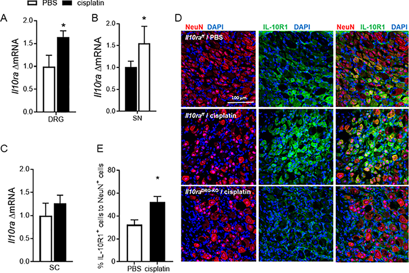
Figure 5.
Cisplatin increases IL-10R1 expression on sensory neurons A. mRNA levels of Il10ra in dorsal root ganglia (DRG) (n=8/group) unpaired t test, F(6,7) = 2.91 p=0.03, B sciatic nerve (SN) (n=8/group) unpaired t test, F(7,5) = 10.8 andC, spinal cord (SC) (n=9/group) on day 8 after start of cisplatin treatment.D, Immunofluorescence analysis of IL-10R1 on DRG neurons. Representative images of DRG sections labeled with anti-IL-10R1 (green) and anti-NeuN antibodies (red) to identify neuronal cells in control (Il10rafloxflox) and Il10DRG-KO mice on day 25 after cisplatin or PBS administration. E. Quantification of the percentage of IL-10R1- and NeuN-positive cells (n= 3F+3M/group) Unpaired t test, F (3,3) = 1.56 p=0.021. Data are shown as mean ± SEM.

