PLOS ONE logoLink to PLOS ONE
. 2021 Mar 16;16(3):e0248730. doi: 10.1371/journal.pone.0248730

Variability in digestive and respiratory tract Ace2 expression is associated with the microbiome

Sean T Koester 1,#, Naisi Li 1,#, Daniel M Lachance 1,2, Norma M Morella 1, Neelendu Dey 1,3,4,*
Editor: Jane Foster5
PMCID: PMC7963026  PMID: 33725024

Abstract

COVID-19 (coronavirus disease 2019) patients exhibiting gastrointestinal symptoms are reported to have worse prognosis. Ace2 (angiotensin-converting enzyme 2), the gene encoding the host protein to which SARS-CoV-2 spike proteins bind, is expressed in the gut and therefore may be a target for preventing or reducing severity of COVID-19. Here we test the hypothesis that Ace2 expression in the gastrointestinal and respiratory tracts is modulated by the microbiome. We used quantitative PCR to profile Ace2 expression in germ-free mice, conventional raised specific pathogen-free mice, and gnotobiotic mice colonized with different microbiota. Intestinal Ace2 expression levels were significantly higher in germ-free mice compared to conventional mice. A similar trend was observed in the respiratory tract. Intriguingly, microbiota depletion via antibiotics partially recapitulated the germ-free phenotype, suggesting potential for microbiome-mediated regulation of Ace2 expression. Variability in intestinal Ace2 expression was observed in gnotobiotic mice colonized with different microbiota, partially attributable to differences in microbiome-encoded proteases and peptidases. Together, these data suggest that the microbiome may be one modifiable factor determining COVID-19 infection risk and disease severity.

Introduction

Epidemiologic studies have reported variable rates of gastrointestinal symptoms among COVID-19 (coronavirus disease 2019) patients [1,2]. This variability may relate to disease outcomes, as gastrointestinal symptoms portend worse prognosis [1]. Although fecal-oral transmission of SARS-CoV-2 as a widespread infectious mechanism is debated, live virus has been recovered from fecal samples, suggesting the gut may serve as a viral reservoir. Ace2 (angiotensin-converting enzyme 2), the gene encoding the host protein to which SARS-CoV-2 spike proteins bind, is expressed in the gut, where it mediates amino acid transport [3]. ACE2 has been linked to the gut microbiome [4,5]; by association, the microbiome may be a target for preventing COVID-19 or mitigating severity thereof.

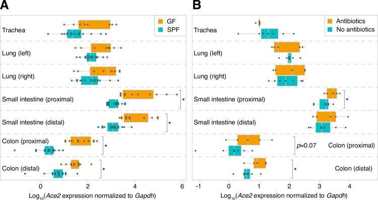
Here we tested the hypothesis that the gut microbiome modulates Ace2 expression in the gastrointestinal and respiratory tracts. We studied germ-free (GF) mice, conventional raised specific pathogen-free (SPF) mice, and gnotobiotic mice colonized with different microbiota. Small intestinal and colonic Ace2 expression levels were significantly higher in GF mice compared to conventional mice; this difference was modest in the respiratory tract. Intriguingly, antibiotic-mediated microbiota depletion in SPF mice partially rescued the phenotype seen in GF mice, suggesting a capacity for microbiome-mediated regulation of Ace2 expression. Variability in intestinal Ace2 expression was observed in gnotobiotic mice colonized with different microbiota, suggesting that population-wide differences in Ace2 expression may in part be attributable to differences in structure and function of the gut microbiome. Together, these data suggest that the microbiome may be one modifiable factor determining COVID-19 disease severity.

Materials and methods

Animal husbandry

Male and female Swiss-Webster and C57BL/6 mice were studied using protocols approved by the Institutional Animal Care and Use Committees of the University of Washington and Fred Hutchinson Cancer Research Center. Gnotobiotic mouse tissue samples were harvested as part of prior experiments unrelated to the present study. The fecal microbiota suspension (derived from fecal pellets collected from conventionally housed wild-type SPF mice) and bacterial consortia used for colonization were prepared in an anaerobic chamber. Defined bacterial consortia were prepared by first anaerobically monoculturing bacterial strains in rich growth media (as previously described [6]) to mid-log phase (optical density at 600 nm of 0.4), combining in equal volumes, and then storing in 25% glycerol at -80°C until use. Mice were colonized via oral gavage by the same individual (N.L.) in all mice in order to minimize variability. Fresh fecal pellets were snap-frozen in liquid nitrogen and stored at -80°C until use. Vancomycin (1 g/L), metronidazole (1 g/L), and neomycin (0.5 g/L) antibiotics (Millipore Sigma, St. Louis, MO) were delivered via drinking water containing 2% sucrose (20 g/L) over 10 days.

Confirmation of microbiota depletion using droplet digital PCR (ddPCR)

Fresh mouse pellets were collected, homogenized via bead-beating in a TissueLyser II (Qiagen, Hilden, Germany; 3 min at frequency 30/sec; tubes contained 0.1 mm Zirconia beads, one 4 mm steel ball, and buffer), and pelleted via centrifugation. The supernatant was diluted 1:10 and used for 16S rRNA PCR reactions (95°C for 5 min; then 40 cycles of 95°C for 30 sec, 60°C for 1 min; then 4°C for 5 min and 90°C for 5 min) prepared in triplicate using BioRad QX200™ ddPCR™ EvaGreen® SuperMix with primers 926F and 1062R [7]. Negative controls were prepared using water as a template. Positive controls were prepared using 1 ng/μl purified bacterial genomic DNA extracted from Clostridium scindens. Droplet preparation and signal acquisition was performed on the QX200™ droplet reader and analyzed using associated QuantaSoft™ software using manufacturer specifications. The threshold for background fluorescence was set using background signal from negative controls. The signal was then normalized to the original fecal pellet weights and corrected for initial dilution (S1 Fig).

RNA isolation

Respiratory and intestinal tract tissues were stored in RNAlater (Thermo Fisher Scientific Inc., Waltham, MA) at 4°C for 24 hours and then -20°C until use. 20 mg of each tissue sample was subjected to bead-beating in a TissueLyser II. RNA was purified using RNeasy Mini kits (Qiagen).

Quantitative reverse transcription PCR

Ace2 and Gapdh expression levels were quantified using the QuantiNova Probe RT-PCR kit and LNA probes (Qiagen), with input of 80 ng RNA/sample. Ace2 expression was normalized to Gapdh expression using the formula 2-ΔCt, where ΔCt is equal to the difference in mean cycle threshold (Ct) values between Ace2 and Gapdh. The relative change in Ace2 expression compared to a baseline state was calculated using the equation 2-ΔΔCt.

Quantification of bacterial genome-encoded proteases and peptidases

All bacterial species used to assemble synthetic consortia were purchased from their respective vendors (American Type Culture Collection (ATCC) or the German Collection of Microorganisms and Cell Culture (DSM)); draft genomes of these type-strains are publicly available via NCBI. The most-recent genome assemblies of all bacteria in synthetic communities were downloaded from the NCBI Prokaryotic RefSeq database and annotated using Prokka (version 1.14.5) [8]. Gene annotations that included “peptidase” or “protease” were counted, with sums represented in S2 Table.

Estimation of microbiome-encoded proteases and peptidases in a metagenomic dataset

Metagenomic sequencing data (quality-filtered and human-genome-filtered FASTQ files generated from 15 COVID-19 patients, 6 non-COVID-19 pneumonia patients, and 15 healthy controls for a total of 36 fecal samples) were downloaded from the NCBI Sequence Read Archive (accession PRJNA624223). One million reads were randomly selected from each sample and mapped using the DIAMOND aligner (diamond blastx—id 60—max-target-seqs 5—evalue .00001) to a reference database comprising 277,176 bacterial protease and peptidase sequences representing 57 gut bacterial genera that was downloaded from the MEROPS database [9] (https://www.ebi.ac.uk/merops/download_list.shtml). Total reads strongly aligning to proteases and peptidases (60% amino acid identity; E-value ≤ 10−5) were compared between COVID-19 cases and pneumonia/healthy controls, with statistical significance calculated using the Kruskal-Wallis test.

Data analysis

Statistical comparisons were performed in R (version 4.0.0). Figures were generated using R using native functions and ggplot2 (version 3.3.0), and then assembled in Adobe Illustrator.

Results

The microbiome is associated with reduced Ace2 expression

We performed quantitative reverse transcription PCR (RT-qPCR) to assess Ace2 expression levels in the trachea, left and right lungs, proximal and distal small intestine, and proximal and distal colon harvested from SPF and GF mice (n = 11-15/cohort). The variability in Ace2 expression that we observed is consistent with a prior study assessing expression in different tissues [10]. Both the microbiome and sample type were significant determinants of Ace2 expression (p<10−17, F1 = 94 [microbiome] and p<10−50, F6 = 98 [sample type], two-way ANOVA of log10-tranformed normalized Ace2 expression levels; S1 Table). Compared to SPF mice, GF mice had significantly higher levels of Ace2 throughout the small intestine and colon (Fig 1A). Similar but non-significant trends were seen in the trachea and lungs. To our knowledge, there are no prior data linking the microbiome to respiratory tract Ace2 expression. Intriguingly, SPF mice subjected to microbiota depletion via antibiotic administration for 10 days had higher intestinal Ace2 expression compared to mice that did not receive antibiotics (n = 5-7/cohort, Fig 1B). Nonetheless, Ace2 transcript levels were not as high after antibiotics as in the GF state (p = 0.08 and p = 0.04 in proximal and distal colon, respectively, two-tailed Student’s t-test). This partial recapitulation of the GF phenotype in the gut, together with the absence of a measurable effect of antibiotics in the respiratory tract, suggests that antibiotic use may not have sizable immediate effects on host Ace2 expression.

Fig 1. The microbiome is associated with reduced gastrointestinal and respiratory tract Ace2 expression.

Fig 1

A. Ace2 expression in GF and SPF mice. B. Antibiotics can modulate intestinal Ace2 expression. Statistical significance was determined using a two-tailed Student’s t-test in (A) and one-tailed Student’s t-test in (B); *, p<0.05.

Microbiota-dependent variability in intestinal Ace2 expression

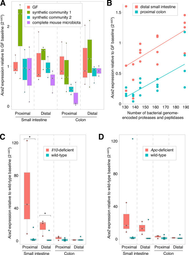
Tissues harvested as part of prior gnotobiotic experiments enabled us to assess whether Ace2 expression varies in different host and environmental contexts. Gnotobiotic mice colonized with one of two synthetic 6-member communities (S2 Table) or with a complete mouse microbiota (via fecal microbiota transplantation using SPF mouse donors) had microbiota-dependent regional variability in gut Ace2 expression (p<0.01, F3 = 5 [microbiota] and p<10−21, F3 = 246 [sample type], two-way ANOVA of log10-tranformed normalized Ace2 expression levels), with the greatest effects seen in the distal small intestine and proximal colon (Fig 2A).

Fig 2. Variability in intestinal Ace2 expression in mice.

Fig 2

Gut Ace2 expression varied in relation to (A) different microbiota, (B) microbially-encoded proteases and peptidases, (C) Il10-deficiency, and (D) Apc-deficiency. Statistical significance was determined using a two-tailed Student’s t-test; *, p<0.05.

Given ACE2’s role in intestinal amino acid transport, we postulated that gut microbial proteases and peptidases may explain this observed variation in Ace2 expression. Consistent with this notion, a prior report demonstrated that dietary supplementation of free amino acids induced greater Ace2 expression in the distal small intestine [11]. Therefore, we hypothesized that gut microbial communities with greater proteolytic and peptidase activities would proportionally induce intestinal Ace2 expression. To test this hypothesis, we quantified Ace2 expression in the distal small intestine and proximal colon in gnotobiotic mice colonized with one of five different 5-6-member bacterial consortia whose metagenomes encoded between 131 and 189 proteases and peptidases (S2 Table). Indeed, we observed a significant correlation between encoded proteases/peptidases and both small intestinal and colonic Ace2 expression (Spearman correlation rho = 0.53, p = 0.02 and rho = 0.73, p = 0.0005, respectively; Fig 2B).

We next assessed whether human gut microbiome-encoded proteases/peptidases differ between healthy individuals and COVID-19 patients. We reanalyzed recently published data generated from 15 COVID-19 patients and two sets of controls (6 patients with pneumonia and 15 healthy individuals) [12]. We did not observe a significant relationship between proportions of total reads mapping to proteases/peptidases and disease status (p>0.05, Kruskal-Wallis test).

Host and dietary factors

A mutation in Il10, which predisposes to intestinal inflammation in humans and mouse models, was associated with significantly increased small intestinal Ace2 expression in GF mice (p = 0.005, Student’s two-tailed t-test; Fig 2C). These findings are consistent with a study that found greater Ace2 tissue concentrations in patients with inflammatory bowel diseases [13]. Several variables were not significant determinants of gut or lung Ace2 expression: genetic background (C57BL/6 versus Swiss-Webster mice); Ret (gene critical to enteric nervous system development); Apc (tumor suppressor commonly mutated in colorectal cancer; Fig 2D); gender; age (comparing 9-16-week-old SPF wild-type mice); or a high-fat diet (p>0.05, one-way ANOVA tests performed for each sample tissue type for each variable; specific comparisons denoted in S1 Table).

Discussion

Here we report an association between the gut microbiome and Ace2 expression in the respiratory and gastrointestinal tracts, and a correlation between gut microbiome-encoded proteases/peptidases and intestinal Ace2 expression in mice. Although it is unclear whether the observed effect size of the microbiome is clinically meaningful, it is comparable to the effect size of cigarette smoking [14,15], a risk factor for more severe disease among individuals with COVID-19. Thus, the microbiome could theoretically also impact COVID-19 severity.

Our results suggest that gut microbial protein digestion and amino acid liberation may modulate intestinal Ace2 expression. However, our findings do not explain why GF mice, which have no bacterial proteases/peptidases, have higher Ace2 expression. One possible explanation is cytokines regulate the expression of Ace2. Higher intestinal Ace2 mRNA levels among Il10 mutant mice supports this hypothesis. In this scenario, Ace2 expression is an indirect effect of microbial depletion. Other mechanisms may explain this observation, such as lower expression of peptidase inhibitors in GF mice [16].

In an analysis of fecal metagenomic data generated from a recent study of COVID-19 patients and controls, we found that abundances of microbiome-encoded proteases/peptidases were not significantly different. This finding could potentially be confounded by variation in diet and regulators of gene expression, and our findings are limited by the small study size. In addition, fecal sampling prior to SARS-CoV-2 infection would be more informative in assessing the gut microbiome’s relationship to COVID-19 susceptibility, as Ace2 expression can change after infection. However, a caveat to this analysis is that core metabolic pathways are relatively evenly encoded in the human gut microbiome [17]; therefore, any variation in expressed proteases and peptidases that might influence host Ace2 expression is not captured in our analysis.

Additional studies are warranted to further delineate mechanisms through which the microbiome regulates Ace2 expression in the respiratory and gastrointestinal tracts. A recent study identified transcriptional factors regulating Ace2 expression in the gut [18], including Gata4, which is known to be regulated by the microbiota [19]. The partial recapitulation of the GF phenotype that we observed with antibiotics further raises the prospect of differential temporal dynamics of the relationships between the microbiome and the lung versus gut with respect to Ace2 expression. Perhaps early-life or longer-term microbial exposures are necessary for modulating Ace2 expression in the lung. Understanding the kinetics and magnitudes of these effects may have implications for antibiotic (and probiotic) use. Protein source and diet composition may be risk-modifying variables. Understanding these links could explain variability in COVID-19 severity and motivate strategies for modulating Ace2 in order to decrease susceptibility to infection (e.g. upregulating Ace2 in the elderly, in whom it is suspected that ACE2 levels become dangerously low [20]).

Supporting information

S1 Fig. Microbiota depletion verification through ddPCR.

Copies of 16S rRNA genes per μl of homogenate per mg of feces (means of the three technical replicates ± standard deviations; each dot represents a single mouse; values plotted along y-axis in log scale). Positive and negative controls similarly represent means (thick horizontal lines) ± standard deviations (pair of thin horizontal lines above and below each thick line).

(TIF)

S1 Table. Ace2 expression levels and associated metadata.

(XLSX)

S2 Table. Microbiota used in mouse studies.

(XLSX)

Data Availability

All relevant data are within the paper and its Supporting Information files.

Funding Statement

This work was supported by grants from the NIH (NIDDK K08 DK111941 and NCI Cancer Center Support Grant P30 CA015704 to ND); institutional funds from the Fred Hutchinson Cancer Research Center (Pathogen-Associated Malignancies Integrated Research Center, Microbiome Research Initiative, and donor-sponsored COVID-19 Evergreen pilot study funds to ND); and postdoctoral fellowship support from the Washington Research Foundation (NMM).

References

  • 1.Pan L, Mu M, Yang P, Sun Y, Wang R, Yan J, et al. Clinical Characteristics of COVID-19 Patients With Digestive Symptoms in Hubei, China: A Descriptive, Cross-Sectional, Multicenter Study. Am J Gastroenterol. 2020; 1. 10.14309/ajg.0000000000000620 [DOI] [PMC free article] [PubMed] [Google Scholar]
  • 2.Huang C, Wang Y, Li X, Ren L, Zhao J, Hu Y, et al. Clinical features of patients infected with 2019 novel coronavirus in Wuhan, China. The Lancet. 2020;395: 497–506. 10.1016/S0140-6736(20)30183-5 [DOI] [PMC free article] [PubMed] [Google Scholar]
  • 3.Camargo SMR, Singer D, Makrides V, Huggel K, Pos KM, Wagner CA, et al. Tissue-Specific Amino Acid Transporter Partners ACE2 and Collectrin Differentially Interact With Hartnup Mutations. Gastroenterology. 2009;136: 872–882.e3. 10.1053/j.gastro.2008.10.055 [DOI] [PMC free article] [PubMed] [Google Scholar]
  • 4.Hashimoto T, Perlot T, Rehman A, Trichereau J, Ishiguro H, Paolino M, et al. ACE2 links amino acid malnutrition to microbial ecology and intestinal inflammation. Nature. 2012;487: 477–481. 10.1038/nature11228 [DOI] [PMC free article] [PubMed] [Google Scholar]
  • 5.Yang T, Chakraborty S, Saha P, Mell B, Cheng X, Yeo J-Y, et al. Gnotobiotic Rats Reveal That Gut Microbiota Regulates Colonic mRNA of Ace2, the Receptor for SARS-CoV-2 Infectivity. Hypertension. 2020;76. 10.1161/HYPERTENSIONAHA.120.15360 [DOI] [PMC free article] [PubMed] [Google Scholar]
  • 6.Dey N, Wagner VE, Blanton LV, Cheng J, Fontana L, Haque R, et al. Regulators of Gut Motility Revealed by a Gnotobiotic Model of Diet-Microbiome Interactions Related to Travel. Cell. 2015;163: 95–107. 10.1016/j.cell.2015.08.059 [DOI] [PMC free article] [PubMed] [Google Scholar]
  • 7.Yang Y-W, Chen M-K, Yang B-Y, Huang X-J, Zhang X-R, He L-Q, et al. Use of 16S rRNA Gene-Targeted Group-Specific Primers for Real-Time PCR Analysis of Predominant Bacteria in Mouse Feces. Griffiths MW, editor. Appl Environ Microbiol. 2015;81: 6749–6756. 10.1128/AEM.01906-15 [DOI] [PMC free article] [PubMed] [Google Scholar]
  • 8.Seemann T. Prokka: rapid prokaryotic genome annotation. Bioinformatics. 2014;30: 2068–2069. 10.1093/bioinformatics/btu153 [DOI] [PubMed] [Google Scholar]
  • 9.Rawlings ND, Barrett AJ, Thomas PD, Huang X, Bateman A, Finn RD. The MEROPS database of proteolytic enzymes, their substrates and inhibitors in 2017 and a comparison with peptidases in the PANTHER database. Nucleic Acids Res. 2018;46: D624–D632. 10.1093/nar/gkx1134 [DOI] [PMC free article] [PubMed] [Google Scholar]
  • 10.Li M-Y, Li L, Zhang Y, Wang X-S. Expression of the SARS-CoV-2 cell receptor gene ACE2 in a wide variety of human tissues. Infect Dis Poverty. 2020;9: 45. 10.1186/s40249-020-00662-x [DOI] [PMC free article] [PubMed] [Google Scholar]
  • 11.Jando J, Camargo SMR, Herzog B, Verrey F. Expression and regulation of the neutral amino acid transporter B0AT1 in rat small intestine. Boudko D, editor. PLOS ONE. 2017;12: e0184845. 10.1371/journal.pone.0184845 [DOI] [PMC free article] [PubMed] [Google Scholar]
  • 12.Zuo T, Zhang F, Lui GCY, Yeoh YK, Li AYL, Zhan H, et al. Alterations in Gut Microbiota of Patients With COVID-19 During Time of Hospitalization. Gastroenterology. 2020;159: 944–955.e8. 10.1053/j.gastro.2020.05.048 [DOI] [PMC free article] [PubMed] [Google Scholar]
  • 13.Garg M, Royce SG, Tikellis C, Shallue C, Batu D, Velkoska E, et al. Imbalance of the renin–angiotensin system may contribute to inflammation and fibrosis in IBD: a novel therapeutic target? Gut. 2020;69: 841–851. 10.1136/gutjnl-2019-318512 [DOI] [PubMed] [Google Scholar]
  • 14.Saheb Sharif-Askari N, Saheb Sharif-Askari F, Alabed M, Temsah M-H, Al Heialy S, Hamid Q, et al. Airways Expression of SARS-CoV-2 Receptor, ACE2, and TMPRSS2 Is Lower in Children Than Adults and Increases with Smoking and COPD. Mol Ther—Methods Clin Dev. 2020;18: 1–6. 10.1016/j.omtm.2020.05.013 [DOI] [PMC free article] [PubMed] [Google Scholar]
  • 15.Li G, He X, Zhang L, Ran Q, Wang J, Xiong A, et al. Assessing ACE2 expression patterns in lung tissues in the pathogenesis of COVID-19. J Autoimmun. 2020;112: 102463. 10.1016/j.jaut.2020.102463 [DOI] [PMC free article] [PubMed] [Google Scholar]
  • 16.Janeckova L, Kostovcikova K, Svec J, Stastna M, Strnad H, Kolar M, et al. Unique Gene Expression Signatures in the Intestinal Mucosa and Organoids Derived from Germ-Free and Monoassociated Mice. Int J Mol Sci. 2019;20: 1581. 10.3390/ijms20071581 [DOI] [PMC free article] [PubMed] [Google Scholar]
  • 17.Turnbaugh PJ, Hamady M, Yatsunenko T, Cantarel BL, Duncan A, Ley RE, et al. A core gut microbiome in obese and lean twins. Nature. 2009;457: 480–484. 10.1038/nature07540 [DOI] [PMC free article] [PubMed] [Google Scholar]
  • 18.Chen L, Marishta A, Ellison CE, Verzi MP. Identification of Transcription Factors Regulating SARS-CoV-2 Entry Genes in the Intestine. Cell Mol Gastroenterol Hepatol. 2020. [cited 16 Oct 2020]. 10.1016/j.jcmgh.2020.08.005 [DOI] [PMC free article] [PubMed] [Google Scholar]
  • 19.Shulzhenko N, Morgun A, Hsiao W, Battle M, Yao M, Gavrilova O, et al. Crosstalk between B lymphocytes, microbiota and the intestinal epithelium governs immunity versus metabolism in the gut. Nat Med. 2011;17: 1585–1593. 10.1038/nm.2505 [DOI] [PMC free article] [PubMed] [Google Scholar]
  • 20.AlGhatrif M, Cingolani O, Lakatta EG. The Dilemma of Coronavirus Disease 2019, Aging, and Cardiovascular Disease: Insights From Cardiovascular Aging Science. JAMA Cardiol. 2020;5: 747. 10.1001/jamacardio.2020.1329 [DOI] [PMC free article] [PubMed] [Google Scholar]

Decision Letter 0

Jane Foster

Transfer Alert

This paper was transferred from another journal. As a result, its full editorial history (including decision letters, peer reviews and author responses) may not be present.

4 Feb 2021

PONE-D-20-36549

Respiratory and digestive tract variability in Ace2 expression associated with the microbiome

PLOS ONE

Dear Dr. Dey,

Thank you for submitting your manuscript to PLOS ONE. After careful consideration, we feel that it has merit but does not fully meet PLOS ONE’s publication criteria as it currently stands. Therefore, we invite you to submit a revised version of the manuscript that addresses the points raised during the review process.

Please submit your revised manuscript by Mar 19 2021 11:59PM. If you will need more time than this to complete your revisions, please reply to this message or contact the journal office at plosone@plos.org. When you're ready to submit your revision, log on to https://www.editorialmanager.com/pone/ and select the 'Submissions Needing Revision' folder to locate your manuscript file.

Please include the following items when submitting your revised manuscript:

  • A rebuttal letter that responds to each point raised by the academic editor and reviewer(s). You should upload this letter as a separate file labeled 'Response to Reviewers'.

  • A marked-up copy of your manuscript that highlights changes made to the original version. You should upload this as a separate file labeled 'Revised Manuscript with Track Changes'.

  • An unmarked version of your revised paper without tracked changes. You should upload this as a separate file labeled 'Manuscript'.

If you would like to make changes to your financial disclosure, please include your updated statement in your cover letter. Guidelines for resubmitting your figure files are available below the reviewer comments at the end of this letter.

If applicable, we recommend that you deposit your laboratory protocols in protocols.io to enhance the reproducibility of your results. Protocols.io assigns your protocol its own identifier (DOI) so that it can be cited independently in the future. For instructions see: http://journals.plos.org/plosone/s/submission-guidelines#loc-laboratory-protocols

We look forward to receiving your revised manuscript.

Kind regards,

Jane Foster, PhD

Academic Editor

PLOS ONE

Journal Requirements:

When submitting your revision, we need you to address these additional requirements.

1. Please ensure that your manuscript meets PLOS ONE's style requirements, including those for file naming. The PLOS ONE style templates can be found at

https://journals.plos.org/plosone/s/file?id=wjVg/PLOSOne_formatting_sample_main_body.pdf and

https://journals.plos.org/plosone/s/file?id=ba62/PLOSOne_formatting_sample_title_authors_affiliations.pdf

[Note: HTML markup is below. Please do not edit.]

Reviewers' comments:

Reviewer's Responses to Questions

Comments to the Author

1. Is the manuscript technically sound, and do the data support the conclusions?

The manuscript must describe a technically sound piece of scientific research with data that supports the conclusions. Experiments must have been conducted rigorously, with appropriate controls, replication, and sample sizes. The conclusions must be drawn appropriately based on the data presented.

Reviewer #1: Yes

Reviewer #2: Yes

Reviewer #3: Yes

**********

2. Has the statistical analysis been performed appropriately and rigorously?

Reviewer #1: Yes

Reviewer #2: I Don't Know

Reviewer #3: No

**********

3. Have the authors made all data underlying the findings in their manuscript fully available?

The PLOS Data policy requires authors to make all data underlying the findings described in their manuscript fully available without restriction, with rare exception (please refer to the Data Availability Statement in the manuscript PDF file). The data should be provided as part of the manuscript or its supporting information, or deposited to a public repository. For example, in addition to summary statistics, the data points behind means, medians and variance measures should be available. If there are restrictions on publicly sharing data—e.g. participant privacy or use of data from a third party—those must be specified.

Reviewer #1: Yes

Reviewer #2: Yes

Reviewer #3: Yes

**********

4. Is the manuscript presented in an intelligible fashion and written in standard English?

PLOS ONE does not copyedit accepted manuscripts, so the language in submitted articles must be clear, correct, and unambiguous. Any typographical or grammatical errors should be corrected at revision, so please note any specific errors here.

Reviewer #1: Yes

Reviewer #2: Yes

Reviewer #3: Yes

**********

5. Review Comments to the Author

Please use the space provided to explain your answers to the questions above. You may also include additional comments for the author, including concerns about dual publication, research ethics, or publication ethics. (Please upload your review as an attachment if it exceeds 20,000 characters)

Reviewer #1: Koester et al. looked into the association between GI and respiratory tract microbiome and Ace2 expression in those sites. The manuscript is well written and clearly shows that microbiota can affect Ace2 expression in different tissues. My comments on this manuscript

Title

I would suggest the title could be Respiratory and …….is associated with microbiome

Introduction section

In paragraph 2, line 6 it is unclear what the author meant to say. Do they mean antibiotics treatment in germ free mice or SPF mice improve their phenotype? Please correct this accordingly.

Reviewer #2: Dear Authors,

This is a concise paper describing experiments completed assessing the expression of Ace2, primarily among mice, related to microbiome expression. It is interesting work that has potential to contribute to the body of work related to severity of COVID-19 infections, particularly among people with gut symptoms. From the data presented, it is difficult to know if increased expression of Ace2 in the gut would be associated with (worse) gut symptoms of those infected. Conceptually it is interesting and would be interesting to follow-up and see if increased expression is associated with increased risk and/or severity of COVID-19 infection. I have some relatively minor comment that I think would improve the paper. Mostly, I think the authors should be careful not to overstate results and add a little more detail to some of the methods and results. Thank you authors for your work and interesting paper.

I think respiratory should be removed from the title, as it seems only the gut results were significant.

The last statement in the introduction may be overstating the results, related to COVID-19 infection risk. Seems that the upregulation of expression of Ace2 may modulate severity, not necessarily risk for acquiring COVID-19. There is no data here that suggests a lower expression of Ace2 in the gut, since respiratory expression changes were modest, decreases risk of acquiring the disease. Recommend changing “infection risk and disease severity” to just disease severity, unless there is data that evaluates exposure to the virus and subsequent infection related to Ace2 expression.

After antibiotic depletion of the SPF mice, were the Ace2 expression levels the same as the GF mice or were they still lower than GF mice? Follow up question (if this wasn’t done, no need to add the experiment for this paper, but would be a cool follow-up study), did you look in a group of mice that were treated with antibiotics, but waited for the antibiotics to washout to see if the change in expression persists? Conversely, perhaps when the gut microbiota reverts back, Ace2 expression also reverts to pre-antibiotic levels? This could have interesting implications for individuals that had antibiotics prescribed early in the course of COVID-19 infection.

Regarding methods, it seems that the authors only counted genes annotated as peptidase or protease. Genes present don’t always associate with expression or protein levels – this should be addressed/made clear that this is what was correlated somewhere in the results/discussion.

Why were GF and SPF mice given different microbiome standard communities? This should be explained or at least noted in the main text, particularly since the GF mice had communities with a higher number of annotated genes. Was the fact they were given difference communities accounted for in the analysis? This could confound the results.

Given the genetic background of mice seems to be unequal for GF and SPF and SPF mice appear to be older than the GF mice, the authors should include the results that support these differences did not influence the results in the supplementary data file. The supplementary file just notes which mice were include in which paragraph and reads that each variable was analyzed separately. Since they are all together in one table, it’s really difficult to validate or decipher which animals go with which experiments/tests.

This first sentence in the discussion should include “in mice” at the end, since microbiome proteases/peditases did not evaluate Ace2 expression in human samples – only disease status, and there was no significant relationship reported.

In the conclusion paragraph, I think the authors should stop after strategies for downregulating Ace2, which may decrease susceptibility. There is no data presented that suggests upregulating Ace2 would facilitate COVID-19 recovery.

In supplementary table 2, the total Metagenome-encoded proteases/peptidases for communities 1 and 2 are reversed (i.e., the total for 2 is the sum of genes for community 1 and vice versa)

Reviewer #3: The authors have performed a straightforward and important study to determine whether microbial factors regulate ACE2 expression at different tissues. They used germ-free, gnotobiotic, and specific pathogen free (SPF) mice in the presence and absence of antibiotic perturbation of the host microbiome. ACE2 expression was measured via qPCR. Their results demonstrate an association between microbial colonization and ACE2 expression in the GI tract, and suggest that perturbation to a healthy community in the gut may increase ACE2 gene expression. They observed an interesting significant correlation between peptidase/protease-encoding bacterial taxa and ACE2 expression in the gut, which may relate to the role of ACE2 in amino acid transport. Overall, this is a significant and interesting study, and I have few minor comments.

Comments.

The description of methods are scant and would benefit from more detail in reference to the FMT, metagenome, and statistical analysis. Depending on the distribution of the annotated metagenome data, one-way ANOVA may not be appropriate.

It’s not clear where the taxa within each synthetic community originate (from ATCC, but unclear if they originate from human isolates), and whether the whole genome analysis confirming peptidase-encoding genes is from the isolates, sequenced directly, or from related bacteria from a public database.

That GF mice have higher ACE2 expression is puzzling and touched on briefly in the discussion. This merits further discussion, including perhaps being regulated by cytokines that may be aberrant in GF mice due to a lack of microbiota (so, ACE2 expression as in indirect effect of microbiome depletion/perturbation). This was partially addressed using il10-deficient mice.

One interesting finding was that a perturbation to the microbiome seemed to increase ACE2 expression, at least in the gut. Some discussion on whether diseases associated with microbiota perturbations may strengthen the impact of this manuscript.

**********

6. PLOS authors have the option to publish the peer review history of their article (what does this mean?). If published, this will include your full peer review and any attached files.

If you choose “no”, your identity will remain anonymous but your review may still be made public.

Do you want your identity to be public for this peer review? For information about this choice, including consent withdrawal, please see our Privacy Policy.

Reviewer #1: Yes: Saroj Khatiwada

Reviewer #2: No

Reviewer #3: No

[NOTE: If reviewer comments were submitted as an attachment file, they will be attached to this email and accessible via the submission site. Please log into your account, locate the manuscript record, and check for the action link "View Attachments". If this link does not appear, there are no attachment files.]

While revising your submission, please upload your figure files to the Preflight Analysis and Conversion Engine (PACE) digital diagnostic tool, https://pacev2.apexcovantage.com/. PACE helps ensure that figures meet PLOS requirements. To use PACE, you must first register as a user. Registration is free. Then, login and navigate to the UPLOAD tab, where you will find detailed instructions on how to use the tool. If you encounter any issues or have any questions when using PACE, please email PLOS at figures@plos.org. Please note that Supporting Information files do not need this step.

PLoS One. 2021 Mar 16;16(3):e0248730. doi: 10.1371/journal.pone.0248730.r002

Author response to Decision Letter 0


2 Mar 2021

March 1, 2021

Jane Foster, PhD

Academic Editor

PLOS ONE

Dear Dr. Foster and colleagues,

We thank you for your thoughtful reviews and for the opportunity to improve our manuscript (PONE-D-20-36549). Our responses to the very helpful comments are below, organized by reviewer and addressed in the order of the original comments:

Reviewer #1

1) I would suggest the title could be Respiratory and …….is associated with microbiome

In order to address both this comment and comment #1 from Reviewer 2, we changed the title to, “Variability in digestive and respiratory tract Ace2 expression is associated with the microbiome.”

2) In paragraph 2, line 6 it is unclear what the author meant to say. Do they mean antibiotics treatment in germ free mice or SPF mice improve their phenotype? Please correct this accordingly.

We agree with this reviewer’s comment and modified this line in order to clarify and disambiguate: “Intriguingly, antibiotic-mediated microbiota depletion in SPF mice partially rescued the phenotype seen in GF mice, suggesting a capacity for microbiome-mediated regulation of Ace2 expression.”

Reviewer #2

1) I think respiratory should be removed from the title, as it seems only the gut results were significant.

In order to address both this comment and comment #1 from Reviewer 1, we changed the title to, “Variability in digestive and respiratory tract Ace2 expression is associated with the microbiome.” As we do not claim significance of respiratory Ace2 expression variation in the title, we hope that by leading with the word “variability,” the extent of our claims is clearer. Since the trend of higher respiratory tract Ace2 expression in germ-free mice was consistent and reproducible across multiple experiments, we feel that it is justified to include “respiratory” in the title; however, if the Editor feels that we should remove “respiratory” or edit the title otherwise, we would be happy to oblige.

2) The last statement in the introduction may be overstating the results, related to COVID-19 infection risk. Seems that the upregulation of expression of Ace2 may modulate severity, not necessarily risk for acquiring COVID-19. There is no data here that suggests a lower expression of Ace2 in the gut, since respiratory expression changes were modest, decreases risk of acquiring the disease. Recommend changing "infection risk and disease severity" to just disease severity, unless there is data that evaluates exposure to the virus and subsequent infection related to Ace2 expression.

We agree and removed “infection risk” from the final line of the introduction.

3) After antibiotic depletion of the SPF mice, were the Ace2 expression levels the same as the GF mice or were they still lower than GF mice? Follow up question (if this wasn't done, no need to add the experiment for this paper, but would be a cool follow-up study), did you look in a group of mice that were treated with antibiotics, but waited for the antibiotics to washout to see if the change in expression persists? Conversely, perhaps when the gut microbiota reverts back, Ace2 expression also reverts to pre-antibiotic levels? This could have interesting implications for individuals that had antibiotics prescribed early in the course of COVID-19 infection.

This is a really interesting comment. To answer the question, Ace2 expression levels were lower in antibiotics-treated SPF mice than in GF mice. We modified the end of the first paragraph of Results: “Intriguingly, SPF mice subjected to microbiota depletion via antibiotic administration for 10 days had higher intestinal Ace2 expression compared to mice that did not receive antibiotics (n=5-7/cohort, Fig 1B). Nonetheless, Ace2 transcript levels were not as high after antibiotics as in the GF state (p=0.08 and p=0.04 in proximal and distal colon, respectively, two-tailed Student’s t-test). This partial recapitulation of the GF phenotype in the gut, together with the absence of a measurable effect of antibiotics in the respiratory tract, suggests that antibiotic use may not have sizable immediate effects on host Ace2 expression.”

We agree the suggested follow-up studies would be quite interesting and perhaps expand the clinical implications of our study. We touch on this point in more general terms through the addition of the following sentence to the final paragraph of the Discussion: “Understanding the kinetics and magnitudes of these effects may have implications for antibiotic (and probiotic) use.”

4) Regarding methods, it seems that the authors only counted genes annotated as peptidase or protease. Genes present don't always associate with expression or protein levels – this should be addressed/made clear that this is what was correlated somewhere in the results/discussion.

We agree with this comment. We have edited the text in two areas:

- To the penultimate paragraph of the Discussion, we added, “However, a caveat to this analysis is that core metabolic pathways are relatively evenly encoded in the human gut microbiome [17]; therefore, any variation in expressed proteases and peptidases that might influence host Ace2 expression is not captured in our analysis.”

- We added the phrase “regulators of gene expression” to the second sentence of the third paragraph of Discussion, in which we describe confounders to our inability to correlate microbiome-encoded peptidases/proteases with Ace2 expression.

5) Why were GF and SPF mice given different microbiome standard communities? This should be explained or at least noted in the main text, particularly since the GF mice had communities with a higher number of annotated genes. Was the fact they were given difference communities accounted for in the analysis? This could confound the results.

We had initially only stated in Methods, “Gnotobiotic mouse tissue samples were harvested as part of prior experiments unrelated to the present study.” To clarify this point within the main text, we added the following sentence to the second paragraph of the Results section to address this comment: “Tissues harvested as part of prior gnotobiotic experiments enabled us to assess whether Ace2 expression varies in different host and environmental contexts.”

6) Given the genetic background of mice seems to be unequal for GF and SPF and SPF mice appear to be older than the GF mice, the authors should include the results that support these differences did not influence the results in the supplementary data file. The supplementary file just notes which mice were include in which paragraph and reads that each variable was analyzed separately. Since they are all together in one table, it's really difficult to validate or decipher which animals go with which experiments/tests.

We address the point that variables such as age did not influence results in the following sentence in the lone paragraph of “Host and dietary factors” (final sub-section of the Results section): “Several variables were not significant determinants of gut or lung Ace2 expression: genetic background (C57BL/6 versus Swiss-Webster mice); Ret (gene critical to enteric nervous system development); Apc (tumor suppressor commonly mutated in colorectal cancer; Figure 2D); gender; age (comparing 9-16-week-old SPF wild-type mice); or a high-fat diet (p>0.05, one-way ANOVA tests performed for each sample tissue type for each variable; specific comparisons denoted in Table S1).”

In Table S1, we re-organized the data as follows: first all wild-type mice, then mutant mice grouped by genotype; within the wild-type mice, we present GF, then SPF, then GF colonized with different microbial communities (grouped by each microbiota), then SPF mice treated with antibiotics, and finally SPF mice treated with antibiotics and re-colonized with defined communities. “MICE INCLUDED IN STATISTICAL CALCULATIONS PERFORMED IN THIS REPORT, STRATIFIED BY SPECIFIC COMPARISON” columns show an “X” in the row of each mouse included in a given comparison.

7) This first sentence in the discussion should include "in mice" at the end, since microbiome proteases/peditases [sic] did not evaluate Ace2 expression in human samples – only disease status, and there was no significant relationship reported.

We agree and added “in mice” to the end of the first sentence in the discussion.

8) In the conclusion paragraph, I think the authors should stop after strategies for downregulating Ace2, which may decrease susceptibility. There is no data presented that suggests upregulating Ace2 would facilitate COVID-19 recovery.

We have modified the final line to read as follows: “Understanding these links could explain variability in COVID-19 severity and motivate strategies for modulating Ace2 in order to decrease susceptibility to infection (e.g. upregulating Ace2 in the elderly, in whom it is suspected that ACE2 levels become dangerously low [20]).” The issue of whether downregulation or upregulation is beneficial may be age-dependent (as shown below in a Figure from AlGhatrif and colleagues (reference 20) that we have copied and pasted here, in which it is proposed that critically low ACE2 levels can render elderly individuals to more severe COVID-19 infection. As such, upregulating Ace2 expression may be helpful in elderly populations.

9) In supplementary table 2, the total Metagenome-encoded proteases/peptidases for communities 1 and 2 are reversed (i.e., the total for 2 is the sum of genes for community 1 and vice versa)

Thank you for astutely catching this error. Following review of our original analysis, we determined that during preparation of Table S2, we inadvertently swapped the numbers of genome-encoded proteases/peptidases for bacterial strains in communities 1 & 2. (The total numbers for each community are correct.) This error has been fixed accordingly.

Reviewer #3

1) The description of methods are scant and would benefit from more detail in reference to the FMT, metagenome, and statistical analysis. Depending on the distribution of the annotated metagenome data, one-way ANOVA may not be appropriate.

We have expanded upon the following sub-sections of the Methods section:

- We have added details regarding microbiota transplantation to the “Animal husbandry” sub-section describing preparation of inoculum, delivery of oral gavages, and duration of gut bacterial colonization.

- We have included additional details regarding ddPCR, and we introduce a Supplemental Figure showing these data.

- We have added details regarding genomic analysis to the “Quantification of bacterial genome-encoded proteases and peptidases” sub-section (see comment #2).

- We have added information about the sequencing data and our read alignment strategy to the “Estimation of microbiome-encoded proteases and peptidases in a metagenomic dataset” sub-section.

- We have added a sub-section to the methods called, “Data analysis,” and we have ensured that all statistical tests and results are clearly stated in the main text. In our review, we identified one instance in which the test we used (ANOVA) was not ideal for the data being analyzed, and instead we performed the Kruskal-Wallis test (penultimate paragraph of Results, immediately preceding “Host and dietary factors”).

2) It's not clear where the taxa within each synthetic community originate (from ATCC, but unclear if they originate from human isolates), and whether the whole genome analysis confirming peptidase-encoding genes is from the isolates, sequenced directly, or from related bacteria from a public database.

To clarify this point, we added the following lines to the “Quantification of bacterial genome-encoded proteases and peptidases” sub-section of Methods: “All bacterial species used to assemble synthetic consortia were purchased from their respective vendors (American Type Culture Collection (ATCC) or the German Collection of Microorganisms and Cell Culture (DSM)); draft genomes of these type-strains are publicly available via NCBI. The most-recent genome assemblies of all bacteria in synthetic communities were downloaded from the NCBI Prokaryotic RefSeq database and annotated using Prokka (version 1.14.5) [8]. Gene annotations that included “peptidase” or “protease” were counted, with sums represented in Table S2.”

3) That GF mice have higher ACE2 expression is puzzling and touched on briefly in the discussion. This merits further discussion, including perhaps being regulated by cytokines that may be aberrant in GF mice due to a lack of microbiota (so, ACE2 expression as in indirect effect of microbiome depletion/perturbation). This was partially addressed using il10-deficient mice.

We agree with this reviewer that GF mice having higher levels of Ace2 is intriguing and added the following lines to the second paragraph of the discussion to address this. “One possible explanation is cytokines regulate the expression of Ace2. Furthermore, higher intestinal Ace2 mRNA levels among Il10 mutant mice supports this hypothesis. In this scenario, Ace2 expression is an indirect effect of microbial depletion.”

4) One interesting finding was that a perturbation to the microbiome seemed to increase ACE2 expression, at least in the gut. Some discussion on whether diseases associated with microbiota perturbations may strengthen the impact of this manuscript.

Whether differences in COVID-19 severity in individuals with diseases characterized by alterations in the gut microbiota (e.g. inflammatory bowel disease, obesity, metabolic syndrome, etc) are attributable to the gut microbiota is unclear. We hesitate to include this line in the Discussion, however, as it is highly speculative.

We again thank you and the reviewers for their thorough and very helpful critiques of the paper. In addition to addressing these comments, we have reformatted the manuscript to conform to PLOS ONE style requirements.

Sincerely,

Neelendu Dey

Attachment

Submitted filename: Response_to_Reviewers.Koester_Li_et_al.v210302.docx

Decision Letter 1

Jane Foster

4 Mar 2021

Variability in digestive and respiratory tract Ace2 expression is associated with the microbiome

PONE-D-20-36549R1

Dear Dr. Dey,

We’re pleased to inform you that your manuscript has been judged scientifically suitable for publication and will be formally accepted for publication once it meets all outstanding technical requirements.

Within one week, you’ll receive an e-mail detailing the required amendments. When these have been addressed, you’ll receive a formal acceptance letter and your manuscript will be scheduled for publication.

An invoice for payment will follow shortly after the formal acceptance. To ensure an efficient process, please log into Editorial Manager at http://www.editorialmanager.com/pone/, click the 'Update My Information' link at the top of the page, and double check that your user information is up-to-date. If you have any billing related questions, please contact our Author Billing department directly at authorbilling@plos.org.

If your institution or institutions have a press office, please notify them about your upcoming paper to help maximize its impact. If they’ll be preparing press materials, please inform our press team as soon as possible -- no later than 48 hours after receiving the formal acceptance. Your manuscript will remain under strict press embargo until 2 pm Eastern Time on the date of publication. For more information, please contact onepress@plos.org.

Kind regards,

Jane Foster, PhD

Academic Editor

PLOS ONE

Acceptance letter

Jane Foster

8 Mar 2021

PONE-D-20-36549R1

Variability in digestive and respiratory tract Ace2 expression is associated with the microbiome

Dear Dr. Dey:

I'm pleased to inform you that your manuscript has been deemed suitable for publication in PLOS ONE. Congratulations! Your manuscript is now with our production department.

If your institution or institutions have a press office, please let them know about your upcoming paper now to help maximize its impact. If they'll be preparing press materials, please inform our press team within the next 48 hours. Your manuscript will remain under strict press embargo until 2 pm Eastern Time on the date of publication. For more information please contact onepress@plos.org.

If we can help with anything else, please email us at plosone@plos.org.

Thank you for submitting your work to PLOS ONE and supporting open access.

Kind regards,

PLOS ONE Editorial Office Staff

on behalf of

Dr. Jane Foster

Academic Editor

PLOS ONE

Associated Data

    This section collects any data citations, data availability statements, or supplementary materials included in this article.

    Supplementary Materials

    S1 Fig. Microbiota depletion verification through ddPCR.

    Copies of 16S rRNA genes per μl of homogenate per mg of feces (means of the three technical replicates ± standard deviations; each dot represents a single mouse; values plotted along y-axis in log scale). Positive and negative controls similarly represent means (thick horizontal lines) ± standard deviations (pair of thin horizontal lines above and below each thick line).

    (TIF)

    S1 Table. Ace2 expression levels and associated metadata.

    (XLSX)

    S2 Table. Microbiota used in mouse studies.

    (XLSX)

    Attachment

    Submitted filename: Response_to_Reviewers.Koester_Li_et_al.v210302.docx

    Data Availability Statement

    All relevant data are within the paper and its Supporting Information files.


    Articles from PLoS ONE are provided here courtesy of PLOS

    RESOURCES