. 2021 Mar 15;10:e59390. doi: 10.7554/eLife.59390

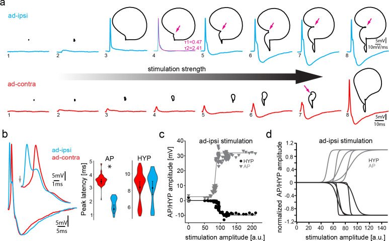
Figure 3. Antidromic stimulation activates motoneuronal hyperpolarization (HYP) that depends on stimulation amplitude.

(a) Intracellular record of antidromically activated motoneuron upon ipsi- (blue) and contralateral (red) activation with increasing stimulation amplitude (schematized by big horizontal arrow). Black lines show phase plane plots, and magenta arrows indicate additional depolarization prior to HYP onset. Magenta line in fourth trial represents exponential fit for this recording, with the constants indicated. Scale bars on top row for phase plane plots, bottom row for color traces. (b) Overlay of ipsi- and contralateral stimulation of neuron shown in (a). Violin plots show HYP and action potential (AP) peak latencies. (c) AP (gray) and HYP (black) peak amplitude of VMN neuron response upon ipsilateral antidromic stimulation of variable amplitude (arbitrary units) and corresponding sigmoid fits (color coded lines). (d) Normalized sigmoid fits of different neurons showing differences in recruitment threshold by antidromic stimulation, but similar time courses.

Figure 3—source data 1. Comparison between motoneuron action potential and hyperpolarization latency during midbrain-evoked vocal and antidromic stimulation.

Figure 3.

Figure 3—figure supplement 1. Collision experiments reveal gap junctional coupling in vocal motoneurons.
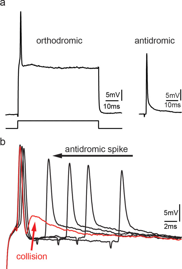
Figure 3—figure supplement 1.

(a) Action potential firing in a vocal motoneuron after intracellular current injection (left) and antidromic stimulation via the vocal nerve (right). (b) Combination of intracellular current injection and antidromic stimulation at different latencies (black arrow) reveal a depolarizing potential when action potentials collide (red trace and arrow).
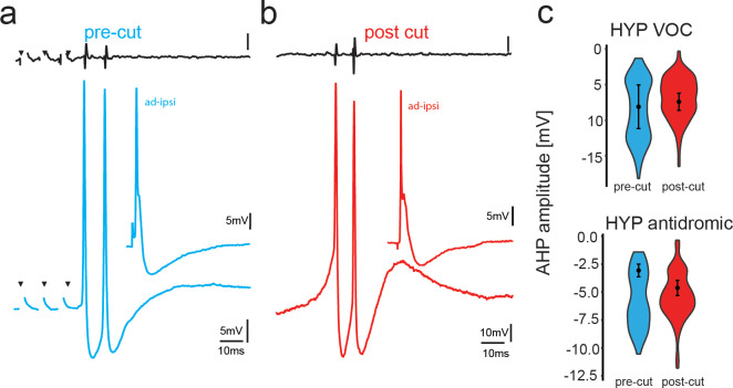
Figure 3—figure supplement 2. Potential reafferent input via the dorsal roots does not contribute to the motoneuronal hyperpolarization (HYP).

Figure 3—figure supplement 2.

(a,b) Midbrain-evoked VOC recording from vocal nerve (top trace, black) and intracellular recording in a vocal motoneuron before (blue) and in another motoneuron in the same fish after transection of the dorsal root input to the hindbrain (red). Inset shows antidromic stimulation of the respective motoneurons. (c) Box and whisker plots of hyperpolarization amplitude during vocal activity (VOC) and antidromic stimulation. Pre-cut (blue) and post-cut (red) conditions are indicated.
Figure 3—figure supplement 2—source data 1. Comparison between motoneuron action potential and hyperpolarization amplitude during baseline conditions and after cutting the dorsal roots.
Figure 3—figure supplement 3. Motoneurons are able to repetitively fire under a pulse train condition, but fail in response to a permanent current injection, revealing the necessity of a hyperpolarization for correct vocal patterning.
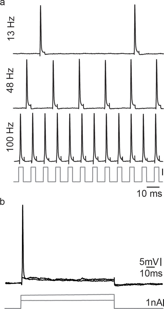
Figure 3—figure supplement 3.

(a) Action potential firing after short intracellular current pulse injection into a vocal motoneuron (at different stimulation frequencies). (b) Intracellular long-lasting current injection into the same vocal motoneuron shows the necessity of repeated pulse stimulation to repetitively elicit action potentials in vocal motoneurons.