Twenty-four patients with recurrent high-grade gliomas were scanned before and during bevacizumab treatment with standard and delayed-contrast MRI. The mean change in lesion volumes of responders (overall survival, >1 year) and nonresponders (overall survival, <1 year) was evaluated. Treatment-response-assessment maps (TRAMs) were calculated by subtracting conventional T1WI (acquired a few minutes postcontrast) from delayed T1WI (acquired with a delay of >1 hour postcontrast). These maps depict the spatial distribution of contrast accumulation and clearance. At progression, the increase in lesion volumes in delayed-contrast MR imaging was 37.5% higher than the increase in conventional T1WI. The authors conclude that the benefit of standard and delayed-contrast MRI for assessing and predicting the response to bevacizumab was demonstrated and that the increased sensitivity of delayed-contrast MRI reflects its potential contribution to the management of bevacizumab-treated patients with recurrent HGG.
Abstract
BACKGROUND AND PURPOSE:
The interpretation of the radiologic response of bevacizumab-treated patients with recurrent high-grade gliomas represents a unique challenge. Delayed-contrast MR imaging was recently introduced for calculating treatment-response-assessment maps in patients with brain tumors, providing clear separation between active tumor and treatment effects. We studied the application of standard and delayed-contrast MR imaging for assessing and predicting the response to bevacizumab.
MATERIALS AND METHODS:
Twenty-four patients with recurrent high-grade gliomas were scanned before and during bevacizumab treatment by standard and delayed-contrast MR imaging. The mean change in lesion volumes of responders (overall survival, ≥1 year) and nonresponders (overall survival, <1 year) was studied. The lesion volumes at baseline and the changes in lesion volumes 1 month after treatment initiation, calculated from standard and delayed-contrast MRIs, were studied as possible predictors of outcome. In scans acquired at progression, the average change in lesion volume from previous follow-up in standard and delayed-contrast MRIs was compared.
RESULTS:
Response and progression patterns were identified from the mean change in lesion volumes, depicted from conventional T1WI, delayed contrast-enhanced MR imaging, and DSC MR imaging. Thresholds for early prediction of response were calculated by using these sequences. For each predictor, sensitivity, specificity, positive predictive values, and negative predictive values were calculated, reaching 85.7%, 87.5%, 75%, and 93.3% for conventional T1WI; 100%, 87.5%, 77.8%, and 100% for delayed-contrast MR imaging; and 75%, 78.6%, 50%, and 91.7% for DSC MR imaging. The benefit of delayed-contrast MR imaging in separating responders and nonresponders was further confirmed by using log-rank tests (conventional T1WI, P = .0022; delayed-contrast MR imaging, P < .0001; DSC MR imaging, P = .0232) and receiver operating characteristic analyses. At progression, the increase in lesion volumes in delayed-contrast MR imaging was 37.5% higher than the increase in conventional T1WI (P < .01); these findings suggest that progression may be depicted more effectively in treatment-response-assessment maps.
CONCLUSIONS:
The benefit of contrast-enhanced MR imaging for assessing and predicting the response to bevacizumab was demonstrated. The increased sensitivity of the treatment-response-assessment maps reflects their potential contribution to the management of bevacizumab-treated patients with recurrent high-grade glioma.
Bevacizumab is an antiangiogenic drug, FDA-approved for patients with recurrent glioblastoma multiforme. Bevacizumab commonly results in prolonged progression-free survival (PFS) and faster reduction of corticosteroid treatment; however, the role of bevacizumab in overall survival (OS) remains controversial.1–4 Radiologically, bevacizumab treatment is often accompanied by a dramatic decrease in contrast enhancement due to vascular normalization, causing unique challenges in interpreting the radiographic response.5–7
Several studies have demonstrated that treatment-response criteria and changes on contrast-enhanced T1WI may serve as predictors of PFS and OS in patients treated with bevacizumab,8–12 while other studies have identified only weak relationships between imaging and OS. This difference may be explained by the difficulty in defining subtle enhancing tumor boundaries after the start of bevacizumab therapy.12 In addition, several physiologic parameters such as relative CBV and hyperperfusion volume (HPV, the fraction of contrast-enhancing volume with relative CBV above a predetermined threshold), calculated from DSC MR imaging, and ADC histogram analysis and functional diffusion maps, calculated from DWI, were also shown to be associated with outcome.13–16 The clinical utility of these physiologic imaging techniques has not yet been confirmed, and methodic concerns such as standardization of measurement parameters, artifact minimization, and improvement of spatial resolution remain unresolved.6,7
Treatment-response-assessment maps (TRAMs) were recently introduced, providing high-resolution differentiation between tumor and nontumor tissues (such as radionecrosis and pseudoprogression) in patients with high-grade gliomas (HGGs) and those with brain metastases undergoing standard treatment.17,18 TRAMs are calculated by subtracting conventional T1WI (acquired a few minutes postcontrast) from delayed T1WI (acquired with a delay of >1 hour postcontrast). These maps depict the spatial distribution of contrast accumulation and clearance.
This model-independent technique is based on robust T1WI sequences, enabling separation between active tumor (contrast clearance at the delayed time point, blue in the TRAMs) and treatment effects (contrast accumulation, red). The TRAMs were validated histologically in 51 patients having undergone resection, resulting in 100% sensitivity and 92% positive predictive value to active tumor. One explanation for the difference between the 2 populations may be found in the vessel morphology typically present in these regions17,18: In blue tumor regions, vessel lumens were viable and undamaged, while vessels in the red regions presented different stages of vessel necrosis. Here, we studied the application of standard and delayed-contrast MR imaging for assessing and predicting the response to bevacizumab.
Materials and Methods
Patients and Treatment
This prospective study was conducted after approval of the local ethics committee at Sheba Medical Center. Written informed consent was obtained from all patients.
Included were patients with recurrent HGG who failed the standard first-line therapy (maximal surgical resection, radiation therapy, and concomitant and adjuvant chemotherapy with temozolomide) and were candidates for bevacizumab, older than 18 years of age, and willing to sign the informed consent form. Exclusion criteria were World Health Organization performance status of ≤3, contraindications to undergoing MR imaging, and contraindications to bevacizumab administration.
Twenty-four patients with HGG who underwent standard chemoradiation treatment and had progressed were recruited and scanned before and periodically after the initiation of bevacizumab treatment (10 mg/kg every 14 days). Six were women, and the mean age at recruitment was 54 ± 13 years, ranging from 25 to 73 years. At recruitment, 15 patients had undergone gross total resection; 2, subtotal resections; and 7, biopsies. HGG histology included the following: 17 with World Health Organization grade IV (16 with glioblastoma multiforme; 1 gliosarcoma) and 7 grade III (3 with anaplastic astrocytomas; 3 with anaplastic oligodendroglioma; 1 with anaplastic oligoastrocytoma).
MR Imaging: Data Acquisition
Patients underwent MR imaging before treatment (following progression), 1 month posttreatment, and every 2–3 months thereafter or earlier according to their clinical condition. Patients were scanned between 2 and 8 times, up to 94 imaging sessions. All patients had a pretreatment scan, acquired 15 ± 9 days before treatment, and a 1-month follow–up scan, acquired 36 ± 9 days posttreatment.
The MRIs were acquired by using 1.5T and 3T MR imaging systems (Optima MR450w and Signa HD; GE Healthcare, Milwaukee, Wisconsin). The patients were scanned up to ∼30 minutes after contrast injection by using the hospital standard brain tumor protocol, which included DSC MR imaging, FSE T2WI, pre- and postcontrast T2-FLAIR imaging, EPI-based DWI, SWI, and high-resolution spin-echo T1WI, which were acquired before and 2 minutes 54 seconds ± 1 minute 24 seconds, on average, after contrast injection (immediately after DSC MR imaging). The patients were then taken out of the MR imaging system and were asked to return for a short scan performed 75 minutes 12 seconds ± 6 minutes 6 seconds after contrast injection, which included the same high-resolution, spin-echo T1WI sequence. T1WIs were acquired with TE = 22 ms, TR = 240 ms, FOV = 26 × 19.5 cm, section thickness of 5 mm with a 0.5-mm gap, and 512 × 512 pixels. DSC MRIs were acquired with TE = 50 ms, TR = 2000 ms, flip angle = 70°, FOV = 26 × 19.5 cm, 5/0.5-mm section thickness, and 96 × 128 pixels. A standard single dose (0.1 mmol/kg) of Gd-DOTA (Dotarem, 0.5 mmol/mL; Guerbet, Aulnay-sous-Bois, France) was injected intravenously by using an automatic injection system 6 seconds after starting DSC MR imaging.
MR Imaging: Data Analysis
All image analysis was performed by using Matlab (Version R2010a; MathWorks, Natick, Massachusetts).
The TRAMs were calculated as previously described,17,18 and several parameters were calculated from conventional and delayed-contrast MR imaging, as defined below:
Enhancing volume calculated from contrast-enhanced T1WI (T1GdV)
FLAIR hyperintense volume calculated from precontrast FLAIR MRI (FLAIRV)
Blue volume calculated from the TRAMs (BlueV), representing efficient clearance of contrast from the tissue
Hyperperfused volume calculated from DSC-MRI (HPV)
Mean ADC value calculated from DWI (mean ADC).
T1GdVs, FLAIRVs, and BlueVs were calculated by using a semiautomatic segmentation algorithm. A detailed description of this algorithm and the calculations of HPV and mean ADC are presented in On-line Appendix A.
Assessment of Progression
For each follow-up scan, radiologic outcome was assessed from the change in T1GdVs and FLAIRVs from the previous scan, by using the same thresholds for 2D-T1WI prescribed by the Response Assessment in Neuro-Oncology (RANO) group guidelines.5 In short, a decrease of ≥50% in T1GdV with stable or reduced FLAIRV was considered a response; an increase of ≥25% in T1GdV or an increase of >25% in FLAIRV, not attributed to other causes, or the appearance of any new lesion was considered progression. PFS was calculated as the time difference between initiation of bevacizumab treatment and the acquisition of the first MR imaging scan indicating progression.
Separating Responders and Nonresponders
Patients with an OS of ≥1 year were considered responders, and those with an OS of ≤1 year, nonresponders.
Response Patterns
In an attempt to identify reliable imaging parameters for early assessment of response and nonresponse to bevacizumab, the mean values of the changes in lesion volumes were plotted separately for responders and nonresponders as a function of time for the following parameters: T1GdVs, FLAIRVs, BlueVs, HPVs, and mean ADCs. The separation between responders and nonresponders was studied for the different parameters at the 1-month follow-up.
Early Assessment of Response to Bevacizumab Treatment
To determine the threshold for each predictor, we plotted the logarithmic values of patients' PFS as a function of the logarithmic values of the change (ratio) in lesion volume after 1 month of treatment for T1GdVs, BlueVs, and HPVs. A linear function was fitted to each plot (log-log plots of PFS versus volume change) and thresholds for differentiating responders and nonresponders were determined for each of the 3 parameters by calculating the change (ratio) in lesion volume corresponding to a PFS of 6 months.
To establish the validity of these predictors for early (1-month) assessment of response to bevacizumab, we divided the patients into 2 groups defined by the thresholds of each of the 3 predictors determined above (T1GdV, BlueV, and HPV): Responders were below the threshold; nonresponders, above it. The median OS of the groups determined by these thresholds was calculated and compared by using log-rank analysis.
Receiver operating characteristic analysis was performed for comparing the ability of the 1-month change in T1GdV, BlueV, and HPV to aid early prediction of PFS and OS, by using the area under the curve as a measure of performance.
Statistical Analysis
Statistical analysis was performed by using GraphPad InStat (Version 3.05; GraphPad Software, San Diego, California).
The median OS of the responders and nonresponders was calculated and compared by using log-rank analysis. Comparison of the unpaired differences between responders and nonresponders and comparison of the baseline radiologic parameters of the responders with those of the nonresponders were performed by using an unpaired t test with a Welch correction. The correlation between patients' PFS and OS was studied by using linear regression.
In all follow-up scans for which progression was determined, the change (ratio) in T1GdVs since the previous follow-up was compared with that of BlueVs and HPVs by using Wilcoxon matched-pairs signed ranks. This method was also used to compare FLAIRVs and mean ADC values before and after treatment.
In all analyses, P < .05 was considered a significant difference.
Results
Separating Responders and Nonresponders
Seven of the 24 recruited patients (29.2%, of which 57.1% were grade IV; 42.9%, grade III; 28.6% underwent biopsy; 14.3%, subtotal resection; 57.1%, gross total resection) were responders, and 17 (70.8%, of which 76.5% were grade IV; 23.5%, grade III; 29.4% underwent biopsy; 11.8%, subtotal resection; 58.8%, gross total resection) were nonresponders.
The median PFS of all patients was 3.5 months (95% CI, 2.3–7.6 months). Eight of the 24 patients had a PFS of ≥6 months, and 16 had a PFS of <6 months. The median OS of all patients was 9.2 months (95% CI, 8.2–11.6 months). The median OS of the responders was significantly higher than that of the nonresponders: 24.1 months (95% CI, 14.8–34.4 months) versus 8.7 months (95% CI, 5.4–9.2 months) (P < .0001). Significant correlation was found between patients' PFS and OS (r2 = 0.94, P < .0001). Seven of the 8 patients with a PFS of ≥6 months reached an OS of ≥1 year (3 were alive at the time of analysis), and all patients with a PFS of <6 months did not.
At 1 month, the response was determined in 15 (62.5%) patients. However, 8 of them showed only short-term benefit (OS of <1 year, with a median OS of 6.9 months; 95% CI, 5.2–11.4 months), while only 7, as mentioned above (less than half of the initially responding patients), showed a long-term response.
Treatment Outcome is Independent of Pretreatment Radiologic Markers
No statistically significant differences were found between any of the baseline parameters of the responding and nonresponding patients: T1GdV: 32.9 ± 13.4/29.4 ± 4.6 mL, P = .81; BlueVs: 17.3 ± 7.7/15.1 ± 2.2 mL, P = .80; FLAIRVs: 156.5 ± 34.3/127.7 ± 15.0 mL, P = .46; HPV: 14.2 ± 5.1/12.6 ± 2.2 mL, P = .79; mean ADCs: 9.8 ± 1.0/1.1 ± 0.6 × 10⁻³ mm2/s, P = .57.
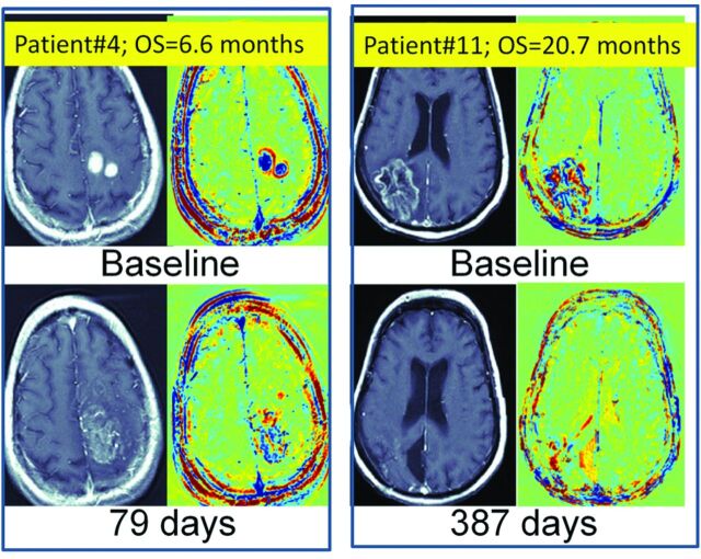
Examples demonstrating that the response to bevacizumab is independent of pretreatment tumor volumes are given in Fig 1.
Fig 1.
Examples demonstrating that the response to bevacizumab is independent of pretreatment tumor volumes: Shown are axial contrast-enhanced T1-weighted MRIs and TRAMs calculated 5 days before bevacizumab treatment and 79 days post-initiation of treatment of a nonresponding patient (left) with a relatively small pretreatment tumor. Also shown are contrast-enhanced T1-weighted MRIs and TRAMs calculated 7 days before bevacizumab treatment and 387 days post-initiation of treatment of a responding patient (right) with a relatively large pretreatment tumor. One month after the initiation of treatment, the nonresponding patient's tumor volumes increased (blue up to 131% of initial volume, and T1, up to 103%). Despite the large initial tumor volume, the responding patient showed significant reduction in tumor volume (blue down to 21% of initial volume, and T1, down to 25%), which remained low for >15 months posttherapy. Both patients showed decreased FLAIR volumes.
Response Patterns
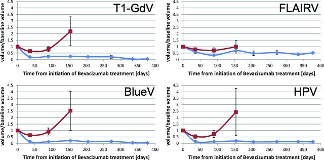
Responders showed a significant decrease in lesion volumes at the 1-month follow-up (T1GdVs: decrease to 20.1% ± 3.8% of baseline volume; BlueVs: 13.7% ± 2.3%; HPVs: 15.1% ± 5.5%), followed by decreased and stable lesion volumes on the next follow-ups. Nonresponders showed a smaller decrease at the 1-month follow-up (T1GdVs: 64.7% ± 9.3%; BlueVs: 63.3% ± 9.4%; HPVs: 53.4% ± 13.4%), followed by a significant increase in the next follow-ups (Fig 2).
Fig 2.
Response patterns. The mean values of the changes in lesion volumes relative to baseline were plotted separately for the responders (blue) and nonresponders (red) as a function of time (days) after initiation of bevacizumab treatment for the following parameters: enhancing volumes on contrast-enhanced T1-weighted MR imaging, FLAIR hyperintensity volumes on precontrast FLAIR, blue volumes on the TRAMs, and hyperperfused volumes on perfusion-weighted MR imaging–based maps.
The separation between responders and nonresponders was studied at the 1-month follow-up and was found significant for the following: T1GdVs: P = .0003; BlueVs: P < .0001; HPVs: P = .017.
When we compared FLAIRVs and mean ADCs before and after 1 month of treatment for all patients (responders and nonresponders), the decrease after the initiation of treatment was significant (FLAIRVs: P = .001; mean ADCs: P = .0002), but there was no significant difference between responders and nonresponders (FLAIRVs: P = .29 and ADCs: P = .49).
Early Assessment of Response to Bevacizumab Treatment
When plotted on a log-log scale, PFS showed significant linear correlation with the change in lesion volume for all 3 predictors: The correlation coefficient of BlueV (r2 = 0.80; P < .0001) was found to be higher than that of T1GdV (r2 = 0.58; P = .0002), and HPV (r2 = 0.55; P = .0015).
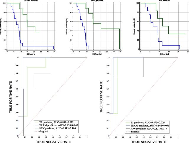
The thresholds for differentiating responders and nonresponders calculated from this fit analysis were 0.32 for T1GdV, 0.29 for BlueV, and 0.19 for HPV. Accordingly, if the change in lesion volume 1 month after initiation of bevacizumab is lower than these thresholds (for example, if T1GdV decreases to below 29% of its pretreatment volume, ie, < 0.29, the patient is predicted to be a responder; if the change is above these thresholds, the patient is predicted to be a nonresponder. The Table summarizes the median OS of the groups determined by these thresholds and the P values resulting from log-rank analysis. Kaplan-Maier curves are presented in Fig 3.
Log-rank analysis of suggested predictorsa
| Method | Median PFS (95% CI) (mo) (Responders/Nonresponders) | P Value (Log-Rank Analysis) | Median OS (95% CI) (mo) (Responders/Nonresponders) | P Value (Log-Rank Analysis) |
|---|---|---|---|---|
| T1GdV | 11.3 (5.6–19.6) | .0112 | 14.8 (11.6–20.7) | .0022 |
| 2.8 (1.8–4.5) | 8.2 (5.4–9.2) | |||
| BlueV | 15.4 (9.1–31.4) | <.0001 | 20.7 (14.6–34.4) | <.0001 |
| 2.8 (1.3–3.5) | 6.9 (5.2–9.2) | |||
| HPV | 5.6 (3.3–9.1) | .0837 | 11.6 (9.0–14.8) | .0232 |
| 2.8 (1.8–4.5) | 6.6 (5.2–9.2) |
The patients were divided into responders and nonresponders using predictors from T1GdV, BlueV, and HPV. The median PFS and OS values of the responders and nonresponders as determined by these thresholds are presented and compared with log-rank analysis.
Fig 3.
Kaplan-Maier curves demonstrating the application of 3 MR imaging–based predictors for separating responders to bevacizumab from nonresponders. Shown are Kaplan-Maier curves by using 1-month change (ratio) in T1GdV ≤ 0.32 (upper left), BlueV ≤ 0.29 (upper middle), and HPV ≤ 0.19 (upper right) as predictors for OS. Receiver operating characteristic curves of the 3 predictors applied for prediction of 6-month PFS (left) and 1-year OS (right) are presented below.
Using the thresholds determined from PFS values, we calculated the sensitivity, specificity, positive predictive values, and negative predictive values to the response (OS of ≥1 year) for each of the 3 predictors reaching 100%, 87.5%, 77.8%, and 100% for TRAMs; 85.7%, 87.5%, 75%, and 93.3% for contrast-enhanced MR imaging; and 75%, 78.6%, 50%, and 91.7% for DSC MRI.
Receiver operating characteristic analysis applied to predefined clinical end points of PFS at ≥6 months and OS at ≥1 year demonstrated the added value of the TRAMs (Fig 3): For prediction of PFS, the areas under the curve were the following: 0.831 ± 0.099, P = .03 for T1GdV; 0.938 ± 0.062, P = .005 for BlueV; and 0.815 ± 0.106, P = .04 for HPV. For OS of ≥1 year, the areas under the curve were the following: 0.893 ± 0.079, P = .02 for T1GdV; 0.946 ± 0.056, P = .008 for BlueV; and 0.821 ± 0.119, P = .06 for HPV.
Sensitivity to Progression
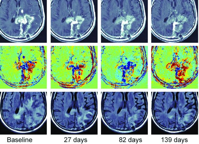
Progression was determined in 13 of the 94 imaging sessions. When we compared the change in T1GdVs relative to the previous follow-up with that of the BlueVs, the increase in BlueVs was found to be 37.5% higher than the increase in T1GdVs (95% CI, 6%–81%, P = .013), suggesting that progression may be depicted more effectively in the TRAMs. An example is shown in Fig 4.
Fig 4.
An example of a patient progressing under bevacizumab and responding to re-irradiation under bevacizumab: Shown are axial contrast-enhanced T1WI (upper row), TRAMs (middle row), and T2 FLAIR images (lower row), at baseline, 27 days, 82 days, and 139 days after initiation of bevacizumab treatment. Lesion volumes at baseline were the following: enhancing T1: 54.1 mL; blue: 21.2 mL; and FLAIR hyperintensity: 165.9 mL. At 1 month, a reduction was seen in all: T1 reached 59% of its baseline volume; blue, 79%; and FLAIR, 43%. At day 83, progression was determined, consistent with the patient's clinical deterioration. T1 reached 84% of its baseline volume (but an increase to 142% relative to the previous scan volume), and blue reached 140% of its baseline volume (an increase to 177% relative to the previous scan), reflecting the stronger sensitivity of TRAMs to progression. FLAIR hyperintensity continued to decrease (25%). At this point, it was decided that the patient should undergo re-irradiation and continue bevacizumab treatment. The next follow-up, at day 139, was acquired 1 month after the initiation of re-radiation. Compared with the previous examination, there was a dramatic increase in FLAIR (172%), as can be expected postirradiation; no change in T1 (99.7%); and a dramatic decrease in blue (61%). In addition, hyperperfused volume increased to 132%; and average apparent diffusion coefficient, to 108% (data not shown). The patient was clinically stable after re-irradiation and lived 5 more months.
When we compared the change in T1GdVs relative to the previous follow-up with that of the HPVs, the difference in the increase was not significant (P = .91).
Comparison of the TRAMs with conventional MR imaging can be found in On-line Appendix B. The effects of re-irradiation during bevacizumab treatment can be found in On-line Appendix C.
Discussion
The application of the TRAMs for differentiating tumor and nontumor tissues in patients with brain tumor following conventional treatment was recently demonstrated and validated histologically.17,18 Unlike other methods (such as PWI), the TRAMs present a model-independent approach with minimal sensitivity to susceptibility artifacts. We applied the TRAMs to monitor 24 patients before and during bevacizumab treatment. The primary end point was to assess whether the TRAMs provide additional information regarding a recurrent HGG response to bevacizumab treatment over conventional MR imaging. Response and progression patterns were identified from the mean change in lesion volumes with time, depicted from conventional T1WI, delayed contrast-enhanced MR imaging, and DSC MR imaging. Thresholds for early (1 month) prediction of response were calculated by using these sequences. The predictor calculated from the TRAMs demonstrated higher sensitivity, specificity, and positive and negative predictive values. The benefit of delayed-contrast MR imaging in separating responders and nonresponders was further confirmed by using log-rank and receiver operating characteristic analyses, showing improved performance as measured by the area under the curve for prediction of PFS at ≥6 months compared with T1GdV and HPV. For prediction of OS at ≥1 year, both BlueV and T1GdV seem to be strong predictors. At progression, the increase in lesion volumes in delayed-contrast MR imaging was significantly higher than the increase in conventional T1WI; these findings suggest that progression may be better depicted in the TRAMs.
Despite 62.5% of the recruited patients showing a positive radiologic response to bevacizumab at the 1-month standard MR imaging, only 29.2% demonstrated a long-term response. These numbers suggest that a more reliable tool for early prediction of long-term response to bevacizumab is required. When studying the response pattern to bevacizumab, we noted that responders presented an initial sharp decrease in tumor volume, which persisted for a prolonged time. Although an initial decrease in tumor volume was also evident in the nonresponding group, it was significantly less and was followed by significant growth, occurring ∼3 months after initiation of treatment, signifying tumor progression. This difference between responders and nonresponders depicted in Fig 2 for T1GdVs, BlueVs, and HPVs suggested that these parameters may be strong predictors of long-term response.
It is claimed19 that bevacizumab may reduce the enhancing volumes in patients with recurrent HGG by reducing treatment effects and not necessarily by antitumor effects. Here, the ability of the TRAMs to differentiate tumor and treatment effects pretreatment is applied to demonstrate the antitumor effects of bevacizumab. All 7 responders had significant BlueVs in the pretreatment TRAMs. This finding, together with significant correlations between the reduction in BlueVs and outcome, suggests that bevacizumab not only reduces the treatment effects, but induces antitumor effects. Most interesting, the response to bevacizumab showed no correlation with initial tumor volumes or any other baseline radiologic parameter.
The estimated median OS in our study (9.2 months) is in agreement with that in previous publications.2,8 Specifically, the Bevacizumab and Irinotecan or Temozolomide in Treating Patients With Recurrent or Refractory Glioblastoma Multiforme or Gliosarcoma (ACRIN/RTOG) study presented a similar OS of 270 days for 123 patients with recurrent HGG treated with bevacizumab.8 Similarly, the authors applied contrast-enhanced T1 as a predictor for OS. However, they used the RANO cutoff as a threshold to demonstrate the prognostic value of early radiologic progression in OS, while we derived a threshold by using PFS data, which are based on the extent of early response. The percentage of early progression at 1-month follow-ups in our data (12.5%) is also consistent with the early progression rate seen at 8 weeks in their study (12%). However, our predictors separated responders and nonresponders efficiently at 1 month, while in the ACRIN/RTOG results, the difference in median OS between initially responding patients and those with stable disease (by using the RANO cutoff at 50%) was not found to be statistically significant.
We noted that the mean ADC of all patients decreased slightly (but statistically significantly) posttreatment. This decrease may be explained by the reduction of extracellular fluids after vessel wall normalization. Still, our ADC data did not provide predictive information regarding response and nonresponse to bevacizumab.
A significant disadvantage of the TRAMs is their inability to depict nonenhancing tumor components. In our experience, images in most patients depicted a decrease in tumor enhancement during bevacizumab treatment, which may be due to blood vessel normalization; however, vessel normalization does not seem complete because most cases had some level of contrast leakage, enabling us to study its late clearance and accumulation by using the TRAMs. The low number of responding patients (n = 7) requires additional studies to establish the results demonstrated here. We also assumed that similar to standard treatments, blue in the TRAMs represents active tumor, while red represents nontumor tissues, considering that nonenhancing regions may consist of additional tumor tissues. Histologic validation of these assumptions is yet to be performed.
Generally, PFS is considered a good surrogate for OS. However, in the case of recurrent HGG treated by bevacizumab, determination of radiologic progression is challenging and 6-month PFS values may not be reliable. In this study, response was determined by using OS, also a reliable measure of clinical outcome in the case of bevacizumab treatment, and predictors of response were confirmed to provide significant separation in OS by log-rank analysis. Moreover, the calculated PFS by using the method described here was significantly correlated with OS.
The studied cohort of patients was heterogeneous with respect to histology, previous resections, and re-irradiation treatment received after failure of bevacizumab, which can potentially give rise to confounding results. However, the median OS of patients with grades III and IV was not found to be statistically different, and the percentages of patients previously undergoing gross total resection and subtotal resection and biopsy in the responding and nonresponding groups were similar; this finding suggests no significant bias due to these considerations. The 7 patients who underwent re-irradiation were all from the nonresponding group, suggesting no significant bias toward the response due to this difference as well.
Conclusions
The benefit of standard and delayed-contrast MR imaging for assessing and predicting the response to bevacizumab was demonstrated. The increased sensitivity of delayed-contrast MR imaging reflects its potential contribution to the management of bevacizumab-treated patients with recurrent HGG.
Supplementary Material
ABBREVIATIONS:
- BlueV
blue volume calculated from the TRAMs
- FLAIRV
FLAIR hyperintense volume calculated from precontrast FLAIR MRI
- HGG
high-grade glioma
- HPV
hyperperfusion volume calculated from DSC MRI
- OS
overall survival
- PFS
progression-free survival
- RANO
Response Assessment in Neuro-Oncology
- T1GdV
enhancing volume calculated from contrast-enhanced T1WI
- TRAM
treatment-response-assessment map
Footnotes
Disclosures: Dianne Daniels—RELATED: Grant: Joseph Sagol PhD Scholarship*; UNRELATED: Patents (planned, pending or issued): I am an inventor on pending patents, some licensed to Brainlab AG. Deborah T. Blumenthal—UNRELATED: Board Membership: Vascular Biogenics (Medical Advisory Board). Yael Mardor—RELATED: Grant: from Brainlab AG,* donation from Roche Pharmaceuticals*; Consulting Fee or Honorarium: Brainlab AG; Support for Travel to Meetings for the Study or Other Purposes: Brainlab AG; UNRELATED: Consultancy: Brainlab AG; Grants/Grants Pending: Israel Science Foundation and Israeli Office of Commerce; Patents (planned, pending or issued): I am an inventor on pending patents, some licensed to Brainlab AG; Travel/Accommodations/Meeting Expenses Unrelated to Activities Listed: Brainlab AG. David Guez—UNRELATED: Patents (planned, pending or issued): I am an inventor on pending patents, some licensed to Brainlab AG. David Last—UNRELATED: Patents (planned, pending or issued): I am an inventor on pending patents, some licensed to Brainlab AG. Leor Zach—UNRELATED: Patents (planned, pending or issued): I am an inventor on pending patents, some licensed to Brainlab AG. *Money paid to the institution.
This work was performed in partial fulfillment of the requirements for a PhD degree by Dianne Daniels at the Sackler Faculty of Medicine, Tel Aviv University, Israel.
This work was supported by a generous donation from Roche Pharmaceuticals, the Joseph Sagol PhD Scholarship for Dianne Daniels, and a research grant from Brainlab AG.
Paper previously presented, in part or whole, at: Annual Meeting of the European Association of Neuro-oncology, October 9–12, 2014; Turin, Italy; Annual Meeting of the American Society of Functional Neuroradiology, March 18–20, 2015; Tucson, Arizona; and Annual Meeting of the American Society for Radiation Oncology, October 18–21, 2015; San Antonio, Texas.
References
- 1. Friedman HS, Prados MD, Wen PY, et al. Bevacizumab alone and in combination with irinotecan in recurrent glioblastoma. J Clin Oncol 2009;27:4733–40 10.1200/JCO.2008.19.8721 [DOI] [PubMed] [Google Scholar]
- 2. Chamberlain MC. Bevacizumab for the treatment of recurrent glioblastoma. Clin Med Insights Oncol 2011;5:117–29 10.4137/CMO.S7232 [DOI] [PMC free article] [PubMed] [Google Scholar]
- 3. Le Rhun E, Rhun EL, Taillibert S, et al. The future of high-grade glioma: where we are and where are we going. Surg Neurol Int 2015;6(suppl 1):S9–S44 10.4103/2152-7806.153100 [DOI] [PMC free article] [PubMed] [Google Scholar]
- 4. Khasraw M, Ameratunga MS, Grant R, et al. Antiangiogenic therapy for high-grade glioma. Cochrane Database Syst Rev 2014;9:CD008218 10.1002/14651858.CD008218.pub3 [DOI] [PubMed] [Google Scholar]
- 5. Wen PY, Macdonald DR, Reardon DA, et al. Updated response assessment criteria for high-grade gliomas: Response Assessment in Neuro-Oncology Working Group. J Clin Oncol 2010;28:1963–72 10.1200/JCO.2009.26.3541 [DOI] [PubMed] [Google Scholar]
- 6. Pope WB, Young JR, Ellingson BM. Advances in MRI assessment of gliomas and response to anti-VEGF therapy. Curr Neurol Neurosci Rep 2011;11:336–44 10.1007/s11910-011-0179-x [DOI] [PMC free article] [PubMed] [Google Scholar]
- 7. Hygino da Cruz LC Jr, Rodriguez I, Domingues RC, et al. Pseudoprogression and pseudoresponse: imaging challenges in the assessment of posttreatment glioma. AJNR Am J Neuroradiol 2011;32:1978–85 10.3174/ajnr.A2397 [DOI] [PMC free article] [PubMed] [Google Scholar]
- 8. Boxerman JL, Zhang Z, Safriel Y, et al. Early post-bevacizumab progression on contrast-enhanced MRI as a prognostic marker for overall survival in recurrent glioblastoma: results from the ACRIN 6677/RTOG 0625 Central Reader Study. Neuro Oncol 2013;15:945–54 10.1093/neuonc/not049 [DOI] [PMC free article] [PubMed] [Google Scholar]
- 9. Prados M, Cloughesy T, Samant M, et al. Response as a predictor of survival in patients with recurrent glioblastoma treated with bevacizumab. Neuro Oncol 2011;13:143–51 10.1093/neuonc/noq151 [DOI] [PMC free article] [PubMed] [Google Scholar]
- 10. Ellingson BM, Cloughesy TF, Lai A, et al. Quantitative volumetric analysis of conventional MRI response in recurrent glioblastoma treated with bevacizumab. Neuro Oncol 2011;13:401–09 10.1093/neuonc/noq206 [DOI] [PMC free article] [PubMed] [Google Scholar]
- 11. Huang RY, Rahman R, Hamdan A, et al. Recurrent glioblastoma: volumetric assessment and stratification of patient survival with early posttreatment magnetic resonance imaging in patients treated with bevacizumab. Cancer 2013;119:3479–88 10.1002/cncr.28210 [DOI] [PubMed] [Google Scholar]
- 12. Ellingson BM, Kim HJ, Woodworth DC, et al. Recurrent glioblastoma treated with bevacizumab: contrast enhanced T1-weighted subtraction maps improve tumor delineation and aid prediction of survival in a multicenter clinical trial. Radiology 2014;271:200–10 10.1148/radiol.13131305 [DOI] [PMC free article] [PubMed] [Google Scholar]
- 13. Schmainda KM, Prah M, Connelly J, et al. Dynamic-susceptibility contrast agent MRI measures of relative cerebral blood volume predict response to bevacizumab in recurrent high-grade glioma. Neuro Oncol 2014;16:880–88 10.1093/neuonc/not216 [DOI] [PMC free article] [PubMed] [Google Scholar]
- 14. Aquino D, Di Stefano AL, Scotti A, et al. Parametric response maps of perfusion MRI may identify recurrent glioblastomas responsive to bevacizumab and irinotecan. PLoS One 2014;9:e90535 10.1371/journal.pone.0090535 [DOI] [PMC free article] [PubMed] [Google Scholar]
- 15. Sawlani RN, Raizer J, Horowitz SW, et al. Glioblastoma: a method for predicting response to antiangiogenic chemotherapy by using MR perfusion imaging: pilot study. Radiology 2010;255:622–28 10.1148/radiol.10091341 [DOI] [PMC free article] [PubMed] [Google Scholar]
- 16. Ellingson BM, Sahebjam S, Kim HJ, et al. Pretreatment ADC histogram analysis is a predictive imaging biomarker for bevacizumab treatment but not chemotherapy in recurrent glioblastoma. AJNR Am J Neuroradiol 2014;35:673–79 10.3174/ajnr.A3748 [DOI] [PMC free article] [PubMed] [Google Scholar]
- 17. Zach L, Guez D, Last D, et al. Delayed contrast extravasation MRI: a new paradigm in neuro-oncology. Neuro Oncol 2015;17:457–65 10.1093/neuonc/nou230 [DOI] [PMC free article] [PubMed] [Google Scholar]
- 18. Zach L, Guez D, Last D, et al. Delayed contrast extravasation MRI for depicting tumor and non-tumoral tissues in primary and metastatic brain tumors. PLoS One 2012;7:e52008 10.1371/journal.pone.0052008 [DOI] [PMC free article] [PubMed] [Google Scholar]
- 19. Taal W, Oosterkamp HM, Walenkamp AM, et al. Single-agent bevacizumab or lomustine versus a combination of bevacizumab plus lomustine in patients with recurrent glioblastoma (BELOB trial): a randomised controlled phase 2 trial. Lancet Oncol 2014;15:943–53 10.1016/S1470-2045(14)70314-6 [DOI] [PubMed] [Google Scholar]
Associated Data
This section collects any data citations, data availability statements, or supplementary materials included in this article.


